 Movie
Movie Controller

[English] 日本語
 Yorodumi
- EMDB-64873: The transition-state structure of the Argonaute protein from a Ve... -
+ Open data

- Basic information
| Entry |  | |||||||||
|---|---|---|---|---|---|---|---|---|---|---|
| Title | The transition-state structure of the Argonaute protein from a Verrucomicrobia bacterium in complex with guide DNA and target RNA. | |||||||||
 Map data Map data | ||||||||||
 Sample Sample |
| |||||||||
 Keywords Keywords | mesophilic prokaryotic Argonaute / RNA BINDING PROTEIN/DNA/RNA / RNA BINDING PROTEIN-DNA-RNA complex | |||||||||
| Biological species |  Verrucomicrobiota bacterium (bacteria) Verrucomicrobiota bacterium (bacteria) | |||||||||
| Method | single particle reconstruction / cryo EM / Resolution: 2.93 Å | |||||||||
 Authors Authors | Wu S | |||||||||
| Funding support |  China, 1 items China, 1 items
| |||||||||
 Citation Citation | Journal: Nat Commun / Year: 2026 Title: Structural insights into C-terminus-mediated RNA target cleavage by a mesophilic prokaryotic argonaute. Authors: Taiyu Chen / Xin Tao / Shunshun Li / Qingmiao Duanmu / Jiening Wang / Kai Chen / Kangle Mu / Hong Yang / Yu Li / Yang Liu / Lixin Ma / Shan Wu / Abstract: Prokaryotic Argonaute proteins (pAgos) are programmable nucleases that always utilize DNA guides to cleave DNA targets. Recent studies show that some pAgos preferentially utilize DNA guides to cleave ...Prokaryotic Argonaute proteins (pAgos) are programmable nucleases that always utilize DNA guides to cleave DNA targets. Recent studies show that some pAgos preferentially utilize DNA guides to cleave RNA targets rather than DNA targets. VbAgo, derived from a Verrucomicrobia bacterium, is a nuclease capable of specifically cleaving single-stranded RNA and highly structured RNA substrates at 37 °C, making it an ideal candidate for developing RNA manipulation toolkits. An in-depth investigation of its mechanism contributes to understanding the functional characteristics of gDtR-type Ago proteins. Here, we present cryo-electron microscopy structures of VbAgo, the VbAgo-guide DNA binary complex, multiple wild-type VbAgo-guide DNA-target RNA ternary complexes, and the catalytically inactive mutant (VbAgo-DM) guide DNA-target RNA ternary complex, with resolutions ranging from 2.5 to 3.2 Å. By integrating these cryo-EM structures with biochemical data, we elucidate the entire catalytic process of VbAgo, revealing its unique C-terminal regulatory mechanism. Specifically, in its apo state, VbAgo's C-terminus occupies the nucleic acid binding channel, partially impeding its catalytic activity while enhancing its stability. The binding of guide DNA displaces the C-terminus, and subsequent binding of target RNA, along with conformational changes in the N-terminal and PAZ domains, facilitates VbAgo dimerization. Following this, the C-terminus transitions from a loop to a helix, enabling maturation of the catalytic center and inducing movements in the MID-PIWI' interactions at the dimer interfaces, ultimately leading to dimer dissociation. Concurrently, cleavage of the target RNA and subsequent product release occur, after which VbAgo reverts to its binary state to initiate the next cleavage cycle. Moreover, we demonstrate that VbAgo exhibits guide DNA mediated RNA knockdown activity in mammalian cells. In summary, our study provides a comprehensive understanding of the molecular mechanisms governing self-inhibition, guide binding, target recognition, and product release in VbAgo. These findings offer valuable insights into the diverse mechanisms of pAgos, broadening their functional scope and enhancing the biotechnological potential of pAgo proteins. | |||||||||
| History |
|
- Structure visualization
| Supplemental images |
|---|

- Downloads & links
-EMDB archive
| Map data | emd_64873.map.gz | 59.7 MB |  EMDB map data format EMDB map data format | |
|---|---|---|---|---|
| Header (meta data) | emd-64873-v30.xmlemd-64873.xml | 19.6 KB 19.6 KB | Display Display | EMDB header |
| FSC (resolution estimation) | emd_64873_fsc.xml | 8.4 KB | Display | FSC data file |
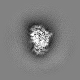
| Images |  emd_64873.png emd_64873.png | 63 KB | ||
| Filedesc metadata | emd-64873.cif.gz | 6.6 KB | ||
| Others | emd_64873_half_map_1.map.gzemd_64873_half_map_2.map.gz | 59.5 MB 59.5 MB | ||
| Archive directory |  http://ftp.pdbj.org/pub/emdb/structures/EMD-64873ftp://ftp.pdbj.org/pub/emdb/structures/EMD-64873 http://ftp.pdbj.org/pub/emdb/structures/EMD-64873ftp://ftp.pdbj.org/pub/emdb/structures/EMD-64873 | HTTPS FTP |
-Related structure data
| Related structure data |  9v9iMC  9v65C  9v66C  9v7sC  9v7zC  9v8aC 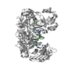 9v93C  9v9gC M: atomic model generated by this map C: citing same article ( |
|---|


-Links
| EMDB pages | EMDB (EBI/PDBe) / EMDataResource |
|---|
-Map
| File | Download / File: emd_64873.map.gz / Format: CCP4 / Size: 64 MB / Type: IMAGE STORED AS FLOATING POINT NUMBER (4 BYTES) | ||||||||||||||||||||||||||||||||||||
|---|---|---|---|---|---|---|---|---|---|---|---|---|---|---|---|---|---|---|---|---|---|---|---|---|---|---|---|---|---|---|---|---|---|---|---|---|---|
| Projections & slices | Image control
Images are generated by Spider. | ||||||||||||||||||||||||||||||||||||
| Voxel size | X=Y=Z: 0.851 Å | ||||||||||||||||||||||||||||||||||||
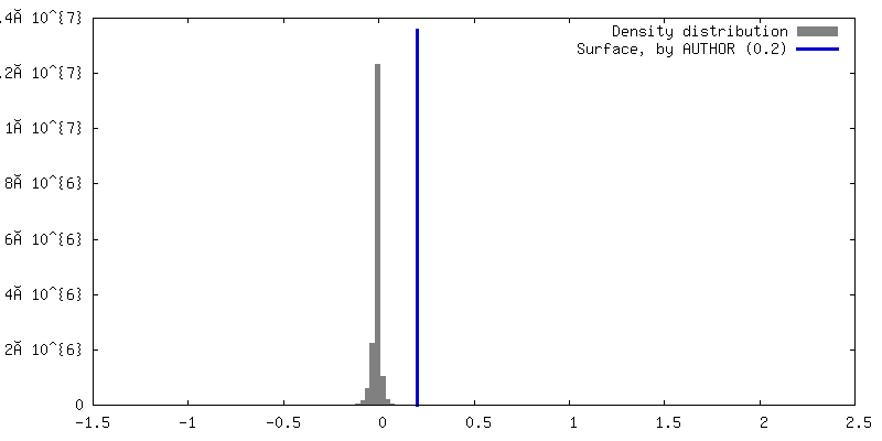
| Density |
| ||||||||||||||||||||||||||||||||||||
| Symmetry | Space group: 1 | ||||||||||||||||||||||||||||||||||||
| Details | EMDB XML:
|
 Z (Sec.)
 Y (Row.)
 X (Col.)


-Supplemental data
-Half map: #2
| File | emd_64873_half_map_1.map | ||||||||||||
|---|---|---|---|---|---|---|---|---|---|---|---|---|---|
| Projections & Slices |
| ||||||||||||
| Density Histograms |


-Half map: #1
| File | emd_64873_half_map_2.map | ||||||||||||
|---|---|---|---|---|---|---|---|---|---|---|---|---|---|
| Projections & Slices |
| ||||||||||||
| Density Histograms |


- Sample components
-Entire : VbAgo ternary complex
| Entire | Name: VbAgo ternary complex |
|---|---|
| Components |
|
-Supramolecule #1: VbAgo ternary complex
| Supramolecule | Name: VbAgo ternary complex / type: complex / ID: 1 / Parent: 0 / Macromolecule list: #1-#3 |
|---|---|
| Source (natural) | Organism: Verrucomicrobiota bacterium (bacteria) |
-Macromolecule #1: VbAgo
| Macromolecule | Name: VbAgo / type: protein_or_peptide / ID: 1 / Number of copies: 1 / Enantiomer: LEVO |
|---|---|
| Source (natural) | Organism: Verrucomicrobiota bacterium (bacteria) |
| Molecular weight | Theoretical: 88.46332 KDa |
| Recombinant expression | Organism: |
| Sequence | String: MSEKQLGATL FPITGLPAQA FRLRVLRVRE TIPMDTQTPV RLNRWATQLW KELKQAVVPT GRFEWPAFLT PDVESLTVGR VLTVQDVPD REYSIEVIGE TVEVNPASAS SEELQLAGEM IKRAISDAFG RNSDKYWRKH WNLYFRLEPE NLQDRRDRVF A YRGLKFSV ...String: MSEKQLGATL FPITGLPAQA FRLRVLRVRE TIPMDTQTPV RLNRWATQLW KELKQAVVPT GRFEWPAFLT PDVESLTVGR VLTVQDVPD REYSIEVIGE TVEVNPASAS SEELQLAGEM IKRAISDAFG RNSDKYWRKH WNLYFRLEPE NLQDRRDRVF A YRGLKFSV VFLGDKPWLA ADILTTYHGQ HALSEYSSEQ RQRELHFHVS ERIEADDRAM FLRDNGKIKI PCRFVGSTGK TV TQYTFPI NGGQKNVREY YEQRYGIRVP ENDEAVFVRD REGCDSWPVP ASRLFPLFTT EYDEVRNCSV VPQMPPDERV ETI RAFLND LRDVSFAGST LAIGHSHFQT AERSVFPAPA LEFGNGQTLT VDASLPIEEG YNRYRQGKMT MLYEHGPFSS QSLP DLVLL YPDNLDRNAR EKLRQRLGEE IKELCGVAPR IARQISYPLG KQPHAGAGLL AAADELVRNN DGTFLPVIVL ADALR EHIY DLLKRRLSSL ASQCVRERTV ARVARDEQAV GGSRLRNLAL GILTAAGLQP WVLAKPLHYD FYMGVDLLAN QVIYVF VCG KGGRNVWVQR GDQLRRRGIT EKIDRVQLAD QFKTGVREAK RLGVPLNSLV VHRDGRWWSN EDLAITEAVA ELQGDGT LS KDCQVGVVEV RKSHLPVRLF SVLNATKGSL ENPMPGSHLI LNNTEAILTP TGQPGRWDKQ GRTAGTLLLR ITRNPNGS P LDIRKIAEDA YGLTHLNWNA PDIEISLPVT IRWSDERLRE IVTNPSATDD TEVEPQETCI V |
-Macromolecule #2: DNA (5'-D(P*TP*GP*AP*GP*GP*TP*AP*GP*TP*AP*GP*GP*TP*TP*GP*TP*AP*T)-3')
| Macromolecule | Name: DNA (5'-D(P*TP*GP*AP*GP*GP*TP*AP*GP*TP*AP*GP*GP*TP*TP*GP*TP*AP*T)-3') type: dna / ID: 2 / Number of copies: 1 / Classification: DNA |
|---|---|
| Source (natural) | Organism: Verrucomicrobiota bacterium (bacteria) |
| Molecular weight | Theoretical: 5.64166 KDa |
| Sequence | String: (DT)(DG)(DA)(DG)(DG)(DT)(DA)(DG)(DT)(DA) (DG)(DG)(DT)(DT)(DG)(DT)(DA)(DT) |
-Macromolecule #3: RNA (5'-R(*UP*AP*UP*AP*CP*AP*AP*CP*CP*UP*AP*CP*UP*AP*CP*CP*UP*CP*...
| Macromolecule | Name: RNA (5'-R(*UP*AP*UP*AP*CP*AP*AP*CP*CP*UP*AP*CP*UP*AP*CP*CP*UP*CP*A)-3') type: rna / ID: 3 / Number of copies: 1 |
|---|---|
| Source (natural) | Organism: Verrucomicrobiota bacterium (bacteria) |
| Molecular weight | Theoretical: 5.926584 KDa |
| Sequence | String: UAUACAACCU ACUACCUCA |
-Macromolecule #4: MAGNESIUM ION
| Macromolecule | Name: MAGNESIUM ION / type: ligand / ID: 4 / Number of copies: 2 / Formula: MG |
|---|---|
| Molecular weight | Theoretical: 24.305 Da |
-Macromolecule #5: water
| Macromolecule | Name: water / type: ligand / ID: 5 / Number of copies: 8 / Formula: HOH |
|---|---|
| Molecular weight | Theoretical: 18.015 Da |
| Chemical component information |  ChemComp-HOH: |
-Experimental details
-Structure determination
| Method | cryo EM |
|---|---|
 Processing Processing | single particle reconstruction |
| Aggregation state | particle |
-Sample preparation
| Buffer | pH: 7.5 |
|---|---|
| Vitrification | Cryogen name: ETHANE |
- Electron microscopy
| Microscope | TFS KRIOS |
|---|---|
| Image recording | Film or detector model: GATAN K3 BIOQUANTUM (6k x 4k) / Average electron dose: 54.0 e/Å2 |
| Electron beam | Acceleration voltage: 300 kV / Electron source:  FIELD EMISSION GUN FIELD EMISSION GUN |
| Electron optics | Illumination mode: FLOOD BEAM / Imaging mode: BRIGHT FIELD / Nominal defocus max: 1.5 µm / Nominal defocus min: 1.0 µm |
| Experimental equipment |  Model: Titan Krios / Image courtesy: FEI Company |

