 Movie
Movie Controller

[English] 日本語
 Yorodumi
- PDB-9v66: Structure of the Argonaute protein from Verrucomicrobia bacterium... -
+ Open data

- Basic information
| Entry | Database: PDB / ID: 9v66 | ||||||||||||||||||||||||
|---|---|---|---|---|---|---|---|---|---|---|---|---|---|---|---|---|---|---|---|---|---|---|---|---|---|
| Title | Structure of the Argonaute protein from Verrucomicrobia bacterium in complex with guide DNA | ||||||||||||||||||||||||
 Components Components |
| ||||||||||||||||||||||||
 Keywords Keywords | RNA BINDING PROTEIN/DNA / mesophilic prokaryotic Argonaute / RNA BINDING PROTEIN-DNA complex | ||||||||||||||||||||||||
| Function / homology | DNA / DNA (> 10) Function and homology information Function and homology information | ||||||||||||||||||||||||
| Biological species |  Verrucomicrobiota bacterium (bacteria) Verrucomicrobiota bacterium (bacteria) | ||||||||||||||||||||||||
| Method | ELECTRON MICROSCOPY / single particle reconstruction / cryo EM / Resolution: 2.95 Å | ||||||||||||||||||||||||
 Authors Authors | Wu, S. | ||||||||||||||||||||||||
| Funding support |  China, 1items China, 1items
| ||||||||||||||||||||||||
 Citation Citation | Journal: Nat Commun / Year: 2026 Title: Structural insights into C-terminus-mediated RNA target cleavage by a mesophilic prokaryotic argonaute. Authors: Taiyu Chen / Xin Tao / Shunshun Li / Qingmiao Duanmu / Jiening Wang / Kai Chen / Kangle Mu / Hong Yang / Yu Li / Yang Liu / Lixin Ma / Shan Wu / Abstract: Prokaryotic Argonaute proteins (pAgos) are programmable nucleases that always utilize DNA guides to cleave DNA targets. Recent studies show that some pAgos preferentially utilize DNA guides to cleave ...Prokaryotic Argonaute proteins (pAgos) are programmable nucleases that always utilize DNA guides to cleave DNA targets. Recent studies show that some pAgos preferentially utilize DNA guides to cleave RNA targets rather than DNA targets. VbAgo, derived from a Verrucomicrobia bacterium, is a nuclease capable of specifically cleaving single-stranded RNA and highly structured RNA substrates at 37 °C, making it an ideal candidate for developing RNA manipulation toolkits. An in-depth investigation of its mechanism contributes to understanding the functional characteristics of gDtR-type Ago proteins. Here, we present cryo-electron microscopy structures of VbAgo, the VbAgo-guide DNA binary complex, multiple wild-type VbAgo-guide DNA-target RNA ternary complexes, and the catalytically inactive mutant (VbAgo-DM) guide DNA-target RNA ternary complex, with resolutions ranging from 2.5 to 3.2 Å. By integrating these cryo-EM structures with biochemical data, we elucidate the entire catalytic process of VbAgo, revealing its unique C-terminal regulatory mechanism. Specifically, in its apo state, VbAgo's C-terminus occupies the nucleic acid binding channel, partially impeding its catalytic activity while enhancing its stability. The binding of guide DNA displaces the C-terminus, and subsequent binding of target RNA, along with conformational changes in the N-terminal and PAZ domains, facilitates VbAgo dimerization. Following this, the C-terminus transitions from a loop to a helix, enabling maturation of the catalytic center and inducing movements in the MID-PIWI' interactions at the dimer interfaces, ultimately leading to dimer dissociation. Concurrently, cleavage of the target RNA and subsequent product release occur, after which VbAgo reverts to its binary state to initiate the next cleavage cycle. Moreover, we demonstrate that VbAgo exhibits guide DNA mediated RNA knockdown activity in mammalian cells. In summary, our study provides a comprehensive understanding of the molecular mechanisms governing self-inhibition, guide binding, target recognition, and product release in VbAgo. These findings offer valuable insights into the diverse mechanisms of pAgos, broadening their functional scope and enhancing the biotechnological potential of pAgo proteins. | ||||||||||||||||||||||||
| History |
|
- Structure visualization
| Structure viewer | Molecule: MolmilJmol/JSmol |
|---|
- Downloads & links
-Download
| PDBx/mmCIF format | 9v66.cif.gz | 154.3 KB | Display | PDBx/mmCIF format |
|---|---|---|---|---|
| PDB format | pdb9v66.ent.gz | 118.2 KB | Display | PDB format |
| PDBx/mmJSON format | 9v66.json.gz | Tree view | PDBx/mmJSON format | |
| Others |  Other downloads Other downloads |
-Validation report
| Arichive directory | https://data.pdbj.org/pub/pdb/validation_reports/v6/9v66ftp://data.pdbj.org/pub/pdb/validation_reports/v6/9v66 | HTTPS FTP |
|---|
-Related structure data
| Related structure data |  64798MC  9v65C  9v7sC  9v7zC  9v8aC 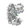 9v93C  9v9gC  9v9iC M: map data used to model this data C: citing same article ( |
|---|---|
| Similar structure data |


-Links
 PDBj
PDBj


- Assembly
| Deposited unit | 
|
|---|---|
| 1 |
|
-Components
| #1: Protein | Mass: 88463.320 Da / Num. of mol.: 1 Source method: isolated from a genetically manipulated source Source: (gene. exp.) Verrucomicrobiota bacterium (bacteria) / Production host: |
|---|---|
| #2: DNA chain | Mass: 5641.660 Da / Num. of mol.: 1 Source method: isolated from a genetically manipulated source Source: (gene. exp.) Verrucomicrobiota bacterium (bacteria) / Production host: |
| #3: Chemical | ChemComp-MG /   Mass: 24.305 Da / Num. of mol.: 1 / Source method: obtained synthetically / Formula: Mg / Feature type: SUBJECT OF INVESTIGATION Mass: 24.305 Da / Num. of mol.: 1 / Source method: obtained synthetically / Formula: Mg / Feature type: SUBJECT OF INVESTIGATION |
| #4: Water | ChemComp-HOH /  Mass: 18.015 Da / Num. of mol.: 8 / Source method: isolated from a natural source / Formula: H2O Mass: 18.015 Da / Num. of mol.: 8 / Source method: isolated from a natural source / Formula: H2O |
| Has ligand of interest | Y |
| Has protein modification | N |
-Experimental details
-Experiment
| Experiment | Method: ELECTRON MICROSCOPY |
|---|---|
| EM experiment | Aggregation state: PARTICLE / 3D reconstruction method: single particle reconstruction |
- Sample preparation
| Component | Name: VbAgo / Type: COMPLEX / Entity ID: #1-#2 / Source: MULTIPLE SOURCES |
|---|---|
| Molecular weight | Experimental value: NO |
| Source (natural) | Organism: Verrucomicrobiota bacterium (bacteria) |
| Source (recombinant) | Organism: |
| Buffer solution | pH: 7.5 |
| Specimen | Embedding applied: NO / Shadowing applied: NO / Staining applied: NO / Vitrification applied: YES |
| Vitrification | Cryogen name: ETHANE |
- Electron microscopy imaging
| Experimental equipment |  Model: Titan Krios / Image courtesy: FEI Company |
|---|---|
| Microscopy | Model: TFS KRIOS |
| Electron gun | Electron source:  FIELD EMISSION GUN / Accelerating voltage: 300 kV / Illumination mode: FLOOD BEAM FIELD EMISSION GUN / Accelerating voltage: 300 kV / Illumination mode: FLOOD BEAM |
| Electron lens | Mode: BRIGHT FIELD / Nominal defocus max: 1500 nm / Nominal defocus min: 1000 nm |
| Image recording | Electron dose: 54 e/Å2 / Film or detector model: GATAN K3 BIOCONTINUUM (6k x 4k) |
- Processing
| EM software |
| ||||||||||||||||||||||||
|---|---|---|---|---|---|---|---|---|---|---|---|---|---|---|---|---|---|---|---|---|---|---|---|---|---|
| CTF correction | Type: PHASE FLIPPING AND AMPLITUDE CORRECTION | ||||||||||||||||||||||||
| 3D reconstruction | Resolution: 2.95 Å / Resolution method: FSC 0.143 CUT-OFF / Num. of particles: 186978 / Symmetry type: POINT | ||||||||||||||||||||||||
| Refinement | Highest resolution: 2.95 Å Stereochemistry target values: REAL-SPACE (WEIGHTED MAP SUM AT ATOM CENTERS) | ||||||||||||||||||||||||
| Refine LS restraints |
|
