 Movie
Movie Controller

[English] 日本語
 Yorodumi
- EMDB-64527: Single-subunit apo Escherichia coli nicotinamide nucleotide trans... -
+ Open data

- Basic information
| Entry |  | |||||||||
|---|---|---|---|---|---|---|---|---|---|---|
| Title | Single-subunit apo Escherichia coli nicotinamide nucleotide transhydrogenase | |||||||||
 Map data Map data | ||||||||||
 Sample Sample |
| |||||||||
 Keywords Keywords | Nicotinamide nucleotide transhydrogenase / Mitochondrial inner membrane / E. coli transhydrogenase / Hydride transfer / Proton motive force / NAD+ / NADH / NADP+ / NADPH / MEMBRANE PROTEIN | |||||||||
| Function / homology |  Function and homology information Function and homology informationproton export across plasma membrane / proton-translocating NAD(P)+ transhydrogenase activity / proton-translocating NAD(P)+ transhydrogenase / NADPH regeneration / NADP binding / oxidoreductase activity / protein dimerization activity / plasma membrane Similarity search - Function | |||||||||
| Biological species |   Escherichia coli K-12 (bacteria) Escherichia coli K-12 (bacteria) | |||||||||
| Method | single particle reconstruction / cryo EM / Resolution: 3.43 Å | |||||||||
 Authors Authors | Zhu J / Gennis RB / Zhang K / Li J | |||||||||
| Funding support |  China, 1 items China, 1 items
| |||||||||
 Citation Citation | Journal: To Be Published Title: Single-subunit apo Escherichia coli nicotinamide nucleotide transhydrogenase Authors: Zhu J / Gennis RB / Zhang K / Li J | |||||||||
| History |
|
- Structure visualization
| Supplemental images |
|---|

- Downloads & links
-EMDB archive
| Map data | emd_64527.map.gz | 10.1 MB | EMDB map data format | |
|---|---|---|---|---|
| Header (meta data) | emd-64527-v30.xmlemd-64527.xml | 14.7 KB 14.7 KB | Display Display | EMDB header |
| Images |  emd_64527.png emd_64527.png | 59.7 KB | ||
| Filedesc metadata | emd-64527.cif.gz | 5.8 KB | ||
| Archive directory |  http://ftp.pdbj.org/pub/emdb/structures/EMD-64527ftp://ftp.pdbj.org/pub/emdb/structures/EMD-64527 http://ftp.pdbj.org/pub/emdb/structures/EMD-64527ftp://ftp.pdbj.org/pub/emdb/structures/EMD-64527 | HTTPS FTP |
-Related structure data
| Related structure data |  9uv7MC  9uv2C  64526 M: atomic model generated by this map C: citing same article ( |
|---|---|
| Similar structure data |


-Links
| EMDB pages | EMDB (EBI/PDBe) / EMDataResource |
|---|---|
| Related items in Molecule of the Month |

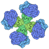
-Map
| File | Download / File: emd_64527.map.gz / Format: CCP4 / Size: 178 MB / Type: IMAGE STORED AS FLOATING POINT NUMBER (4 BYTES) | ||||||||||||||||||||||||||||||||||||
|---|---|---|---|---|---|---|---|---|---|---|---|---|---|---|---|---|---|---|---|---|---|---|---|---|---|---|---|---|---|---|---|---|---|---|---|---|---|
| Projections & slices | Image control
Images are generated by Spider. | ||||||||||||||||||||||||||||||||||||
| Voxel size | X=Y=Z: 0.82 Å | ||||||||||||||||||||||||||||||||||||
| Density |
| ||||||||||||||||||||||||||||||||||||
| Symmetry | Space group: 1 | ||||||||||||||||||||||||||||||||||||
| Details | EMDB XML:
|
 Z (Sec.)
 Y (Row.)
 X (Col.)


-Supplemental data
- Sample components
-Entire : The E. coli transhydrogenase complex.
| Entire | Name: The E. coli transhydrogenase complex. |
|---|---|
| Components |
|
-Supramolecule #1: The E. coli transhydrogenase complex.
| Supramolecule | Name: The E. coli transhydrogenase complex. / type: complex / ID: 1 / Parent: 0 / Macromolecule list: all |
|---|---|
| Source (natural) | Organism: Escherichia coli K-12 (bacteria) |
-Macromolecule #1: NAD(P) transhydrogenase subunit beta
| Macromolecule | Name: NAD(P) transhydrogenase subunit beta / type: protein_or_peptide / ID: 1 / Number of copies: 2 / Enantiomer: LEVO / EC number: proton-translocating NAD(P)+ transhydrogenase |
|---|---|
| Source (natural) | Organism: Escherichia coli K-12 (bacteria) / Strain: K-12 |
| Molecular weight | Theoretical: 48.762746 KDa |
| Recombinant expression | Organism: Escherichia coli (E. coli) |
| Sequence | String: MSGGLVTAAY IVAAILFIFS LAGLSKHETS RQGNNFGIAG MAIALIATIF GPDTGNVGWI LLAMVIGGAI GIRLAKKVEM TEMPELVAI LHSFVGLAAV LVGFNSYLHH DAGMAPILVN IHLTEVFLGI FIGAVTFTGS VVAFGKLCGK ISSKPLMLPN R HKMNLAAL ...String: MSGGLVTAAY IVAAILFIFS LAGLSKHETS RQGNNFGIAG MAIALIATIF GPDTGNVGWI LLAMVIGGAI GIRLAKKVEM TEMPELVAI LHSFVGLAAV LVGFNSYLHH DAGMAPILVN IHLTEVFLGI FIGAVTFTGS VVAFGKLCGK ISSKPLMLPN R HKMNLAAL VVSFLLLIVF VRTDSVGLQV LALLIMTAIA LVFGWHLVAS IGGADMPVVV SMLNSYSGWA AAAAGFMLSN DL LIVTGAL VGSSGAILSY IMCKAMNRSF ISVIAGGFGT DGSSTGDDQE VGEHREITAE ETAELLKNSH SVIITPGYGM AVA QAQYPV AEITEKLRAR GINVRFGIHP VAGRLPGHMN VLLAEAKVPY DIVLEMDEIN DDFADTDTVL VIGANDTVNP AAQD DPKSP IAGMPVLEVW KAQNVIVFKR SMNTGYAGVQ NPLFFKENTH MLFGDAKASV DAILKAL UniProtKB: NAD(P) transhydrogenase subunit beta |
-Macromolecule #2: NAD(P) transhydrogenase subunit alpha
| Macromolecule | Name: NAD(P) transhydrogenase subunit alpha / type: protein_or_peptide / ID: 2 / Number of copies: 2 / Enantiomer: LEVO / EC number: proton-translocating NAD(P)+ transhydrogenase |
|---|---|
| Source (natural) | Organism: Escherichia coli K-12 (bacteria) / Strain: K-12 |
| Molecular weight | Theoretical: 10.742778 KDa |
| Recombinant expression | Organism: Escherichia coli (E. coli) |
| Sequence | String: ILFGWMASVA PKEFLGHFTV FALACVVGYY VVWNVSHALH TPLMSVTNAI SGIIVVGALL QIGQGGWVSF LSFIAVLIAS INIFGGFTV TQRMLKMFKN UniProtKB: NAD(P) transhydrogenase subunit alpha |
-Macromolecule #3: NAD(P) transhydrogenase subunit alpha
| Macromolecule | Name: NAD(P) transhydrogenase subunit alpha / type: protein_or_peptide / ID: 3 / Number of copies: 2 / Enantiomer: LEVO / EC number: proton-translocating NAD(P)+ transhydrogenase |
|---|---|
| Source (natural) | Organism: Escherichia coli K-12 (bacteria) / Strain: K-12 |
| Molecular weight | Theoretical: 40.183902 KDa |
| Recombinant expression | Organism: Escherichia coli (E. coli) |
| Sequence | String: HHGRIGIPRE RLTNETRVAA TPKTVEQLLK LGFTVAVESG AGQLASFDDK AFVQAGAEIV EGNSVWQSEI ILKVNAPLDD EIALLNPGT TLVSFIWPAQ NPELMQKLAE RNVTVMAMDS VPRISRAQSL DALSSMANIA GYRAIVEAAH EFGRFFTGQI T AAGKVPPA ...String: HHGRIGIPRE RLTNETRVAA TPKTVEQLLK LGFTVAVESG AGQLASFDDK AFVQAGAEIV EGNSVWQSEI ILKVNAPLDD EIALLNPGT TLVSFIWPAQ NPELMQKLAE RNVTVMAMDS VPRISRAQSL DALSSMANIA GYRAIVEAAH EFGRFFTGQI T AAGKVPPA KVMVIGAGVA GLAAIGAANS LGAIVRAFDT RPEVKEQVQS MGAEFLELDF KEEAGSGDGY AKVMSDAFIK AE MELFAAQ AKEVDIIVTT ALIPGKPAPK LITREMVDSM KAGSVIVDLA AQNGGNCEYT VPGEIFTTEN GVKVIGYTDL PGR LPTQSS QLYGTNLVNL LKLLCKEKDG NITVDFDDVV IRGVTVIRAG EITWPAPPIQ V UniProtKB: NAD(P) transhydrogenase subunit alpha |
-Experimental details
-Structure determination
| Method | cryo EM |
|---|---|
 Processing Processing | single particle reconstruction |
| Aggregation state | particle |
-Sample preparation
| Buffer | pH: 8 |
|---|---|
| Vitrification | Cryogen name: ETHANE |
- Electron microscopy
| Microscope | TFS KRIOS |
|---|---|
| Image recording | Film or detector model: GATAN K3 (6k x 4k) / Average electron dose: 1.25 e/Å2 |
| Electron beam | Acceleration voltage: 300 kV / Electron source:  FIELD EMISSION GUN FIELD EMISSION GUN |
| Electron optics | Illumination mode: FLOOD BEAM / Imaging mode: BRIGHT FIELD / Nominal defocus max: 2.0 µm / Nominal defocus min: 1.2 µm |
| Experimental equipment |  Model: Titan Krios / Image courtesy: FEI Company |
