 Movie
Movie Controller


+ Open data

- Basic information
| Entry |  | |||||||||
|---|---|---|---|---|---|---|---|---|---|---|
| Title | Cryo-EM structure of the HBsAg dimer and Complex with Fab | |||||||||
 Map data Map data | ||||||||||
 Sample Sample |
| |||||||||
 Keywords Keywords | HBV antigen / dimer / Fab / complex / VIRAL PROTEIN/IMMUNE SYSTEM / VIRAL PROTEIN-IMMUNE SYSTEM complex | |||||||||
| Function / homology | Large envelope protein S / Major surface antigen from hepadnavirus / membrane fusion involved in viral entry into host cell / symbiont entry into host cell / virion attachment to host cell / virion membrane / Middle S protein Function and homology information Function and homology information | |||||||||
| Biological species |  Homo sapiens (human) / Homo sapiens (human) /  hepatitis B virus genotype A hepatitis B virus genotype A | |||||||||
| Method | single particle reconstruction / cryo EM / Resolution: 4.0 Å | |||||||||
 Authors Authors | Liu Y / Liao M / Liu Z / Ju B / Zhang Z | |||||||||
| Funding support | 1 items
| |||||||||
 Citation Citation | Journal: Gut / Year: 2026 Title: Characterisation of plasmablast-derived HBsAg-specific antibody and its structural basis for binding to native HBsAg dimer. Authors: Bin Ju / Zhouqing Liu / Hu Yan / Yong Liu / Lu Zhang / Xiangyang Ge / Xin Wang / Zhu Si / Bing Zhou / Qing Fan / Miao Wang / Yuxiao Li / Wenlong Lai / Jianhui Gan / Haiyan Wang / Juanjuan ...Authors: Bin Ju / Zhouqing Liu / Hu Yan / Yong Liu / Lu Zhang / Xiangyang Ge / Xin Wang / Zhu Si / Bing Zhou / Qing Fan / Miao Wang / Yuxiao Li / Wenlong Lai / Jianhui Gan / Haiyan Wang / Juanjuan Zhao / Yuchen Xia / Maofu Liao / Zheng Zhang /  Abstract: BACKGROUND: Plasmablast-derived HBV surface antigen (HBsAg)-specific monoclonal antibody (mAb) and structural basis for binding to native HBsAg are poorly known. OBJECTIVE: We aimed to identify plasmablast-derived HBsAg-specific mAbs, evaluate their antiviral activities and resolve their structure for binding to native HBsAg. DESIGN: A previously vaccinated volunteer was enrolled in this study, who was boosted with a dose of recombinant hepatitis B vaccine and donated the blood sample. Activated plasmablasts were sorted ...DESIGN: A previously vaccinated volunteer was enrolled in this study, who was boosted with a dose of recombinant hepatitis B vaccine and donated the blood sample. Activated plasmablasts were sorted from fresh peripheral blood mononuclear cells and mAbs were expressed. Their gene features, cross-genotypic binding activities and antiviral functions in vitro and in vivo were comprehensively analysed. The cryo-electron microscopy (cryo-EM) was used to determine the structure of representative mAb bound to the native HBsAg. RESULTS: In this study, we cloned a series of HBsAg-specific mAbs directly from clonally expanded plasmablasts from a vaccinated individual. Most of the mAbs displayed cross-reactivities of binding ...RESULTS: In this study, we cloned a series of HBsAg-specific mAbs directly from clonally expanded plasmablasts from a vaccinated individual. Most of the mAbs displayed cross-reactivities of binding to different genotype HBsAg proteins and antiviral functions such as neutralisation and antibody-dependent cellular phagocytosis. These human anti-HBsAg mAbs, especially SY-4-class and SY-23-class, could be good candidates for antibody drugs. The cryo-EM structure of SY-23 bound to the dimeric HBsAg was determined, revealing its binding mechanism and unprecedented structural detail of the major antigenic loop (AGL) of HBsAg. CONCLUSION: Overall, our work has uncovered the diverse gene features and varied anti-HBV activities of plasmablast-derived mAbs, providing a series of antibody drug candidates and the long-sought- ...CONCLUSION: Overall, our work has uncovered the diverse gene features and varied anti-HBV activities of plasmablast-derived mAbs, providing a series of antibody drug candidates and the long-sought-after atomic model of AGL has paved the way for a wholistic characterisation of the AGL's dynamic conformation during HBV infection and immune response. | |||||||||
| History |
|
- Structure visualization
| Supplemental images |
|---|

- Downloads & links
-EMDB archive
| Map data | emd_64142.map.gz | 59.9 MB | EMDB map data format | |
|---|---|---|---|---|
| Header (meta data) | emd-64142-v30.xmlemd-64142.xml | 20.4 KB 20.4 KB | Display Display | EMDB header |
| FSC (resolution estimation) | emd_64142_fsc.xml | 9.2 KB | Display | FSC data file |
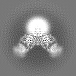
| Images |  emd_64142.png emd_64142.png | 49.7 KB | ||
| Masks | emd_64142_msk_1.map | 64 MB | Mask map | |
| Filedesc metadata | emd-64142.cif.gz | 6.1 KB | ||
| Others | emd_64142_additional_1.map.gzemd_64142_half_map_1.map.gzemd_64142_half_map_2.map.gz | 4.1 MB 48.5 MB 48.5 MB | ||
| Archive directory |  http://ftp.pdbj.org/pub/emdb/structures/EMD-64142ftp://ftp.pdbj.org/pub/emdb/structures/EMD-64142 http://ftp.pdbj.org/pub/emdb/structures/EMD-64142ftp://ftp.pdbj.org/pub/emdb/structures/EMD-64142 | HTTPS FTP |
-Related structure data
| Related structure data |  9ugoMC M: atomic model generated by this map C: citing same article ( |
|---|---|
| Similar structure data |
-Links
| EMDB pages | EMDB (EBI/PDBe) / EMDataResource |
|---|
-Map
| File | Download / File: emd_64142.map.gz / Format: CCP4 / Size: 64 MB / Type: IMAGE STORED AS FLOATING POINT NUMBER (4 BYTES) | ||||||||||||||||||||||||||||||||||||
|---|---|---|---|---|---|---|---|---|---|---|---|---|---|---|---|---|---|---|---|---|---|---|---|---|---|---|---|---|---|---|---|---|---|---|---|---|---|
| Projections & slices | Image control
Images are generated by Spider. | ||||||||||||||||||||||||||||||||||||
| Voxel size | X=Y=Z: 0.855 Å | ||||||||||||||||||||||||||||||||||||
| Density |
| ||||||||||||||||||||||||||||||||||||
| Symmetry | Space group: 1 | ||||||||||||||||||||||||||||||||||||
| Details | EMDB XML:
|
 Z (Sec.)
 Y (Row.)
 X (Col.)


-Supplemental data
-Mask #1
| File | emd_64142_msk_1.map | ||||||||||||
|---|---|---|---|---|---|---|---|---|---|---|---|---|---|
| Projections & Slices |
| ||||||||||||
| Density Histograms |


-Additional map: #1
| File | emd_64142_additional_1.map | ||||||||||||
|---|---|---|---|---|---|---|---|---|---|---|---|---|---|
| Projections & Slices |
| ||||||||||||
| Density Histograms |


-Half map: #2
| File | emd_64142_half_map_1.map | ||||||||||||
|---|---|---|---|---|---|---|---|---|---|---|---|---|---|
| Projections & Slices |
| ||||||||||||
| Density Histograms |


-Half map: #1
| File | emd_64142_half_map_2.map | ||||||||||||
|---|---|---|---|---|---|---|---|---|---|---|---|---|---|
| Projections & Slices |
| ||||||||||||
| Density Histograms |


- Sample components
-Entire : complex of HBsAg dimer with Fab
| Entire | Name: complex of HBsAg dimer with Fab |
|---|---|
| Components |
|
-Supramolecule #1: complex of HBsAg dimer with Fab
| Supramolecule | Name: complex of HBsAg dimer with Fab / type: complex / ID: 1 / Parent: 0 / Macromolecule list: all |
|---|---|
| Source (natural) | Organism: Homo sapiens (human) |
-Macromolecule #1: Fab light chain
| Macromolecule | Name: Fab light chain / type: protein_or_peptide / ID: 1 / Number of copies: 2 / Enantiomer: LEVO |
|---|---|
| Source (natural) | Organism: Homo sapiens (human) |
| Molecular weight | Theoretical: 23.425979 KDa |
| Recombinant expression | Organism: Homo sapiens (human) |
| Sequence | String: EIVMTQSPAT LSVSPGERAT LSCRASQSVS NNLAWYQQKP GQAPRLLIYG ASTRATGIPA RFSGSGSGTE FTLTISSLQS EDSAVYYCQ QHHSWPPFTF GQGTKLEIKR TVAAPSVFIF PPSDEQLKSG TASVVCLLNN FYPREAKVQW KVDNALQSGN S QESVTEQD ...String: EIVMTQSPAT LSVSPGERAT LSCRASQSVS NNLAWYQQKP GQAPRLLIYG ASTRATGIPA RFSGSGSGTE FTLTISSLQS EDSAVYYCQ QHHSWPPFTF GQGTKLEIKR TVAAPSVFIF PPSDEQLKSG TASVVCLLNN FYPREAKVQW KVDNALQSGN S QESVTEQD SKDSTYSLSS TLTLSKADYE KHKVYACEVT HQGLSSPVTK SFNRGEC |
-Macromolecule #2: Fab heavy chain
| Macromolecule | Name: Fab heavy chain / type: protein_or_peptide / ID: 2 / Number of copies: 2 / Enantiomer: LEVO |
|---|---|
| Source (natural) | Organism: Homo sapiens (human) |
| Molecular weight | Theoretical: 24.12116 KDa |
| Recombinant expression | Organism: Homo sapiens (human) |
| Sequence | String: QVQLVQSGAE VKKPGSSVKV YCKASGGTFR SYGLNWVRQA PGQGLEWMGR IVPIFGISNF AQKFQGRFTI TADRSANTPY MELSSLRSE DTAVYYCVRG GAVAGAGSAG EGIFDYWDQG TLVIVSSAST KGPSVFPLAP SSKSTSGGTA ALGCLVKDYF P EPVTVSWN ...String: QVQLVQSGAE VKKPGSSVKV YCKASGGTFR SYGLNWVRQA PGQGLEWMGR IVPIFGISNF AQKFQGRFTI TADRSANTPY MELSSLRSE DTAVYYCVRG GAVAGAGSAG EGIFDYWDQG TLVIVSSAST KGPSVFPLAP SSKSTSGGTA ALGCLVKDYF P EPVTVSWN SGALTSGVHT FPAVLQSSGL YSLSSVVTVP SSSLGTQTYI CNVNHKPSNT KVDKKVEPKS C |
-Macromolecule #3: Middle S protein
| Macromolecule | Name: Middle S protein / type: protein_or_peptide / ID: 3 Details: The sequence of organism hepatitis B virus genotype A is not available during the biocuration, replaced by B8QJB4 temporarily. Number of copies: 2 / Enantiomer: LEVO |
|---|---|
| Source (natural) | Organism: hepatitis B virus genotype A |
| Molecular weight | Theoretical: 11.0082 KDa |
| Recombinant expression | Organism: Yeast centromeric expression vector p416CYC (others) |
| Sequence | String: RWMCLRRFII FLFILLLCLI FLLVLLDYQG MLPVCPLIPG SSTTSTGPCR TCMTTAQGTS MYPSCCCTKP SDGNCTCIPI PSSWAFGKF LWEWASARF UniProtKB: Middle S protein |
-Experimental details
-Structure determination
| Method | cryo EM |
|---|---|
 Processing Processing | single particle reconstruction |
| Aggregation state | particle |
-Sample preparation
| Buffer | pH: 7.5 |
|---|---|
| Vitrification | Cryogen name: ETHANE |
- Electron microscopy
| Microscope | TFS KRIOS |
|---|---|
| Image recording | Film or detector model: GATAN K3 (6k x 4k) / Average electron dose: 50.0 e/Å2 |
| Electron beam | Acceleration voltage: 300 kV / Electron source:  FIELD EMISSION GUN FIELD EMISSION GUN |
| Electron optics | Illumination mode: FLOOD BEAM / Imaging mode: BRIGHT FIELD / Nominal defocus max: 2.0 µm / Nominal defocus min: 1.0 µm |
| Experimental equipment |  Model: Titan Krios / Image courtesy: FEI Company |

