[English] 日本語
Yorodumi
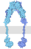
- EMDB-57339: Insulin receptor ecto-domain in inverted-U apo-conformation -

+
Open data


ID or keywords:

Loading...

-
Basic information

Entry
Database: EMDB / ID: EMD-57339
TitleInsulin receptor ecto-domain in inverted-U apo-conformation
Map datainverted-U apo-IR
Sample
  • Complex: insulin receptor ecto-domain in inverted-U apo-conformation
    • Protein or peptide: Insulin receptor
    • Protein or peptide: Insulin receptor
Keywordscomplex / IR / apo-form / inverted-U / MEMBRANE PROTEIN
Function / homology
Function and homology information


regulation of female gonad development / positive regulation of meiotic cell cycle / insulin-like growth factor II binding / positive regulation of developmental growth / insulin receptor complex / insulin-like growth factor I binding / insulin receptor activity / positive regulation of protein-containing complex disassembly / adrenal gland development / dendritic spine maintenance ...regulation of female gonad development / positive regulation of meiotic cell cycle / insulin-like growth factor II binding / positive regulation of developmental growth / insulin receptor complex / insulin-like growth factor I binding / insulin receptor activity / positive regulation of protein-containing complex disassembly / adrenal gland development / dendritic spine maintenance / insulin binding / cargo receptor activity / PTB domain binding / Signaling by Insulin receptor / IRS activation / neuronal cell body membrane / positive regulation of respiratory burst / amyloid-beta clearance / insulin receptor substrate binding / positive regulation of receptor internalization / heart morphogenesis / regulation of embryonic development / positive regulation of glycogen biosynthetic process / Signal attenuation / protein kinase activator activity / phosphatidylinositol 3-kinase binding / transport across blood-brain barrier / Insulin receptor recycling / insulin-like growth factor receptor binding / neuron projection maintenance / positive regulation of mitotic nuclear division / receptor-mediated endocytosis / positive regulation of glycolytic process / Insulin receptor signalling cascade / learning / dendrite membrane / positive regulation of D-glucose import across plasma membrane / male gonad development / receptor protein-tyrosine kinase / memory / caveola / cellular response to insulin stimulus / positive regulation of nitric oxide biosynthetic process / glucose homeostasis / insulin receptor signaling pathway / late endosome / protein autophosphorylation / amyloid-beta binding / PI5P, PP2A and IER3 Regulate PI3K/AKT Signaling / protein tyrosine kinase activity / positive regulation of MAPK cascade / positive regulation of phosphatidylinositol 3-kinase/protein kinase B signal transduction / positive regulation of canonical NF-kappaB signal transduction / signaling receptor complex / lysosome / endosome membrane / positive regulation of cell migration / G protein-coupled receptor signaling pathway / external side of plasma membrane / protein domain specific binding / axon / positive regulation of cell population proliferation / symbiont entry into host cell / regulation of DNA-templated transcription / positive regulation of DNA-templated transcription / GTP binding / protein-containing complex binding / extracellular exosome / ATP binding / membrane / identical protein binding / plasma membrane
Similarity search - Function
Insulin receptor, trans-membrane domain / Insulin receptor trans-membrane segment / Tyrosine-protein kinase, insulin-like receptor / Tyrosine-protein kinase, receptor class II, conserved site / Receptor tyrosine kinase class II signature. / Receptor L-domain / Furin-like cysteine-rich domain / Receptor L-domain superfamily / Furin-like cysteine rich region / Receptor L domain ...Insulin receptor, trans-membrane domain / Insulin receptor trans-membrane segment / Tyrosine-protein kinase, insulin-like receptor / Tyrosine-protein kinase, receptor class II, conserved site / Receptor tyrosine kinase class II signature. / Receptor L-domain / Furin-like cysteine-rich domain / Receptor L-domain superfamily / Furin-like cysteine rich region / Receptor L domain / Furin-like repeat / Furin-like repeats / Growth factor receptor cysteine-rich domain superfamily / Fibronectin type 3 domain / : / Fibronectin type-III domain profile. / Fibronectin type III / Fibronectin type III superfamily / Tyrosine-protein kinase, catalytic domain / Tyrosine kinase, catalytic domain / Tyrosine protein kinases specific active-site signature. / Tyrosine-protein kinase, active site / Protein tyrosine and serine/threonine kinase / Serine-threonine/tyrosine-protein kinase, catalytic domain / Protein kinase, ATP binding site / Protein kinases ATP-binding region signature. / Immunoglobulin-like fold / Protein kinase domain profile. / Protein kinase domain / Protein kinase-like domain superfamily
Similarity search - Domain/homology
Biological speciesHomo sapiens (human)
Methodsingle particle reconstruction / cryo EM / Resolution: 7.5 Å
AuthorsPolak M / Novacek J / Jiracek J
Funding support Czech Republic, 2 items
OrganizationGrant numberCountry
Ministry of Education, Youth and Sports of the Czech RepublicLM2023042 Czech Republic
Ministry of Education, Youth and Sports of the Czech RepublicLX22NPO5104 Czech Republic
CitationJournal: To Be Published
Title: Antagonistic insulin mimetics lock the insulin receptor in an alternative apo-state
Authors: Polak M / Pinkas M
History
DepositionApr 1, 2026-
Header (metadata) releaseJul 22, 2026-
Map releaseJul 22, 2026-
UpdateJul 22, 2026-
Current statusJul 22, 2026Processing site: PDBe / Status: Released

-
Structure visualization

Supplemental images

Downloads & links

-
Map

FileDownload / File: emd_57339.map.gz / Format: CCP4 / Size: 10.5 MB / Type: IMAGE STORED AS FLOATING POINT NUMBER (4 BYTES)
Annotationinverted-U apo-IR
Projections & slices

Image control

Size
Brightness
Contrast
Others
AxesZ (Sec.)Y (Row.)X (Col.)
1.67 Å/pix.
x 140 pix.
= 233.408 Å
1.67 Å/pix.
x 140 pix.
= 233.408 Å
1.67 Å/pix.
x 140 pix.
= 233.408 Å

Surface

Projections

Slices (1/3)

Slices (1/2)

Slices (2/3)

Images are generated by Spider.

Voxel sizeX=Y=Z: 1.6672 Å
Density
Contour LevelBy AUTHOR: 0.2
Minimum - Maximum-0.13361453 - 0.56812865
Average (Standard dev.)0.0054971697 (±0.036681905)
SymmetrySpace group: 1
Details

EMDB XML:

Map geometry
Axis orderXYZ
Origin000
Dimensions140140140
Spacing140140140
CellA=B=C: 233.40799 Å
α=β=γ: 90.0 °

-
Supplemental data

-
Half map: half map

Fileemd_57339_half_map_1.map
Annotationhalf map
Projections & Slices
AxesZYX

Projections

Slices (1/2)
Density Histograms

-
Half map: half map

Fileemd_57339_half_map_2.map
Annotationhalf map
Projections & Slices
AxesZYX

Projections

Slices (1/2)
Density Histograms

-
Sample components

-
Entire : insulin receptor ecto-domain in inverted-U apo-conformation

EntireName: insulin receptor ecto-domain in inverted-U apo-conformation
Components
  • Complex: insulin receptor ecto-domain in inverted-U apo-conformation
    • Protein or peptide: Insulin receptor
    • Protein or peptide: Insulin receptor

-
Supramolecule #1: insulin receptor ecto-domain in inverted-U apo-conformation

SupramoleculeName: insulin receptor ecto-domain in inverted-U apo-conformation
type: complex / ID: 1 / Parent: 0 / Macromolecule list: all
Source (natural)Organism: Homo sapiens (human)
Molecular weightTheoretical: 200 KDa

-
Macromolecule #1: Insulin receptor

MacromoleculeName: Insulin receptor / type: protein_or_peptide / ID: 1 / Number of copies: 2 / Enantiomer: LEVO / EC number: receptor protein-tyrosine kinase
Source (natural)Organism: Homo sapiens (human)
Molecular weightTheoretical: 82.551742 KDa
Recombinant expressionOrganism: Homo sapiens (human)
SequenceString: HLYPGEVCPG MDIRNNLTRL HELENCSVIE GHLQILLMFK TRPEDFRDLS FPKLIMITDY LLLFRVYGLE SLKDLFPNLT VIRGSRLFF NYALVIFEMV HLKELGLYNL MNITRGSVRI EKNNELCYLA TIDWSRILDS VEDNYIVLNK DDNEECGDIC P GTAKGKTN ...String:
HLYPGEVCPG MDIRNNLTRL HELENCSVIE GHLQILLMFK TRPEDFRDLS FPKLIMITDY LLLFRVYGLE SLKDLFPNLT VIRGSRLFF NYALVIFEMV HLKELGLYNL MNITRGSVRI EKNNELCYLA TIDWSRILDS VEDNYIVLNK DDNEECGDIC P GTAKGKTN CPATVINGQF VERCWTHSHC QKVCPTICKS HGCTAEGLCC HSECLGNCSQ PDDPTKCVAC RNFYLDGRCV ET CPPPYYH FQDWRCVNFS FCQDLHHKCK NSRRQGCHQY VIHNNKCIPE CPSGYTMNSS NLLCTPCLGP CPKVCHLLEG EKT IDSVTS AQELRGCTVI NGSLIINIRG GNNLAAELEA NLGLIEEISG YLKIRRSYAL VSLSFFRKLR LIRGETLEIG NYSF YALDN QNLRQLWDWS KHNLTITQGK LFFHYNPKLC LSEIHKMEEV SGTKGRQERN DIALKTNGDQ ASCENELLKF SYIRT SFDK ILLRWEPYWP PDFRDLLGFM LFYKEAPYQN VTEFDGQDAC GSNSWTVVDI DPPLRSNDPK SQNHPGWLMR GLKPWT QYA IFVKTLVTFS DERRTYGAKS DIIYVQTDAT NPSVPLDPIS VSNSSSQIIL KWKPPSDPNG NITHYLVFWE RQAEDSE LF ELDYCLKGLK LPSRTWSPPF ESEDSQKHNQ SEYEDSAGEC CSCPKTDSQI LKELEESSFR KTFEDYLHNV VFVPRPS

UniProtKB: Insulin receptor

-
Macromolecule #2: Insulin receptor

MacromoleculeName: Insulin receptor / type: protein_or_peptide / ID: 2 / Number of copies: 1 / Enantiomer: LEVO / EC number: receptor protein-tyrosine kinase
Source (natural)Organism: Homo sapiens (human)
Molecular weightTheoretical: 18.5139 KDa
Recombinant expressionOrganism: Homo sapiens (human)
SequenceString:
HRPFEKVVNK ESLVISGLRH FTGYRIELQA CNQDTPEERC SVAAYVSART MPEAKADDIV GPVTHEIFEN NVVHLMWQEP KEPNGLIVL YEVSYRRYGD EELHLCVSRK HFALERGCRL RGLSPGNYSV RIRATSLAGN GSWTEPTYFY VTDYLDVPSN I AK

UniProtKB: Insulin receptor

-
Experimental details

-
Structure determination

Methodcryo EM
Processingsingle particle reconstruction
Aggregation stateparticle

-
Sample preparation

BufferpH: 7.5
VitrificationCryogen name: ETHANE / Chamber humidity: 90 % / Chamber temperature: 278 K / Instrument: FEI VITROBOT MARK IV

-
Electron microscopy

MicroscopeTFS KRIOS
TemperatureMin: 80.0 K / Max: 80.0 K
Image recordingFilm or detector model: GATAN K3 BIOQUANTUM (6k x 4k) / Average electron dose: 43.2 e/Å2
Electron beamAcceleration voltage: 300 kV / Electron source: FIELD EMISSION GUN
Electron opticsC2 aperture diameter: 30.0 µm / Illumination mode: FLOOD BEAM / Imaging mode: BRIGHT FIELD / Cs: 2.7 mm / Nominal defocus max: 2.4 µm / Nominal defocus min: 1.2 µm / Nominal magnification: 105000
Sample stageSpecimen holder model: FEI TITAN KRIOS AUTOGRID HOLDER / Cooling holder cryogen: NITROGEN
Experimental equipment
Model: Titan Krios / Image courtesy: FEI Company

+
Image processing

CTF correctionType: PHASE FLIPPING ONLY
Startup modelType of model: INSILICO MODEL
Final reconstructionResolution.type: BY AUTHOR / Resolution: 7.5 Å / Resolution method: FSC 0.143 CUT-OFF / Software - Name: cryoSPARC / Number images used: 77249
Initial angle assignmentType: MAXIMUM LIKELIHOOD
Final angle assignmentType: MAXIMUM LIKELIHOOD

-
Atomic model buiding 1

RefinementSpace: REAL / Protocol: FLEXIBLE FIT / Overall B value: 708 / Target criteria: cross-correlation coefficient
Output model

PDB-29rt:
Insulin receptor ecto-domain in inverted-U apo-conformation

+
About Yorodumi

-
News

-
Feb 9, 2022. New format data for meta-information of EMDB entries

New format data for meta-information of EMDB entries

  • Version 3 of the EMDB header file is now the official format.
  • The previous official version 1.9 will be removed from the archive.

Related info.:EMDB header

External links:wwPDB to switch to version 3 of the EMDB data model

-
Aug 12, 2020. Covid-19 info

Covid-19 info

URL: https://pdbj.org/emnavi/covid19.php

New page: Covid-19 featured information page in EM Navigator.

Related info.:Covid-19 info / Mar 5, 2020. Novel coronavirus structure data

+
Mar 5, 2020. Novel coronavirus structure data

Novel coronavirus structure data

Related info.:Yorodumi Speices / Aug 12, 2020. Covid-19 info

External links:COVID-19 featured content - PDBj / Molecule of the Month (242):Coronavirus Proteases

+
Jan 31, 2019. EMDB accession codes are about to change! (news from PDBe EMDB page)

EMDB accession codes are about to change! (news from PDBe EMDB page)

  • The allocation of 4 digits for EMDB accession codes will soon come to an end. Whilst these codes will remain in use, new EMDB accession codes will include an additional digit and will expand incrementally as the available range of codes is exhausted. The current 4-digit format prefixed with “EMD-” (i.e. EMD-XXXX) will advance to a 5-digit format (i.e. EMD-XXXXX), and so on. It is currently estimated that the 4-digit codes will be depleted around Spring 2019, at which point the 5-digit format will come into force.
  • The EM Navigator/Yorodumi systems omit the EMD- prefix.

Related info.:Q: What is EMD? / ID/Accession-code notation in Yorodumi/EM Navigator

External links:EMDB Accession Codes are Changing Soon! / Contact to PDBj

+
Jul 12, 2017. Major update of PDB

Major update of PDB

  • wwPDB released updated PDB data conforming to the new PDBx/mmCIF dictionary.
  • This is a major update changing the version number from 4 to 5, and with Remediation, in which all the entries are updated.
  • In this update, many items about electron microscopy experimental information are reorganized (e.g. em_software).
  • Now, EM Navigator and Yorodumi are based on the updated data.

External links:wwPDB Remediation / Enriched Model Files Conforming to OneDep Data Standards Now Available in the PDB FTP Archive

-
Yorodumi

Thousand views of thousand structures

  • Yorodumi is a browser for structure data from EMDB, PDB, SASBDB, etc.
  • This page is also the successor to EM Navigator detail page, and also detail information page/front-end page for Omokage search.
  • The word "yorodu" (or yorozu) is an old Japanese word meaning "ten thousand". "mi" (miru) is to see.

Related info.:EMDB / PDB / SASBDB / Comparison of 3 databanks / Yorodumi Search / Aug 31, 2016. New EM Navigator & Yorodumi / Yorodumi Papers / Jmol/JSmol / Function and homology information / Changes in new EM Navigator and Yorodumi

Read more