 Movie
Movie Controller

[English] 日本語
 Yorodumi
- EMDB-56971: Molecular basis of ZPD homopolymerization: cryo-EM structure of a... -
+ Open data

- Basic information
| Entry |  | ||||||||||||
|---|---|---|---|---|---|---|---|---|---|---|---|---|---|
| Title | Molecular basis of ZPD homopolymerization: cryo-EM structure of a native vertebrate egg coat filament | ||||||||||||
 Map data Map data | cryoSPARC map | ||||||||||||
 Sample Sample |
| ||||||||||||
 Keywords Keywords | Epidermal growth factor domain / EGF domain / zona pellucida module / zona pellucida domain / ZP module / ZP domain / ZP-N domain / ZP-C domain / interdomain linker / extracellular matrix / glycoprotein / N-glycan / structural protein / protein filament / protein polymerization / fertilization / egg coat | ||||||||||||
| Function / homology |  Function and homology information Function and homology information: / : / ZP-N domain / Zona pellucida, ZP-C domain / ZP-C domain / Zona pellucida (ZP) domain / ZP domain profile. / Zona pellucida domain / EGF-like domain / Epidermal growth factor-like domain. ...: / : / ZP-N domain / Zona pellucida, ZP-C domain / ZP-C domain / Zona pellucida (ZP) domain / ZP domain profile. / Zona pellucida domain / EGF-like domain / Epidermal growth factor-like domain. / EGF-like domain profile. / EGF-like domain signature 1. / EGF-like domain signature 2. / EGF-like domain Similarity search - Domain/homology | ||||||||||||
| Biological species |  | ||||||||||||
| Method | single particle reconstruction / cryo EM / Resolution: 4.6 Å | ||||||||||||
 Authors Authors | Banjara S / Okumura H / Jovine L | ||||||||||||
| Funding support |  Sweden, 3 items Sweden, 3 items
| ||||||||||||
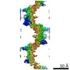
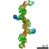
 Citation Citation | Journal: Cell / Year: 2024 Title: ZP2 cleavage blocks polyspermy by modulating the architecture of the egg coat. Authors: Shunsuke Nishio / Chihiro Emori / Benjamin Wiseman / Dirk Fahrenkamp / Elisa Dioguardi / Sara Zamora-Caballero / Marcel Bokhove / Ling Han / Alena Stsiapanava / Blanca Algarra / Yonggang Lu ...Authors: Shunsuke Nishio / Chihiro Emori / Benjamin Wiseman / Dirk Fahrenkamp / Elisa Dioguardi / Sara Zamora-Caballero / Marcel Bokhove / Ling Han / Alena Stsiapanava / Blanca Algarra / Yonggang Lu / Mayo Kodani / Rachel E Bainbridge / Kayla M Komondor / Anne E Carlson / Michael Landreh / Daniele de Sanctis / Shigeki Yasumasu / Masahito Ikawa / Luca Jovine /    Abstract: Following the fertilization of an egg by a single sperm, the egg coat or zona pellucida (ZP) hardens and polyspermy is irreversibly blocked. These events are associated with the cleavage of the N- ...Following the fertilization of an egg by a single sperm, the egg coat or zona pellucida (ZP) hardens and polyspermy is irreversibly blocked. These events are associated with the cleavage of the N-terminal region (NTR) of glycoprotein ZP2, a major subunit of ZP filaments. ZP2 processing is thought to inactivate sperm binding to the ZP, but its molecular consequences and connection with ZP hardening are unknown. Biochemical and structural studies show that cleavage of ZP2 triggers its oligomerization. Moreover, the structure of a native vertebrate egg coat filament, combined with AlphaFold predictions of human ZP polymers, reveals that two protofilaments consisting of type I (ZP3) and type II (ZP1/ZP2/ZP4) components interlock into a left-handed double helix from which the NTRs of type II subunits protrude. Together, these data suggest that oligomerization of cleaved ZP2 NTRs extensively cross-links ZP filaments, rigidifying the egg coat and making it physically impenetrable to sperm. | ||||||||||||
| History |
|
- Structure visualization
| Supplemental images |
|---|

- Downloads & links
-EMDB archive
| Map data | emd_56971.map.gz | 248.3 MB | EMDB map data format | |
|---|---|---|---|---|
| Header (meta data) | emd-56971-v30.xmlemd-56971.xml | 40.9 KB 40.9 KB | Display Display | EMDB header |
| FSC (resolution estimation) | emd_56971_fsc.xml | 23.5 KB | Display | FSC data file |
| Images |  emd_56971.png emd_56971.png | 60.4 KB | ||
| Filedesc metadata | emd-56971.cif.gz | 9.4 KB | ||
| Others | emd_56971_additional_1.map.gzemd_56971_additional_2.map.gzemd_56971_half_map_1.map.gzemd_56971_half_map_2.map.gz | 9 MB 8.7 MB 475.5 MB 475.5 MB | ||
| Archive directory |  http://ftp.pdbj.org/pub/emdb/structures/EMD-56971ftp://ftp.pdbj.org/pub/emdb/structures/EMD-56971 http://ftp.pdbj.org/pub/emdb/structures/EMD-56971ftp://ftp.pdbj.org/pub/emdb/structures/EMD-56971 | HTTPS FTP |
-Related structure data
| Related structure data |  28yjMC M: atomic model generated by this map C: citing same article ( |
|---|---|
| Similar structure data |

-Links
| EMDB pages | EMDB (EBI/PDBe) / EMDataResource |
|---|---|
| Related items in Molecule of the Month |


-Map
| File | Download / File: emd_56971.map.gz / Format: CCP4 / Size: 512 MB / Type: IMAGE STORED AS FLOATING POINT NUMBER (4 BYTES) | ||||||||||||||||||||||||||||||||||||
|---|---|---|---|---|---|---|---|---|---|---|---|---|---|---|---|---|---|---|---|---|---|---|---|---|---|---|---|---|---|---|---|---|---|---|---|---|---|
| Annotation | cryoSPARC map | ||||||||||||||||||||||||||||||||||||
| Projections & slices | Image control
Images are generated by Spider. | ||||||||||||||||||||||||||||||||||||
| Voxel size | X=Y=Z: 0.7336 Å | ||||||||||||||||||||||||||||||||||||
| Density |
| ||||||||||||||||||||||||||||||||||||
| Symmetry | Space group: 1 | ||||||||||||||||||||||||||||||||||||
| Details | EMDB XML:
|
 Z (Sec.)
 Y (Row.)
 X (Col.)


-Supplemental data
-Additional map: LocScale-2.0 feature-enhanced map, boxed around model
| File | emd_56971_additional_1.map | ||||||||||||
|---|---|---|---|---|---|---|---|---|---|---|---|---|---|
| Annotation | LocScale-2.0 feature-enhanced map, boxed around model | ||||||||||||
| Projections & Slices |
| ||||||||||||
| Density Histograms |


-Additional map: EMReady2 postprocessed map, boxed around model
| File | emd_56971_additional_2.map | ||||||||||||
|---|---|---|---|---|---|---|---|---|---|---|---|---|---|
| Annotation | EMReady2 postprocessed map, boxed around model | ||||||||||||
| Projections & Slices |
| ||||||||||||
| Density Histograms |


-Half map: cryoSPARC half-map 1
| File | emd_56971_half_map_1.map | ||||||||||||
|---|---|---|---|---|---|---|---|---|---|---|---|---|---|
| Annotation | cryoSPARC half-map 1 | ||||||||||||
| Projections & Slices |
| ||||||||||||
| Density Histograms |


-Half map: cryoSPARC half-map 2
| File | emd_56971_half_map_2.map | ||||||||||||
|---|---|---|---|---|---|---|---|---|---|---|---|---|---|
| Annotation | cryoSPARC half-map 2 | ||||||||||||
| Projections & Slices |
| ||||||||||||
| Density Histograms |


- Sample components
-Entire : Native chicken ZPD homopolymeric filament
| Entire | Name: Native chicken ZPD homopolymeric filament |
|---|---|
| Components |
|
-Supramolecule #1: Native chicken ZPD homopolymeric filament
| Supramolecule | Name: Native chicken ZPD homopolymeric filament / type: complex / ID: 1 / Parent: 0 / Macromolecule list: all |
|---|---|
| Source (natural) | Organism: Location in cell: Zona pellucida (specialized extracellular matrix surrounding the oocyte) |
-Macromolecule #1: Uromodulin
| Macromolecule | Name: Uromodulin / type: protein_or_peptide / ID: 1 / Number of copies: 4 / Enantiomer: LEVO |
|---|---|
| Source (natural) | Organism: |
| Molecular weight | Theoretical: 36.825234 KDa |
| Sequence | String: NKSELVSPHN SRGRFALRAK RSSDACVPNP CQHHGGCQVI EDRPICSCKP GFTGAFCQDV VLKLACEEEH MKMMVRKEVF ELLKIPREL VHLKNQACKV SEREEEGEMF FAATLTGENH TACGSVIQQN SSHVSYSNII ETGREAHRGV ISRSFQLEVH F SCVYAYEQ ...String: NKSELVSPHN SRGRFALRAK RSSDACVPNP CQHHGGCQVI EDRPICSCKP GFTGAFCQDV VLKLACEEEH MKMMVRKEVF ELLKIPREL VHLKNQACKV SEREEEGEMF FAATLTGENH TACGSVIQQN SSHVSYSNII ETGREAHRGV ISRSFQLEVH F SCVYAYEQ VVKMPFALTP VDKLVQFMVR EGHFNVSMRL YKTASYLEPY DLLTAAVPIT DTLYVMLKIE GQHQLRYFLL SV EDCWATP SADPYQDVLH ELIEQGCPHD ETVTYLNAIG ESTTAKFSFQ MFQFVGYPKV FLHCRVRLCL PDGPEPCAKQ CPT LWRS UniProtKB: Uromodulin |
-Experimental details
-Structure determination
| Method | cryo EM |
|---|---|
 Processing Processing | single particle reconstruction |
| Aggregation state | filament |
-Sample preparation
| Concentration | 0.7 mg/mL |
|---|---|
| Buffer | pH: 7 / Component - Concentration: 10.0 mM / Component - Formula: C8H18N2O4S / Component - Name: HEPES |
| Vitrification | Cryogen name: NITROGEN |
- Electron microscopy
| Microscope | TFS KRIOS |
|---|---|
| Specialist optics | Energy filter - Name: TFS Selectris / Energy filter - Slit width: 10 eV |
| Image recording | Film or detector model: FEI FALCON IV (4k x 4k) / Number real images: 19953 / Average exposure time: 2.75 sec. / Average electron dose: 53.0 e/Å2 |
| Electron beam | Acceleration voltage: 300 kV / Electron source:  FIELD EMISSION GUN FIELD EMISSION GUN |
| Electron optics | C2 aperture diameter: 50.0 µm / Illumination mode: SPOT SCAN / Imaging mode: BRIGHT FIELD / Cs: 2.7 mm / Nominal defocus max: 2.8000000000000003 µm / Nominal defocus min: 0.7000000000000001 µm / Nominal magnification: 165000 |
| Sample stage | Specimen holder model: FEI TITAN KRIOS AUTOGRID HOLDER / Cooling holder cryogen: NITROGEN |
| Experimental equipment |  Model: Titan Krios / Image courtesy: FEI Company |
+Image processing

-Atomic model buiding 1
| Initial model | Chain - Source name: AlphaFold / Chain - Initial model type: in silico model |
|---|---|
| Details | Model building was initiated using a local installation of AlphaFold 3 to predict a minimal filament fragment comprising one full-length subunit (chain A) and two partial subunits (chains B and C). The top-ranked prediction was rigid-body fitted into an initial 8.6 A-resolution map (postprocessed with EMReady2) using UCSF Chimera, followed by flexible fitting with Namdinator. Non-resolved terminal regions were trimmed, and well-defined N-glycan densities were manually built in Coot. The model was refined by real-space refinement in Phenix using NCS constraints and increased non-bonded interaction weights, followed by ADP refinement against the unsharpened map. This model served as a starting point for extension with an additional EGF and ZP-N domain from a fourth subunit (chain D). The extended model was docked into the present 4.6 A-resolution map, manually adjusted, and subjected to flexible fitting using the cryo-EM minimizer from cg2all; subsequently, it was refined using Refmac Servalcat task of CCP-EM Doppio, applying global NCS restraints, ProSMART-derived self-restraints, and increased non-bonded interaction weights. Following additional rounds of manual model rebuilding in Coot and real-space refinement in PHENIX (as described above), with positional refinement performed against a LocScale2-postprocessed map and ADP refinement against the unsharpened map, the model was validated using MolProbity and PHENIX. Note that the EGF domain of chain A (and, to a lesser extent, portions of its ZP-N domain near the postprocessed map boundary and the distal regions of the EGF domains in chains C and D) are weakly defined in the density, consistent with their elevated B-factors. These regions were retained in the model to preserve biological completeness, with their conformations constrained by NCS during refinement. |
| Refinement | Space: REAL / Protocol: FLEXIBLE FIT |
| Output model | PDB-28yj: |
