 Movie
Movie Controller

[English] 日本語
 Yorodumi
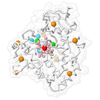
- EMDB-55097: focused structure of regulatory domains of cis-basal conformer of... -
+ Open data

- Basic information
| Entry |  | |||||||||
|---|---|---|---|---|---|---|---|---|---|---|
| Title | focused structure of regulatory domains of cis-basal conformer of human CBS induced by non-activating allosteric SAO ligand - by Helical approach | |||||||||
 Map data Map data | ||||||||||
 Sample Sample |
| |||||||||
 Keywords Keywords | transsulfuration pathway / L-serine hydro-lyase / heme-binding protein / CBS domain / LYASE | |||||||||
| Function / homology |  Function and homology information Function and homology informationCysteine formation from homocysteine / L-homocysteine catabolic process / modified amino acid binding / cystathionine beta-synthase / cystathionine beta-synthase activity / Metabolism of ingested SeMet, Sec, MeSec into H2Se / carbon monoxide binding / hydrogen sulfide biosynthetic process / L-serine catabolic process / homocysteine metabolic process ...Cysteine formation from homocysteine / L-homocysteine catabolic process / modified amino acid binding / cystathionine beta-synthase / cystathionine beta-synthase activity / Metabolism of ingested SeMet, Sec, MeSec into H2Se / carbon monoxide binding / hydrogen sulfide biosynthetic process / L-serine catabolic process / homocysteine metabolic process / L-cysteine catabolic process / L-cysteine biosynthetic process / L-serine metabolic process / transsulfuration / nitric oxide binding / DNA protection / S-adenosyl-L-methionine binding / nitrite reductase (NO-forming) activity / oxygen binding / response to nutrient levels / cellular response to insulin stimulus / pyridoxal phosphate binding / cellular response to hypoxia / ubiquitin protein ligase binding / heme binding / negative regulation of apoptotic process / enzyme binding / protein homodimerization activity / identical protein binding / nucleus / cytosol / cytoplasm Similarity search - Function | |||||||||
| Biological species |  Homo sapiens (human) Homo sapiens (human) | |||||||||
| Method | helical reconstruction / cryo EM / Resolution: 4.04 Å | |||||||||
 Authors Authors | Inayathulla M / Tomas M | |||||||||
| Funding support |  Switzerland, 1 items Switzerland, 1 items
| |||||||||
 Citation Citation | Journal: Nat Commun / Year: 2026 Title: Structural basis for a filamentous morpheein model of human cystathionine beta-synthase. Authors: Inayathulla Mohammed / Ela Mijatovic / Thilo Magnus Philipp / Lucia Janickova / Kelly Ascencao / Francisco J Asturias / Luis Alfonso Martinez-Cruz / Csaba Szabo / Henning Stahlberg / Tomas Majtan /   Abstract: Human cystathionine beta-synthase (CBS) is a vital enzyme that regulates sulfur amino acid metabolism, hydrogen sulfide production, and cellular redox balance. Using a multidisciplinary approach, we ...Human cystathionine beta-synthase (CBS) is a vital enzyme that regulates sulfur amino acid metabolism, hydrogen sulfide production, and cellular redox balance. Using a multidisciplinary approach, we demonstrate that CBS functions as a filamentous morpheein, with its stability, turnover, and activity governed by dynamic quaternary structural transitions. Three distinct filamentous assemblies were resolved by cryo-EM and are mediated by the oligomerization loop (residues 516-525): (i) ligand-free trans-dimers that form trans-basal filaments with basal stability and activity, (ii) adenosylornithine-bound cis-dimers that assemble into stabilized cis-basal filaments and (iii) S-adenosylmethionine-bound allo-dimers, which, together with cis-dimers, form highly stable, allo-activated stacked filaments. These reversible filamentous assemblies redefine CBS biology by integrating oligomerization and allosteric regulation within a morpheein framework. These findings provide a transformative perspective on CBS function and open avenues for pharmacological targeting of dysregulated CBS in various diseases including homocystinuria, cancer, and Down syndrome. | |||||||||
| History |
|
- Structure visualization
| Supplemental images |
|---|

- Downloads & links
-EMDB archive
| Map data | emd_55097.map.gz | 32.9 MB | EMDB map data format | |
|---|---|---|---|---|
| Header (meta data) | emd-55097-v30.xmlemd-55097.xml | 20.6 KB 20.6 KB | Display Display | EMDB header |
| FSC (resolution estimation) | emd_55097_fsc.xml | 9.6 KB | Display | FSC data file |
| Images |  emd_55097.png emd_55097.png | 62 KB | ||
| Masks | emd_55097_msk_1.map | 64 MB | Mask map | |
| Filedesc metadata | emd-55097.cif.gz | 6.3 KB | ||
| Others | emd_55097_additional_1.map.gzemd_55097_half_map_1.map.gzemd_55097_half_map_2.map.gz | 16.6 MB 59.3 MB 59.3 MB | ||
| Archive directory |  https://data.pdbj.org/pub/emdb/structures/EMD-55097ftp://data.pdbj.org/pub/emdb/structures/EMD-55097 https://data.pdbj.org/pub/emdb/structures/EMD-55097ftp://data.pdbj.org/pub/emdb/structures/EMD-55097 | HTTPS FTP |
-Related structure data
| Related structure data |  9spvMC  9shmC  9shnC  9si8C  9smlC  9spwC  9sq0C  9sqqC M: atomic model generated by this map C: citing same article ( |
|---|---|
| Similar structure data |


-Links
| EMDB pages | EMDB (EBI/PDBe) / EMDataResource |
|---|---|
| Related items in Molecule of the Month |
-Map
| File | Download / File: emd_55097.map.gz / Format: CCP4 / Size: 64 MB / Type: IMAGE STORED AS FLOATING POINT NUMBER (4 BYTES) | ||||||||||||||||||||||||||||||||||||
|---|---|---|---|---|---|---|---|---|---|---|---|---|---|---|---|---|---|---|---|---|---|---|---|---|---|---|---|---|---|---|---|---|---|---|---|---|---|
| Projections & slices | Image control
Images are generated by Spider. | ||||||||||||||||||||||||||||||||||||
| Voxel size | X=Y=Z: 1.04 Å | ||||||||||||||||||||||||||||||||||||
| Density |
| ||||||||||||||||||||||||||||||||||||
| Symmetry | Space group: 1 | ||||||||||||||||||||||||||||||||||||
| Details | EMDB XML:
|
 Z (Sec.)
 Y (Row.)
 X (Col.)


-Supplemental data
-Mask #1
| File | emd_55097_msk_1.map | ||||||||||||
|---|---|---|---|---|---|---|---|---|---|---|---|---|---|
| Projections & Slices |
| ||||||||||||
| Density Histograms |


-Additional map: unsharpened map
| File | emd_55097_additional_1.map | ||||||||||||
|---|---|---|---|---|---|---|---|---|---|---|---|---|---|
| Annotation | unsharpened map | ||||||||||||
| Projections & Slices |
| ||||||||||||
| Density Histograms |


-Half map: #2
| File | emd_55097_half_map_1.map | ||||||||||||
|---|---|---|---|---|---|---|---|---|---|---|---|---|---|
| Projections & Slices |
| ||||||||||||
| Density Histograms |


-Half map: #1
| File | emd_55097_half_map_2.map | ||||||||||||
|---|---|---|---|---|---|---|---|---|---|---|---|---|---|
| Projections & Slices |
| ||||||||||||
| Density Histograms |


- Sample components
-Entire : human Cystathionine beta-synthase Oligomeric complex
| Entire | Name: human Cystathionine beta-synthase Oligomeric complex |
|---|---|
| Components |
|
-Supramolecule #1: human Cystathionine beta-synthase Oligomeric complex
| Supramolecule | Name: human Cystathionine beta-synthase Oligomeric complex / type: complex / ID: 1 / Parent: 0 / Macromolecule list: #1 |
|---|---|
| Source (natural) | Organism: Homo sapiens (human) |
-Macromolecule #1: Cystathionine beta-synthase
| Macromolecule | Name: Cystathionine beta-synthase / type: protein_or_peptide / ID: 1 / Number of copies: 10 / Enantiomer: LEVO / EC number: cystathionine beta-synthase |
|---|---|
| Source (natural) | Organism: Homo sapiens (human) |
| Molecular weight | Theoretical: 60.665375 KDa |
| Recombinant expression | Organism:   Escherichia coli BL21(DE3) (bacteria) Escherichia coli BL21(DE3) (bacteria) |
| Sequence | String: MPSETPQAEV GPTGCPHRSG PHSAKGSLEK GSPEDKEAKE PLWIRPDAPS RCTWQLGRPA SESPHHHTAP AKSPKILPDI LKKIGDTPM VRINKIGKKF GLKCELLAKC EFFNAGGSVK DRISLRMIED AERDGTLKPG DTIIEPTSGN TGIGLALAAA V RGYRCIIV ...String: MPSETPQAEV GPTGCPHRSG PHSAKGSLEK GSPEDKEAKE PLWIRPDAPS RCTWQLGRPA SESPHHHTAP AKSPKILPDI LKKIGDTPM VRINKIGKKF GLKCELLAKC EFFNAGGSVK DRISLRMIED AERDGTLKPG DTIIEPTSGN TGIGLALAAA V RGYRCIIV MPEKMSSEKV DVLRALGAEI VRTPTNARFD SPESHVGVAW RLKNEIPNSH ILDQYRNASN PLAHYDTTAD EI LQQCDGK LDMLVASVGT GGTITGIARK LKEKCPGCRI IGVDPEGSIL AEPEELNQTE QTTYEVEGIG YDFIPTVLDR TVV DKWFKS NDEEAFTFAR MLIAQEGLLC GGSAGSTVAV AVKAAQELQE GQRCVVILPD SVRNYMTKFL SDRWMLQKGF LKEE DLTEK KPWWWHLRVQ ELGLSAPLTV LPTITCGHTI EILREKGFDQ APVVDEAGVI LGMVTLGNML SSLLAGKVQP SDQVG KVIY KQFKQIRLTD TLGRLSHILE MDHFALVVHE QIQYHSTGKS SQRQMVFGVV TAIDLLNFVA AQERDQK UniProtKB: Cystathionine beta-synthase |
-Macromolecule #2: SINEFUNGIN
| Macromolecule | Name: SINEFUNGIN / type: ligand / ID: 2 / Number of copies: 8 / Formula: SFG |
|---|---|
| Molecular weight | Theoretical: 381.387 Da |
| Chemical component information | 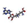 ChemComp-SFG: |
-Experimental details
-Structure determination
| Method | cryo EM |
|---|---|
 Processing Processing | helical reconstruction |
| Aggregation state | filament |
-Sample preparation
| Buffer | pH: 7.4 |
|---|---|
| Vitrification | Cryogen name: ETHANE |
- Electron microscopy
| Microscope | TFS KRIOS |
|---|---|
| Image recording | Film or detector model: FEI FALCON IV (4k x 4k) / Average electron dose: 50.0 e/Å2 |
| Electron beam | Acceleration voltage: 300 kV / Electron source:  FIELD EMISSION GUN FIELD EMISSION GUN |
| Electron optics | Illumination mode: FLOOD BEAM / Imaging mode: BRIGHT FIELD / Nominal defocus max: 2.5 µm / Nominal defocus min: 1.2 µm |
| Experimental equipment |  Model: Titan Krios / Image courtesy: FEI Company |
+Image processing

-Atomic model buiding 1
| Refinement | Space: REAL / Protocol: FLEXIBLE FIT |
|---|---|
| Output model | PDB-9spv: |
