 Movie
Movie Controller


+ Open data

- Basic information
| Entry |  | |||||||||||||||||||||||||||||||||||||||
|---|---|---|---|---|---|---|---|---|---|---|---|---|---|---|---|---|---|---|---|---|---|---|---|---|---|---|---|---|---|---|---|---|---|---|---|---|---|---|---|---|
| Title | Trimeric photosystem I of Acaryochloris marina NIES-2412 | |||||||||||||||||||||||||||||||||||||||
 Map data Map data | ||||||||||||||||||||||||||||||||||||||||
 Sample Sample |
| |||||||||||||||||||||||||||||||||||||||
 Keywords Keywords | Chlorophyll d / photosystem I / ELECTRON TRANSPORT | |||||||||||||||||||||||||||||||||||||||
| Biological species |  Acaryochloris marina (bacteria) / Acaryochloris marina NIES-2412 (bacteria) Acaryochloris marina (bacteria) / Acaryochloris marina NIES-2412 (bacteria) | |||||||||||||||||||||||||||||||||||||||
| Method | single particle reconstruction / cryo EM / Resolution: 2.63 Å | |||||||||||||||||||||||||||||||||||||||
 Authors Authors | Leong HF / Consoli G | |||||||||||||||||||||||||||||||||||||||
| Funding support |  Netherlands, European Union, Netherlands, European Union,  United Kingdom, 12 items United Kingdom, 12 items
| |||||||||||||||||||||||||||||||||||||||
 Citation Citation | Journal: Protein Sci / Year: 2018 Title: UCSF ChimeraX: Meeting modern challenges in visualization and analysis. Authors: Thomas D Goddard / Conrad C Huang / Elaine C Meng / Eric F Pettersen / Gregory S Couch / John H Morris / Thomas E Ferrin /  Abstract: UCSF ChimeraX is next-generation software for the visualization and analysis of molecular structures, density maps, 3D microscopy, and associated data. It addresses challenges in the size, scope, and ...UCSF ChimeraX is next-generation software for the visualization and analysis of molecular structures, density maps, 3D microscopy, and associated data. It addresses challenges in the size, scope, and disparate types of data attendant with cutting-edge experimental methods, while providing advanced options for high-quality rendering (interactive ambient occlusion, reliable molecular surface calculations, etc.) and professional approaches to software design and distribution. This article highlights some specific advances in the areas of visualization and usability, performance, and extensibility. ChimeraX is free for noncommercial use and is available from http://www.rbvi.ucsf.edu/chimerax/ for Windows, Mac, and Linux. | |||||||||||||||||||||||||||||||||||||||
| History |
|
- Structure visualization
| Supplemental images |
|---|

- Downloads & links
-EMDB archive
| Map data | emd_54956.map.gz | 415.3 MB |  EMDB map data format EMDB map data format | |
|---|---|---|---|---|
| Header (meta data) | emd-54956-v30.xmlemd-54956.xml | 33.5 KB 33.5 KB | Display Display | EMDB header |
| FSC (resolution estimation) | emd_54956_fsc.xml | 19.7 KB | Display | FSC data file |
| Images |  emd_54956.png emd_54956.png | 196.5 KB | ||
| Filedesc metadata | emd-54956.cif.gz | 8.8 KB | ||
| Others | emd_54956_half_map_1.map.gzemd_54956_half_map_2.map.gz | 763.6 MB 763.6 MB | ||
| Archive directory |  http://ftp.pdbj.org/pub/emdb/structures/EMD-54956ftp://ftp.pdbj.org/pub/emdb/structures/EMD-54956 http://ftp.pdbj.org/pub/emdb/structures/EMD-54956ftp://ftp.pdbj.org/pub/emdb/structures/EMD-54956 | HTTPS FTP |
-Related structure data


-Links
| EMDB pages | EMDB (EBI/PDBe) / EMDataResource |
|---|
-Map
| File | Download / File: emd_54956.map.gz / Format: CCP4 / Size: 824 MB / Type: IMAGE STORED AS FLOATING POINT NUMBER (4 BYTES) | ||||||||||||||||||||||||||||||||||||
|---|---|---|---|---|---|---|---|---|---|---|---|---|---|---|---|---|---|---|---|---|---|---|---|---|---|---|---|---|---|---|---|---|---|---|---|---|---|
| Projections & slices | Image control
Images are generated by Spider. | ||||||||||||||||||||||||||||||||||||
| Voxel size | X=Y=Z: 1.058 Å | ||||||||||||||||||||||||||||||||||||
| Density |
| ||||||||||||||||||||||||||||||||||||
| Symmetry | Space group: 1 | ||||||||||||||||||||||||||||||||||||
| Details | EMDB XML:
|
 Z (Sec.)
 Y (Row.)
 X (Col.)


-Supplemental data
-Half map: #2
| File | emd_54956_half_map_1.map | ||||||||||||
|---|---|---|---|---|---|---|---|---|---|---|---|---|---|
| Projections & Slices |
| ||||||||||||
| Density Histograms |


-Half map: #1
| File | emd_54956_half_map_2.map | ||||||||||||
|---|---|---|---|---|---|---|---|---|---|---|---|---|---|
| Projections & Slices |
| ||||||||||||
| Density Histograms |


- Sample components
+Entire : Trimeric PSI
+Supramolecule #1: Trimeric PSI
+Macromolecule #1: Photosystem I protein PsaA
+Macromolecule #2: Photosystem I protein PsaB
+Macromolecule #3: Photosystem I protein PsaC
+Macromolecule #4: Photosystem I protein PsaD
+Macromolecule #5: Photosystem I protein PsaE
+Macromolecule #6: Photosystem I protein PsaF
+Macromolecule #7: Photosystem I protein PsaI
+Macromolecule #8: Photosystem I protein PsaJ
+Macromolecule #9: Photosystem I protein PsaK
+Macromolecule #10: Photosystem I protein PsaL
+Macromolecule #11: Photosystem I protein PsaM
+Macromolecule #12: Photosystem I protein PsaX2
+Macromolecule #13: CHLOROPHYLL D ISOMER
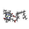
+Macromolecule #14: PHEOPHYTIN A

+Macromolecule #15: CHLOROPHYLL D
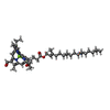
+Macromolecule #16: PHYLLOQUINONE
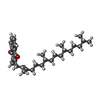
+Macromolecule #17: (6'S)-beta,epsilon-carotene
+Macromolecule #18: 1,2-DIPALMITOYL-PHOSPHATIDYL-GLYCEROLE
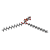
+Macromolecule #19: 1,2-DISTEAROYL-MONOGALACTOSYL-DIGLYCERIDE
+Macromolecule #20: IRON/SULFUR CLUSTER
+Macromolecule #21: water

-Experimental details
-Structure determination
| Method | cryo EM |
|---|---|
 Processing Processing | single particle reconstruction |
| Aggregation state | particle |
-Sample preparation
| Concentration | 1 mg/mL |
|---|---|
| Buffer | pH: 6.5 Details: 50 mM MES, 1 M betaine monohydrate, 20 mM CaCl2, 5 mM MgCl2 and 10% (v/v) glycerol, pH 6.5 adjusted with NaOH |
| Grid | Model: UltrAuFoil / Material: GOLD / Mesh: 300 / Pretreatment - Type: PLASMA CLEANING / Pretreatment - Time: 100 sec. |
| Vitrification | Cryogen name: ETHANE / Chamber humidity: 95 % / Chamber temperature: 277 K / Instrument: FEI VITROBOT MARK IV |
- Electron microscopy
| Microscope | TFS KRIOS |
|---|---|
| Image recording | Film or detector model: GATAN K3 (6k x 4k) / Digitization - Dimensions - Width: 5120 pixel / Digitization - Dimensions - Height: 4096 pixel / Average electron dose: 40.0 e/Å2 |
| Electron beam | Acceleration voltage: 300 kV / Electron source:  FIELD EMISSION GUN FIELD EMISSION GUN |
| Electron optics | Illumination mode: SPOT SCAN / Imaging mode: BRIGHT FIELD / Cs: 2.7 mm / Nominal defocus max: 2.0 µm / Nominal defocus min: 0.8 µm |
| Experimental equipment |  Model: Titan Krios / Image courtesy: FEI Company |

