 Movie
Movie Controller


+ Open data

- Basic information
| Entry |  | |||||||||
|---|---|---|---|---|---|---|---|---|---|---|
| Title | E.coli cytochrome bd-I dimer bound to Aurachin D | |||||||||
 Map data Map data | ||||||||||
 Sample Sample |
| |||||||||
 Keywords Keywords | Respiration / Complex / Cytochrome / OXIDOREDUCTASE | |||||||||
| Function / homology |  Function and homology information Function and homology informationquinol oxidase (electrogenic, proton-motive force generating) / oxidoreductase activity, acting on diphenols and related substances as donors / cytochrome complex / aerobic electron transport chain / outer membrane / oxidoreductase activity, acting on diphenols and related substances as donors, oxygen as acceptor / oxidative phosphorylation / cellular response to hypoxia / electron transfer activity / heme binding ...quinol oxidase (electrogenic, proton-motive force generating) / oxidoreductase activity, acting on diphenols and related substances as donors / cytochrome complex / aerobic electron transport chain / outer membrane / oxidoreductase activity, acting on diphenols and related substances as donors, oxygen as acceptor / oxidative phosphorylation / cellular response to hypoxia / electron transfer activity / heme binding / membrane / metal ion binding / plasma membrane Similarity search - Function | |||||||||
| Biological species |   Escherichia coli (E. coli) / Escherichia coli K-12 (bacteria) Escherichia coli (E. coli) / Escherichia coli K-12 (bacteria) | |||||||||
| Method | single particle reconstruction / cryo EM / Resolution: 2.47 Å | |||||||||
 Authors Authors | van der Velden TT / Kayastha K / Bruenle S / Jeuken LJC | |||||||||
| Funding support |  Netherlands, 1 items Netherlands, 1 items
| |||||||||
 Citation Citation | Journal: To Be Published Title: Molecular basis of quinol turnover and inhibition of a bd-type oxidase Authors: van der Velden TT / Kayastha K / Pelser F / Bruenle S / Jeuken LJC | |||||||||
| History |
|
- Structure visualization
| Supplemental images |
|---|

- Downloads & links
-EMDB archive
| Map data | emd_54812.map.gz | 92 MB | EMDB map data format | |
|---|---|---|---|---|
| Header (meta data) | emd-54812-v30.xmlemd-54812.xml | 25.7 KB 25.7 KB | Display Display | EMDB header |
| FSC (resolution estimation) | emd_54812_fsc.xml | 11.8 KB | Display | FSC data file |
| Images |  emd_54812.png emd_54812.png | 145.7 KB | ||
| Masks | emd_54812_msk_1.map | 178 MB | Mask map | |
| Filedesc metadata | emd-54812.cif.gz | 7.5 KB | ||
| Others | emd_54812_half_map_1.map.gzemd_54812_half_map_2.map.gz | 165.1 MB 165.1 MB | ||
| Archive directory |  http://ftp.pdbj.org/pub/emdb/structures/EMD-54812ftp://ftp.pdbj.org/pub/emdb/structures/EMD-54812 http://ftp.pdbj.org/pub/emdb/structures/EMD-54812ftp://ftp.pdbj.org/pub/emdb/structures/EMD-54812 | HTTPS FTP |
-Related structure data
| Related structure data |  9sejMC  9se4C  9sfhC  9sfjC  9sfvC M: atomic model generated by this map C: citing same article ( |
|---|---|
| Similar structure data |


-Links
| EMDB pages | EMDB (EBI/PDBe) / EMDataResource |
|---|
-Map
| File | Download / File: emd_54812.map.gz / Format: CCP4 / Size: 178 MB / Type: IMAGE STORED AS FLOATING POINT NUMBER (4 BYTES) | ||||||||||||||||||||||||||||||||||||
|---|---|---|---|---|---|---|---|---|---|---|---|---|---|---|---|---|---|---|---|---|---|---|---|---|---|---|---|---|---|---|---|---|---|---|---|---|---|
| Projections & slices | Image control
Images are generated by Spider. | ||||||||||||||||||||||||||||||||||||
| Voxel size | X=Y=Z: 0.88 Å | ||||||||||||||||||||||||||||||||||||
| Density |
| ||||||||||||||||||||||||||||||||||||
| Symmetry | Space group: 1 | ||||||||||||||||||||||||||||||||||||
| Details | EMDB XML:
|
 Z (Sec.)
 Y (Row.)
 X (Col.)


-Supplemental data
-Mask #1
| File | emd_54812_msk_1.map | ||||||||||||
|---|---|---|---|---|---|---|---|---|---|---|---|---|---|
| Projections & Slices |
| ||||||||||||
| Density Histograms |


-Half map: #2
| File | emd_54812_half_map_1.map | ||||||||||||
|---|---|---|---|---|---|---|---|---|---|---|---|---|---|
| Projections & Slices |
| ||||||||||||
| Density Histograms |


-Half map: #1
| File | emd_54812_half_map_2.map | ||||||||||||
|---|---|---|---|---|---|---|---|---|---|---|---|---|---|
| Projections & Slices |
| ||||||||||||
| Density Histograms |


- Sample components
+Entire : E.coli cytochrome bd-I dimer bound to Aurachin D
+Supramolecule #1: E.coli cytochrome bd-I dimer bound to Aurachin D
+Macromolecule #1: Cytochrome bd-I ubiquinol oxidase subunit 1
+Macromolecule #2: Cytochrome bd-I ubiquinol oxidase subunit 2
+Macromolecule #3: Cytochrome bd-I ubiquinol oxidase CydH (Uncharacterized protein YnhF)
+Macromolecule #4: Cytochrome bd-I ubiquinol oxidase subunit X
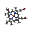
+Macromolecule #5: PROTOPORPHYRIN IX CONTAINING FE
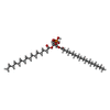
+Macromolecule #6: 2-(HEXADECANOYLOXY)-1-[(PHOSPHONOOXY)METHYL]ETHYL HEXADECANOATE
+Macromolecule #7: Ubiquinone-8

+Macromolecule #8: Aurachin D

+Macromolecule #9: OXYGEN MOLECULE
+Macromolecule #10: TRANS-HEME D HYDROXYCHLORIN GAMMA-SPIROLACTONE
+Macromolecule #11: (1S)-2-{[{[(2R)-2,3-DIHYDROXYPROPYL]OXY}(HYDROXY)PHOSPHORYL]OXY}-...
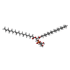
+Macromolecule #12: (2S)-3-(hexadecanoyloxy)-2-[(9Z)-octadec-9-enoyloxy]propyl 2-(tri...
+Macromolecule #13: water

-Experimental details
-Structure determination
| Method | cryo EM |
|---|---|
 Processing Processing | single particle reconstruction |
| Aggregation state | particle |
-Sample preparation
| Concentration | 1.5 mg/mL |
|---|---|
| Buffer | pH: 7.4 |
| Grid | Model: UltrAuFoil R1.2/1.3 / Material: GOLD / Pretreatment - Type: GLOW DISCHARGE |
| Vitrification | Cryogen name: ETHANE / Chamber humidity: 100 % / Instrument: FEI VITROBOT MARK IV |
- Electron microscopy
| Microscope | TFS GLACIOS |
|---|---|
| Image recording | Film or detector model: TFS FALCON 4i (4k x 4k) / Average electron dose: 100.0 e/Å2 |
| Electron beam | Acceleration voltage: 200 kV / Electron source:  FIELD EMISSION GUN FIELD EMISSION GUN |
| Electron optics | Illumination mode: FLOOD BEAM / Imaging mode: BRIGHT FIELD / Cs: 2.7 mm / Nominal defocus max: 2.0 µm / Nominal defocus min: 0.8 µm / Nominal magnification: 130000 |

