quinol oxidase (electrogenic, proton-motive force generating) / oxidoreductase activity, acting on diphenols and related substances as donors / cytochrome complex / aerobic electron transport chain / outer membrane / oxidoreductase activity, acting on diphenols and related substances as donors, oxygen as acceptor / oxidative phosphorylation / cellular response to hypoxia / electron transfer activity / heme binding ...quinol oxidase (electrogenic, proton-motive force generating) / oxidoreductase activity, acting on diphenols and related substances as donors / cytochrome complex / aerobic electron transport chain / outer membrane / oxidoreductase activity, acting on diphenols and related substances as donors, oxygen as acceptor / oxidative phosphorylation / cellular response to hypoxia / electron transfer activity / heme binding / membrane / metal ion binding / plasma membrane Similarity search - Function
: / Membrane protein YnhF-like / Cyd operon protein YbgT / Membrane bound YbgT-like / Membrane bound YbgT-like protein / Cytochrome ubiquinol oxidase subunit 1 / Cytochrome ubiquinol oxidase subunit 2 / Cytochrome bd terminal oxidase subunit I / Cytochrome bd terminal oxidase subunit II Similarity search - Domain/homology
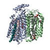
Journal: Sci Adv / Year: 2026 Title: Visualizing the mechanism of quinol oxidation and inhibition of a -type oxidase using cryo-EM. Authors: Tijn T van der Velden / Kanwal Kayastha / Famke Pelser / Steffen Brünle / Lars J C Jeuken / Abstract: Cytochrome is a prokaryotic terminal oxidase recognized as an antibiotic target against various pathogens. Despite its critical role in respiration, failure to capture the mechanism of quinol ...Cytochrome is a prokaryotic terminal oxidase recognized as an antibiotic target against various pathogens. Despite its critical role in respiration, failure to capture the mechanism of quinol oxidation and inhibition prohibits structure guided drug discovery. Here, we present cryo-electron microscopy structures of cytochrome -I in monomeric and dimeric forms, in several quinone and inhibitor-bound states. We identify a dynamic Q-loop lid that undergoes a disorder-to-order transition upon substrate binding to the dimer, completing the active site and enabling catalysis. Structure-guided mutagenesis confirms Tyr243 and Arg298 as conserved catalytic residues only found in long Q-loop oxidases, highlighting evolutionary divergence from other subfamilies. Inhibition by Aurachin D triggers refolding of the active site, occluding substrate access via an Asp239-mediated mechanism. The structural and mechanistic insights presented here establish a comprehensive framework, opening paths for drug discovery against oxidases.
In the structure databanks used in Yorodumi, some data are registered as the other names, "COVID-19 virus" and "2019-nCoV". Here are the details of the virus and the list of structure data.
Jan 31, 2019. EMDB accession codes are about to change! (news from PDBe EMDB page)
EMDB accession codes are about to change! (news from PDBe EMDB page)
The allocation of 4 digits for EMDB accession codes will soon come to an end. Whilst these codes will remain in use, new EMDB accession codes will include an additional digit and will expand incrementally as the available range of codes is exhausted. The current 4-digit format prefixed with “EMD-” (i.e. EMD-XXXX) will advance to a 5-digit format (i.e. EMD-XXXXX), and so on. It is currently estimated that the 4-digit codes will be depleted around Spring 2019, at which point the 5-digit format will come into force.
The EM Navigator/Yorodumi systems omit the EMD- prefix.
Related info.:Q: What is EMD? / ID/Accession-code notation in Yorodumi/EM Navigator
Yorodumi is a browser for structure data from EMDB, PDB, SASBDB, etc.
This page is also the successor to EM Navigator detail page, and also detail information page/front-end page for Omokage search.
The word "yorodu" (or yorozu) is an old Japanese word meaning "ten thousand". "mi" (miru) is to see.
Related info.:EMDB / PDB / SASBDB / Comparison of 3 databanks / Yorodumi Search / Aug 31, 2016. New EM Navigator & Yorodumi / Yorodumi Papers / Jmol/JSmol / Function and homology information / Changes in new EM Navigator and Yorodumi
 Movie
Movie Controller


 Open data
 Basic information
 Map data
 Sample
 Keywords
 Function and homology information
 Authors
 Netherlands, 1 items
  Citation
 Structure visualization
 Downloads & links
 emd_54801.png
 http://ftp.pdbj.org/pub/emdb/structures/EMD-54801


 Z (Sec.)
 Y (Row.)
 X (Col.)


 Sample components


 Processing
 Electron microscopy
 FIELD EMISSION GUN