 Movie
Movie Controller


+ Open data

- Basic information
| Entry |  | |||||||||
|---|---|---|---|---|---|---|---|---|---|---|
| Title | Unliganded dimer - bacterial | |||||||||
 Map data Map data | ||||||||||
 Sample Sample |
| |||||||||
 Keywords Keywords | Glycogen phosphorylase / TRANSFERASE | |||||||||
| Function / homology |  Function and homology information Function and homology informationpeptidyl-histidine phosphorylation / glycogen phosphorylase / glycogen phosphorylase activity / glycogen catabolic process / pyridoxal phosphate binding / protein homodimerization activity / cytoplasm Similarity search - Function | |||||||||
| Biological species |  | |||||||||
| Method | single particle reconstruction / cryo EM / Resolution: 3.1 Å | |||||||||
 Authors Authors | Di Domenico V / Mastrella L / Alcaide-Jimenez A / Villegas-Ruiz JC / D'Angelo C / Cifuente JO / Connell SR / Guerin ME | |||||||||
| Funding support |  Spain, 1 items Spain, 1 items
| |||||||||
 Citation Citation | Journal: Nat Commun / Year: 2026 Title: Structural basis for phosphorylation and allosteric regulation of bacterial glycogen phosphorylase by histidine phosphocarrier protein. Authors: Valerio Di Domenico / Jorick Franceus / Leonardo Mastrella / Emma De Beul / Adrià Alcaide-Jiménez / Francisco Paredes-Martínez / Juan Carlos Villegas-Ruiz / Elena Holden / Alejandro ...Authors: Valerio Di Domenico / Jorick Franceus / Leonardo Mastrella / Emma De Beul / Adrià Alcaide-Jiménez / Francisco Paredes-Martínez / Juan Carlos Villegas-Ruiz / Elena Holden / Alejandro Delgado Rey / Cecilia D'Angelo / Javier O Cifuente / Weston B Struwe / Ricardo M Biondi / Alberto Marina / Sean R Connell / Justin L P Benesch / Patricia Casino / Christophe Colleoni / Tom Desmet / Marcelo E Guerin /     Abstract: Protein phosphorylation is a universal regulatory mechanism, controlling virtually all aspects of bacterial physiology and pathogenesis, yet histidine phosphorylation remains among the least ...Protein phosphorylation is a universal regulatory mechanism, controlling virtually all aspects of bacterial physiology and pathogenesis, yet histidine phosphorylation remains among the least understood. The histidine phosphocarrier protein HPr not only drives bacterial glucose transmembrane uptake through the phosphotransferase system (PTS), but also controls key enzymes for central carbon metabolism, including glycogen phosphorylase (GP). Here we report cryoEM structures of multimeric Escherichia coli GP and their complexes with HPr. The EM maps reveal an unanticipated density at H806 of GP, consistent with histidine phosphorylation within a histidine-rich pocket at the N-terminal domain. Enzymatic assays reveal that HPr transfers a phosphoryl group to the N1 position of a histidine residue in GP. Through an integrative structural, mutational and functional approach, we uncover the molecular basis of HPr- GP selectivity and define the allosteric mechanism by which HPr regulates GP. We establish histidine phosphorylation as a mechanism of GP regulation, expanding the traditional paradigm of glycogen metabolism control in bacteria. | |||||||||
| History |
|
- Structure visualization
| Supplemental images |
|---|

- Downloads & links
-EMDB archive
| Map data | emd_54650.map.gz | 87.9 MB | EMDB map data format | |
|---|---|---|---|---|
| Header (meta data) | emd-54650-v30.xmlemd-54650.xml | 24 KB 24 KB | Display Display | EMDB header |
| Images |  emd_54650.png emd_54650.png | 41.4 KB | ||
| Filedesc metadata | emd-54650.cif.gz | 7.3 KB | ||
| Others | emd_54650_additional_1.map.gzemd_54650_half_map_1.map.gzemd_54650_half_map_2.map.gz | 158 MB 164.8 MB 164.8 MB | ||
| Archive directory |  http://ftp.pdbj.org/pub/emdb/structures/EMD-54650ftp://ftp.pdbj.org/pub/emdb/structures/EMD-54650 http://ftp.pdbj.org/pub/emdb/structures/EMD-54650ftp://ftp.pdbj.org/pub/emdb/structures/EMD-54650 | HTTPS FTP |
-Related structure data
| Related structure data |  9s7vMC  9s86C  9s8bC 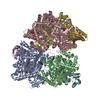 9s8kC M: atomic model generated by this map C: citing same article ( |
|---|---|
| Similar structure data |


-Links
| EMDB pages | EMDB (EBI/PDBe) / EMDataResource |
|---|---|
| Related items in Molecule of the Month |

-Map
| File | Download / File: emd_54650.map.gz / Format: CCP4 / Size: 178 MB / Type: IMAGE STORED AS FLOATING POINT NUMBER (4 BYTES) | ||||||||||||||||||||||||||||||||||||
|---|---|---|---|---|---|---|---|---|---|---|---|---|---|---|---|---|---|---|---|---|---|---|---|---|---|---|---|---|---|---|---|---|---|---|---|---|---|
| Projections & slices | Image control
Images are generated by Spider. | ||||||||||||||||||||||||||||||||||||
| Voxel size | X=Y=Z: 0.645 Å | ||||||||||||||||||||||||||||||||||||
| Density |
| ||||||||||||||||||||||||||||||||||||
| Symmetry | Space group: 1 | ||||||||||||||||||||||||||||||||||||
| Details | EMDB XML:
|
 Z (Sec.)
 Y (Row.)
 X (Col.)


-Supplemental data
-Additional map: Sharpened map
| File | emd_54650_additional_1.map | ||||||||||||
|---|---|---|---|---|---|---|---|---|---|---|---|---|---|
| Annotation | Sharpened map | ||||||||||||
| Projections & Slices |
| ||||||||||||
| Density Histograms |


-Half map: #1
| File | emd_54650_half_map_1.map | ||||||||||||
|---|---|---|---|---|---|---|---|---|---|---|---|---|---|
| Projections & Slices |
| ||||||||||||
| Density Histograms |


-Half map: #2
| File | emd_54650_half_map_2.map | ||||||||||||
|---|---|---|---|---|---|---|---|---|---|---|---|---|---|
| Projections & Slices |
| ||||||||||||
| Density Histograms |


- Sample components
-Entire : Recombinantly purified Glycogen phosphorylase from E. coli
| Entire | Name: Recombinantly purified Glycogen phosphorylase from E. coli |
|---|---|
| Components |
|
-Supramolecule #1: Recombinantly purified Glycogen phosphorylase from E. coli
| Supramolecule | Name: Recombinantly purified Glycogen phosphorylase from E. coli type: complex / ID: 1 / Parent: 0 / Macromolecule list: all |
|---|---|
| Source (natural) | Organism: |
| Molecular weight | Theoretical: 930 KDa |
-Macromolecule #1: Glycogen phosphorylase
| Macromolecule | Name: Glycogen phosphorylase / type: protein_or_peptide / ID: 1 / Number of copies: 2 / Enantiomer: LEVO / EC number: glycogen phosphorylase |
|---|---|
| Source (natural) | Organism: |
| Molecular weight | Theoretical: 95.536625 KDa |
| Recombinant expression | Organism: |
| Sequence | String: MHHHHHHENL YFQGSGAMNA PFTYSSPTLS VEALKHSIAY KLMFTIGKDP VVANKHEWLN ATLFAVRDRL VERWLRSNRA QLSQETRQV YYLSMEFLIG RTLSNAMLSL GIYEDVQGAL EAMGLNLEEL IDEENDPGLG NGGLGRLAAC FLDSLATLGL P GRGYGIRY ...String: MHHHHHHENL YFQGSGAMNA PFTYSSPTLS VEALKHSIAY KLMFTIGKDP VVANKHEWLN ATLFAVRDRL VERWLRSNRA QLSQETRQV YYLSMEFLIG RTLSNAMLSL GIYEDVQGAL EAMGLNLEEL IDEENDPGLG NGGLGRLAAC FLDSLATLGL P GRGYGIRY DYGMFKQNIV NGSQKESPDY WLEYGNPWEF KRHNTRYKVR FGGRIQQEGK KTRWIETEEI LGVAYDQIIP GY DTDATNT LRLWSAQASS EINLGKFNQG DYFAAVEDKN HSENVSRVLY PDDSTYSGRE LRLRQEYFLV SSTIQDILSR HYQ LHKTYD NLADKIAIHL NDTHPVLSIP EMMRLLIDEH QFSWDDAFEV CCQVFSYTNH TLMSEALETW PVDMLGKILP RHLQ IIFEI NDYFLKTLQE QYPNDTDLLG RASIIDESNG RRVRMAWLAV VVSHKVNGVS ELHSNLMVQS LFADFAKIFP GRFTN VTNG VTPRRWLAVA NPSLSAVLDE HLGRNWRTDL SLLNELQQHC DFPMVNHAVH QAKLENKKRL AEYIAQQLNV VVNPKA LFD VQIKRIHEYK RQLMNVLHVI TRYNRIKADP DAKWVPRVNI FGGKAASAYY MAKHIIHLIN DVAKVINNDP QIGDKLK VV FIPNYSVSLA QLIIPAADLS EQISLAGTEA SGTSNM(LLP)FAL NGALTIGTLD GANVEMLDHV GADNIFIFGN TAEE VEELR RQGYKPREYY EKDEELHQVL TQIGSGVFSP EDPGRYRDLV DSLINFGDHY QVLADYRSYV DCQDKVDELY ELQEE WTAK AMLNIANMGY FSSDRTIKEY ADHIWHIDPV RL UniProtKB: Glycogen phosphorylase |
-Experimental details
-Structure determination
| Method | cryo EM |
|---|---|
 Processing Processing | single particle reconstruction |
| Aggregation state | particle |
-Sample preparation
| Concentration | 0.3 mg/mL |
|---|---|
| Buffer | pH: 8 |
| Grid | Model: Quantifoil R1.2/1.3 / Material: COPPER / Mesh: 300 / Support film - Material: CARBON / Support film - topology: CONTINUOUS / Pretreatment - Type: GLOW DISCHARGE |
| Vitrification | Cryogen name: ETHANE |
- Electron microscopy
| Microscope | TFS KRIOS |
|---|---|
| Image recording | Film or detector model: GATAN K3 (6k x 4k) / Average electron dose: 50.0 e/Å2 |
| Electron beam | Acceleration voltage: 300 kV / Electron source:  FIELD EMISSION GUN FIELD EMISSION GUN |
| Electron optics | Illumination mode: OTHER / Imaging mode: DIFFRACTION / Nominal defocus max: 2.0 µm / Nominal defocus min: 0.7000000000000001 µm |
| Experimental equipment |  Model: Titan Krios / Image courtesy: FEI Company |
+Image processing
-Atomic model buiding 1
| Initial model | Chain - Source name: AlphaFold / Chain - Initial model type: in silico model |
|---|---|
| Refinement | Protocol: OTHER |
| Output model | PDB-9s7v: |
