 Movie
Movie Controller

[English] 日本語
 Yorodumi
- PDB-9s8b: Structure of glycogen phosphorylase - dimeric form - in complex w... -
+ Open data

- Basic information
| Entry | Database: PDB / ID: 9s8b | ||||||||||||||||||||||||
|---|---|---|---|---|---|---|---|---|---|---|---|---|---|---|---|---|---|---|---|---|---|---|---|---|---|
| Title | Structure of glycogen phosphorylase - dimeric form - in complex with HPr from Escherichia coli | ||||||||||||||||||||||||
 Components Components |
| ||||||||||||||||||||||||
 Keywords Keywords | TRANSFERASE / Glycogen phosphorylase | ||||||||||||||||||||||||
| Function / homology |  Function and homology information Function and homology informationphosphotransferase activity, nitrogenous group as acceptor / regulation of carbon utilization / antisigma factor binding / peptidyl-histidine phosphorylation / positive regulation of glycogen catabolic process / phosphoenolpyruvate-dependent sugar phosphotransferase system / glycogen phosphorylase / glycogen phosphorylase activity / glycogen catabolic process / enzyme regulator activity ...phosphotransferase activity, nitrogenous group as acceptor / regulation of carbon utilization / antisigma factor binding / peptidyl-histidine phosphorylation / positive regulation of glycogen catabolic process / phosphoenolpyruvate-dependent sugar phosphotransferase system / glycogen phosphorylase / glycogen phosphorylase activity / glycogen catabolic process / enzyme regulator activity / enzyme inhibitor activity / enzyme activator activity / pyridoxal phosphate binding / protein homodimerization activity / cytosol / cytoplasm Similarity search - Function | ||||||||||||||||||||||||
| Biological species |   Escherichia coli (E. coli) Escherichia coli (E. coli) | ||||||||||||||||||||||||
| Method | ELECTRON MICROSCOPY / single particle reconstruction / cryo EM / Resolution: 2.79 Å | ||||||||||||||||||||||||
 Authors Authors | Di Domenico, V. / Mastrella, L. / Alcaide-Jimenez, A. / Villegas-Ruiz, J.C. / D'Angelo, C. / Cifuente, J.O. / Connell, S.R. / Guerin, M.E. | ||||||||||||||||||||||||
| Funding support |  Spain, 1items Spain, 1items
| ||||||||||||||||||||||||
 Citation Citation | Journal: Nat Commun / Year: 2026 Title: Structural basis for phosphorylation and allosteric regulation of bacterial glycogen phosphorylase by histidine phosphocarrier protein. Authors: Valerio Di Domenico / Jorick Franceus / Leonardo Mastrella / Emma De Beul / Adrià Alcaide-Jiménez / Francisco Paredes-Martínez / Juan Carlos Villegas-Ruiz / Elena Holden / Alejandro ...Authors: Valerio Di Domenico / Jorick Franceus / Leonardo Mastrella / Emma De Beul / Adrià Alcaide-Jiménez / Francisco Paredes-Martínez / Juan Carlos Villegas-Ruiz / Elena Holden / Alejandro Delgado Rey / Cecilia D'Angelo / Javier O Cifuente / Weston B Struwe / Ricardo M Biondi / Alberto Marina / Sean R Connell / Justin L P Benesch / Patricia Casino / Christophe Colleoni / Tom Desmet / Marcelo E Guerin /     Abstract: Protein phosphorylation is a universal regulatory mechanism, controlling virtually all aspects of bacterial physiology and pathogenesis, yet histidine phosphorylation remains among the least ...Protein phosphorylation is a universal regulatory mechanism, controlling virtually all aspects of bacterial physiology and pathogenesis, yet histidine phosphorylation remains among the least understood. The histidine phosphocarrier protein HPr not only drives bacterial glucose transmembrane uptake through the phosphotransferase system (PTS), but also controls key enzymes for central carbon metabolism, including glycogen phosphorylase (GP). Here we report cryoEM structures of multimeric Escherichia coli GP and their complexes with HPr. The EM maps reveal an unanticipated density at H806 of GP, consistent with histidine phosphorylation within a histidine-rich pocket at the N-terminal domain. Enzymatic assays reveal that HPr transfers a phosphoryl group to the N1 position of a histidine residue in GP. Through an integrative structural, mutational and functional approach, we uncover the molecular basis of HPr- GP selectivity and define the allosteric mechanism by which HPr regulates GP. We establish histidine phosphorylation as a mechanism of GP regulation, expanding the traditional paradigm of glycogen metabolism control in bacteria. | ||||||||||||||||||||||||
| History |
|
- Structure visualization
| Structure viewer | Molecule: MolmilJmol/JSmol |
|---|
- Downloads & links
-Download
| PDBx/mmCIF format | 9s8b.cif.gz | 353.2 KB | Display | PDBx/mmCIF format |
|---|---|---|---|---|
| PDB format | pdb9s8b.ent.gz | 279 KB | Display | PDB format |
| PDBx/mmJSON format | 9s8b.json.gz | Tree view | PDBx/mmJSON format | |
| Others |  Other downloads Other downloads |
-Validation report
| Arichive directory | https://data.pdbj.org/pub/pdb/validation_reports/s8/9s8bftp://data.pdbj.org/pub/pdb/validation_reports/s8/9s8b | HTTPS FTP |
|---|
-Related structure data
| Related structure data |  54658MC  9s7vC  9s86C 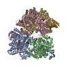 9s8kC C: citing same article ( M: map data used to model this data |
|---|---|
| Similar structure data |


-Links
 PDBj
PDBj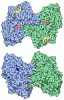
- Assembly
| Deposited unit | 
|
|---|---|
| 1 |
|
-Components
| #1: Protein | Mass: 95536.625 Da / Num. of mol.: 2 Source method: isolated from a genetically manipulated source Source: (gene. exp.) Escherichia coli (E. coli) / Gene: glgP, glgY, b3428, JW3391 / Production host: Escherichia coli (E. coli) / References: UniProt: P0AC86, glycogen phosphorylase#2: Protein | | Mass: 11236.499 Da / Num. of mol.: 1 Source method: isolated from a genetically manipulated source Source: (gene. exp.) Escherichia coli (E. coli) / Gene: ptsH, hpr, b2415, JW2408 / Production host: Escherichia coli (E. coli) / References: UniProt: P0AA04#3: Protein | | Mass: 11156.519 Da / Num. of mol.: 1 Source method: isolated from a genetically manipulated source Source: (gene. exp.) Escherichia coli (E. coli) / Gene: ptsH, hpr, b2415, JW2408 / Production host: Escherichia coli (E. coli) / References: UniProt: P0AA04#4: Water | ChemComp-HOH / |  Mass: 18.015 Da / Num. of mol.: 31 / Source method: isolated from a natural source / Formula: H2O Mass: 18.015 Da / Num. of mol.: 31 / Source method: isolated from a natural source / Formula: H2OHas ligand of interest | Y | Has protein modification | Y | |
|---|
-Experimental details
-Experiment
| Experiment | Method: ELECTRON MICROSCOPY |
|---|---|
| EM experiment | Aggregation state: PARTICLE / 3D reconstruction method: single particle reconstruction |
- Sample preparation
| Component | Name: Recombinantly purified Glycogen phosphorylase in complex with the phosphocarrier protein HPr from E. coli Type: COMPLEX / Entity ID: #1-#2 / Source: RECOMBINANT |
|---|---|
| Molecular weight | Value: 0.228 MDa / Experimental value: YES |
| Source (natural) | Organism: Escherichia coli (E. coli) |
| Source (recombinant) | Organism: Escherichia coli (E. coli) |
| Buffer solution | pH: 8 |
| Specimen | Conc.: 0.3 mg/ml / Embedding applied: NO / Shadowing applied: NO / Staining applied: NO / Vitrification applied: YES |
| Specimen support | Grid material: COPPER / Grid mesh size: 300 divisions/in. / Grid type: Quantifoil R1.2/1.3 |
| Vitrification | Instrument: FEI VITROBOT MARK IV / Cryogen name: ETHANE / Humidity: 90 % / Chamber temperature: 277.15 K |
- Electron microscopy imaging
| Experimental equipment |  Model: Titan Krios / Image courtesy: FEI Company |
|---|---|
| Microscopy | Model: TFS KRIOS |
| Electron gun | Electron source:  FIELD EMISSION GUN / Accelerating voltage: 300 kV / Illumination mode: OTHER FIELD EMISSION GUN / Accelerating voltage: 300 kV / Illumination mode: OTHER |
| Electron lens | Mode: BRIGHT FIELD / Nominal magnification: 130000 X / Nominal defocus max: 2000 nm / Nominal defocus min: 700 nm |
| Image recording | Electron dose: 50 e/Å2 / Film or detector model: GATAN K3 (6k x 4k) |
- Processing
| EM software |
| |||||||||||||||||||||||||||||||||||
|---|---|---|---|---|---|---|---|---|---|---|---|---|---|---|---|---|---|---|---|---|---|---|---|---|---|---|---|---|---|---|---|---|---|---|---|---|
| Image processing |
| |||||||||||||||||||||||||||||||||||
| CTF correction |
| |||||||||||||||||||||||||||||||||||
| Symmetry |
| |||||||||||||||||||||||||||||||||||
| 3D reconstruction |
| |||||||||||||||||||||||||||||||||||
| Atomic model building | Protocol: OTHER | |||||||||||||||||||||||||||||||||||
| Atomic model building | PDB-ID: 9S7V Accession code: 9S7V / Source name: PDB / Type: experimental model | |||||||||||||||||||||||||||||||||||
| Refinement | Highest resolution: 2.79 Å |
