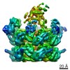
- EMDB-5271: High resolution helical reconstruction of the bacterial p-type AT... -
+
データを開く
IDまたはキーワード:
読み込み中...
-
基本情報
登録情報
データベース: EMDB / ID: EMD-5271
タイトル
High resolution helical reconstruction of the bacterial p-type ATPase copper transporter CopA.
マップデータ
This is a cubic section encompassing the unit cell of a Fourier-Bessel reconstructed map of tubular vesicles - crystals of deltaC-CopA from Archaeoglobus fulgidus.
ジャーナル: Structure / 年: 2011 タイトル: The architecture of CopA from Archeaoglobus fulgidus studied by cryo-electron microscopy and computational docking. 著者: Gregory S Allen / Chen-Chou Wu / Tim Cardozo / David L Stokes / 要旨: CopA uses ATP to pump Cu(+) across cell membranes. X-ray crystallography has defined atomic structures of several related P-type ATPases. We have determined a structure of CopA at 10 Å resolution ...CopA uses ATP to pump Cu(+) across cell membranes. X-ray crystallography has defined atomic structures of several related P-type ATPases. We have determined a structure of CopA at 10 Å resolution by cryo-electron microscopy of a new crystal form and used computational molecular docking to study the interactions between the N-terminal metal-binding domain (NMBD) and other elements of the molecule. We found that the shorter-chain lipids used to produce these crystals are associated with movements of the cytoplasmic domains, with a novel dimer interface and with disordering of the NMBD, thus offering evidence for the transience of its interaction with the other cytoplasmic domains. Docking identified a binding site that matched the location of the NMBD in our previous structure by cryo-electron microscopy, allowing a more detailed view of its binding configuration and further support for its role in autoinhibition.
ダウンロード / ファイル: emd_5271.map.gz / 形式: CCP4 / 大きさ: 789.1 KB / タイプ: IMAGE STORED AS FLOATING POINT NUMBER (4 BYTES)
注釈
This is a cubic section encompassing the unit cell of a Fourier-Bessel reconstructed map of tubular vesicles - crystals of deltaC-CopA from Archaeoglobus fulgidus.
ボクセルのサイズ
X=Y=Z: 2 Å
密度
表面レベル
登録者による: 130.0 / ムービー #1: 130
最小 - 最大
-30.964200000000002 - 199.592999999999989
平均 (標準偏差)
104.631 (±38.0715)
対称性
空間群: 1
詳細
EMDB XML:
マップ形状
Axis order
X
Y
Z
Origin
0
0
0
サイズ
51
50
81
Spacing
51
50
81
セル
A: 100 Å / B: 102 Å / C: 162 Å α=β=γ: 90 °
CCP4マップ ヘッダ情報:
mode
Image stored as Reals
Å/pix. X/Y/Z
2
2
2
M x/y/z
50
51
81
origin x/y/z
0.000
0.000
0.000
length x/y/z
100.000
102.000
162.000
α/β/γ
90.000
90.000
90.000
start NX/NY/NZ
-62
-62
-62
NX/NY/NZ
125
125
125
MAP C/R/S
1
2
3
start NC/NR/NS
0
0
0
NC/NR/NS
50
51
81
D min/max/mean
-30.964
199.593
104.631
-
添付データ
-
試料の構成要素
-
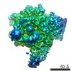
全体 : deltaC-CopA in DMPC-DOPE lipids
全体
名称: deltaC-CopA in DMPC-DOPE lipids
要素
試料: deltaC-CopA in DMPC-DOPE lipids
タンパク質・ペプチド: membrane protein膜タンパク質
-
超分子 #1000: deltaC-CopA in DMPC-DOPE lipids
超分子
名称: deltaC-CopA in DMPC-DOPE lipids / タイプ: sample / ID: 1000 詳細: deltaC-CopA tubular crystals were grown with a 4-to-1 mixture of DMPC-DOPE at a protein concentration of 1 mg per mL and at a lipid-to-protein weight ratio of 0.4. Dialysis was carried out ...詳細: deltaC-CopA tubular crystals were grown with a 4-to-1 mixture of DMPC-DOPE at a protein concentration of 1 mg per mL and at a lipid-to-protein weight ratio of 0.4. Dialysis was carried out for 5 days in 50 ul dialysis buttons at 30 degrees C against 500 mL of 50 mM MES, pH 6.1, 25 mM Na2SO4, 25 mM K2SO4, 200 uM BCDS, 10 mM MgSO4, and 2 mM beta-mercaptoethanol. Stock solutions of lipid were made in dodecyl octaethylene glycol ether (C12E8) at 1 mg lipid per 2 mg detergent. Number unique components: 1
 ムービー
ムービー コントローラー


 データを開く
 基本情報
基本情報 マップデータ
 試料
試料 キーワード
キーワード p-type ATPase / copper transporter / CopA / Adenosine Triphosphatases /
  機能・相同性情報

 データ登録者
 引用
引用
 構造の表示
構造の表示
 ダウンロードとリンク
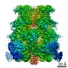
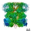
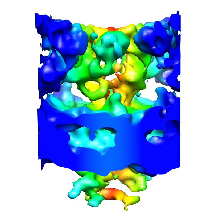 emd_5271_1.png
 http://ftp.pdbj.org/pub/emdb/structures/EMD-5271


 試料の構成要素
 解析
解析 電子顕微鏡法