DNA topoisomerase type II (double strand cut, ATP-hydrolyzing) activity / DNA topoisomerase (ATP-hydrolysing) / DNA topological change / chromosome / DNA replication / magnesium ion binding / DNA binding / ATP binding Similarity search - Function
DNA topoisomerase VI, subunit B, C-terminal / Type 2 DNA topoisomerase 6 subunit B C-terminal domain / DNA topoisomerase VI, subunit A / : / All-beta domain in DNA topoisomerase VI alpha subunit / Spo11/DNA topoisomerase VI subunit A / Spo11/DNA topoisomerase VI, subunit A, N-terminal / Topoisomerase 6 subunit A/Spo11, TOPRIM domain / Spo11/DNA topoisomerase VI subunit A superfamily / Type IIB DNA topoisomerase ...DNA topoisomerase VI, subunit B, C-terminal / Type 2 DNA topoisomerase 6 subunit B C-terminal domain / DNA topoisomerase VI, subunit A / : / All-beta domain in DNA topoisomerase VI alpha subunit / Spo11/DNA topoisomerase VI subunit A / Spo11/DNA topoisomerase VI, subunit A, N-terminal / Topoisomerase 6 subunit A/Spo11, TOPRIM domain / Spo11/DNA topoisomerase VI subunit A superfamily / Type IIB DNA topoisomerase / Topoisomerase 6 subunit A/Spo11, Toprim domain / Topoisomerase (Topo) IIB-type catalytic domain profile. / DNA topoisomerase VI, subunit B / DNA topoisomerase VI, subunit B, transducer / Topoisomerase VI B subunit, transducer / Histidine kinase-, DNA gyrase B-, and HSP90-like ATPase / Histidine kinase-like ATPases / Histidine kinase/HSP90-like ATPase / Histidine kinase/HSP90-like ATPase superfamily / Ribosomal protein S13-like, H2TH / Ribosomal protein S5 domain 2-type fold, subgroup / Ribosomal protein S5 domain 2-type fold / Winged helix-like DNA-binding domain superfamily Similarity search - Domain/homology
National Institutes of Health/National Cancer Institute (NIH/NCI)
5R35 CA263778
United States
National Institutes of Health/National Institute of General Medical Sciences (NIH/NIGMS)
1F32 GM128269
United States
Citation
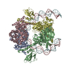
Journal: Nat Commun / Year: 2026 Title: Supercoiled DNA recognition and cleavage control in topoisomerase VI. Authors: Daniel E Richman / Timothy J Wendorff / Fahad Rashid / Curtis Beck / Qianyun Yan / Haley R Johnson / Ryan A Eckerty / Jonathan M Fogg / Matthew L Baker / Lynn Zechiedrich / James M Berger / Abstract: Type II topoisomerases modulate DNA supercoiling and resolve chromosome entanglements. Type IIB topoisomerases, exemplified by DNA topoisomerase VI (Top6), are used by plants and archaea to support ...Type II topoisomerases modulate DNA supercoiling and resolve chromosome entanglements. Type IIB topoisomerases, exemplified by DNA topoisomerase VI (Top6), are used by plants and archaea to support endoreduplication and cell proliferation, respectively; homologs of Top6 further serve to initiate meiotic recombination in eukaryotes and constitute the nuclease portion of MksBEFG/Wadjet/Gabija bacterial defense systems. To understand how such factors act upon DNA, we determine structures of Top6 bound to supercoiled minicircles in cleaved and uncleaved states using single-particle electron cryo-microscopy. The structures show that Top6 binds a curved 74 bp region of the supercoiled minicircle DNA and that it cuts at a distinct deformability motif, explaining its preference for supercoiled substrates and highlighting the role of DNA plasticity in cleavage site selection. Dynamic protein-DNA interactions and an unanticipated tension sensor help recognize bent DNA and couple ATPase disposition to cleavage state activation. Our observations explain how DNA recognition and cleavage by type II topoisomerases are regulated by interdependent structural changes in DNA and the enzyme.
Name: 306 bp minicircle DNA / type: complex / ID: 3 / Parent: 1 / Macromolecule list: #3-#6 Details: Approximately 80 bp of the 306 bp minicircle are visible in EM imaging of the Top6-DNA complex
Source (natural)
Organism: unidentified (others)
+
Macromolecule #1: Type 2 DNA topoisomerase 6 subunit A
Macromolecule
Name: Type 2 DNA topoisomerase 6 subunit A / type: protein_or_peptide / ID: 1 / Number of copies: 2 / Enantiomer: LEVO / EC number: DNA topoisomerase (ATP-hydrolysing)
Macromolecule #2: Type 2 DNA topoisomerase 6 subunit B
Macromolecule
Name: Type 2 DNA topoisomerase 6 subunit B / type: protein_or_peptide / ID: 2 / Number of copies: 2 / Enantiomer: LEVO / EC number: DNA topoisomerase (ATP-hydrolysing)
Name: POTASSIUM ION / type: ligand / ID: 9 / Number of copies: 2 / Formula: K
Molecular weight
Theoretical: 39.098 Da
-
Experimental details
-
Structure determination
Method
cryo EM
Processing
single particle reconstruction
Aggregation state
particle
-
Sample preparation
Concentration
0.1 mg/mL
Buffer
pH: 7.5 Component:
Concentration
Formula
Name
20.0 mM
C8H18N2O4S
HEPES
50.0 mM
C5H8KNO4
potassium glutamate
10.0 mM
CaCl2
calcium chloride
1.0 percent (v/v)
C3H8O3
glycerol
1.0 mM
C9H15O6P
TCEP
0.25 mM
C29H32O13
etoposide
1.0 mM
C10H17N6O12P3
ADPNP
Details: Etoposide was not observed in the structure
Grid
Model: Au-flat 1.2/1.3 / Material: GOLD / Mesh: 300 / Support film - #0 - Film type ID: 1 / Support film - #0 - Material: CARBON / Support film - #0 - topology: CONTINUOUS / Support film - #0 - Film thickness: 4 / Support film - #1 - Film type ID: 2 / Support film - #1 - Material: GOLD / Support film - #1 - topology: HOLEY / Support film - #1 - Film thickness: 45 / Pretreatment - Type: GLOW DISCHARGE / Pretreatment - Time: 15 sec. / Pretreatment - Atmosphere: AIR / Pretreatment - Pressure: 0.035 kPa / Details: Current set to 15 milliamp
Vitrification
Cryogen name: ETHANE / Chamber humidity: 100 % / Chamber temperature: 283 K / Instrument: FEI VITROBOT MARK IV / Details: Vitrification carried out in air.
Details
Summary of order of assembly: minicircle DNA, Top6, then additional buffer components, ending with ADPNP. Sample was soluble and monodisperse.
-
Electron microscopy
Microscope
TFS KRIOS
Specialist optics
Energy filter - Name: TFS Selectris / Energy filter - Slit width: 10 eV
Image recording
Film or detector model: TFS FALCON 4i (4k x 4k) / Digitization - Dimensions - Width: 4096 pixel / Digitization - Dimensions - Height: 4096 pixel / Number grids imaged: 1 / Number real images: 26728 / Average exposure time: 4.0 sec. / Average electron dose: 30.0 e/Å2
Electron beam
Acceleration voltage: 300 kV / Electron source: FIELD EMISSION GUN
In the structure databanks used in Yorodumi, some data are registered as the other names, "COVID-19 virus" and "2019-nCoV". Here are the details of the virus and the list of structure data.
Jan 31, 2019. EMDB accession codes are about to change! (news from PDBe EMDB page)
EMDB accession codes are about to change! (news from PDBe EMDB page)
The allocation of 4 digits for EMDB accession codes will soon come to an end. Whilst these codes will remain in use, new EMDB accession codes will include an additional digit and will expand incrementally as the available range of codes is exhausted. The current 4-digit format prefixed with “EMD-” (i.e. EMD-XXXX) will advance to a 5-digit format (i.e. EMD-XXXXX), and so on. It is currently estimated that the 4-digit codes will be depleted around Spring 2019, at which point the 5-digit format will come into force.
The EM Navigator/Yorodumi systems omit the EMD- prefix.
Related info.:Q: What is EMD? / ID/Accession-code notation in Yorodumi/EM Navigator
Yorodumi is a browser for structure data from EMDB, PDB, SASBDB, etc.
This page is also the successor to EM Navigator detail page, and also detail information page/front-end page for Omokage search.
The word "yorodu" (or yorozu) is an old Japanese word meaning "ten thousand". "mi" (miru) is to see.
Related info.:EMDB / PDB / SASBDB / Comparison of 3 databanks / Yorodumi Search / Aug 31, 2016. New EM Navigator & Yorodumi / Yorodumi Papers / Jmol/JSmol / Function and homology information / Changes in new EM Navigator and Yorodumi
 Movie
Movie Controller

 Yorodumi
 Open data
 Basic information
 Map data
 Sample
 Keywords
 Function and homology information
 Methanosarcina mazei Go1 (archaea) / unidentified (others)
 Authors
 United States, 2 items
  Citation
 Structure visualization
 Downloads & links
 emd_49972.png
 http://ftp.pdbj.org/pub/emdb/structures/EMD-49972


 Z (Sec.)
 Y (Row.)
 X (Col.)


 Sample components

 Processing
 Electron microscopy
 FIELD EMISSION GUN


