 Movie
Movie Controller

[English] 日本語
 Yorodumi
- PDB-9o0g: CryoEM structure of M. mazei topoisomerase VI(A-E342Q)-minicircle... -
+ Open data

- Basic information
| Entry | Database: PDB / ID: 9o0g | ||||||||||||||||||||||||||||||
|---|---|---|---|---|---|---|---|---|---|---|---|---|---|---|---|---|---|---|---|---|---|---|---|---|---|---|---|---|---|---|---|
| Title | CryoEM structure of M. mazei topoisomerase VI(A-E342Q)-minicircle DNA complex in cleavage state | ||||||||||||||||||||||||||||||
 Components Components |
| ||||||||||||||||||||||||||||||
 Keywords Keywords | ISOMERASE/DNA / TOPRIM / GHKL / minicircle DNA / Spo11 / ISOMERASE-DNA complex | ||||||||||||||||||||||||||||||
| Function / homology |  Function and homology information Function and homology informationDNA topoisomerase type II (double strand cut, ATP-hydrolyzing) activity / DNA topoisomerase (ATP-hydrolysing) / DNA topological change / chromosome / DNA replication / magnesium ion binding / DNA binding / ATP binding Similarity search - Function | ||||||||||||||||||||||||||||||
| Biological species |  Methanosarcina mazei Go1 (archaea) Methanosarcina mazei Go1 (archaea)unidentified (others) | ||||||||||||||||||||||||||||||
| Method | ELECTRON MICROSCOPY / single particle reconstruction / cryo EM / Resolution: 2.5 Å | ||||||||||||||||||||||||||||||
 Authors Authors | Richman, D.E. / Berger, J.M. | ||||||||||||||||||||||||||||||
| Funding support |  United States, 2items United States, 2items
| ||||||||||||||||||||||||||||||
 Citation Citation | Journal: Nat Commun / Year: 2026 Title: Supercoiled DNA recognition and cleavage control in topoisomerase VI. Authors: Daniel E Richman / Timothy J Wendorff / Fahad Rashid / Curtis Beck / Qianyun Yan / Haley R Johnson / Ryan A Eckerty / Jonathan M Fogg / Matthew L Baker / Lynn Zechiedrich / James M Berger / Abstract: Type II topoisomerases modulate DNA supercoiling and resolve chromosome entanglements. Type IIB topoisomerases, exemplified by DNA topoisomerase VI (Top6), are used by plants and archaea to support ...Type II topoisomerases modulate DNA supercoiling and resolve chromosome entanglements. Type IIB topoisomerases, exemplified by DNA topoisomerase VI (Top6), are used by plants and archaea to support endoreduplication and cell proliferation, respectively; homologs of Top6 further serve to initiate meiotic recombination in eukaryotes and constitute the nuclease portion of MksBEFG/Wadjet/Gabija bacterial defense systems. To understand how such factors act upon DNA, we determine structures of Top6 bound to supercoiled minicircles in cleaved and uncleaved states using single-particle electron cryo-microscopy. The structures show that Top6 binds a curved 74 bp region of the supercoiled minicircle DNA and that it cuts at a distinct deformability motif, explaining its preference for supercoiled substrates and highlighting the role of DNA plasticity in cleavage site selection. Dynamic protein-DNA interactions and an unanticipated tension sensor help recognize bent DNA and couple ATPase disposition to cleavage state activation. Our observations explain how DNA recognition and cleavage by type II topoisomerases are regulated by interdependent structural changes in DNA and the enzyme. | ||||||||||||||||||||||||||||||
| History |
|
- Structure visualization
| Structure viewer | Molecule: MolmilJmol/JSmol |
|---|
- Downloads & links
-Download
| PDBx/mmCIF format | 9o0g.cif.gz | 1 MB | Display | PDBx/mmCIF format |
|---|---|---|---|---|
| PDB format | pdb9o0g.ent.gz | 687.4 KB | Display | PDB format |
| PDBx/mmJSON format | 9o0g.json.gz | Tree view | PDBx/mmJSON format | |
| Others |  Other downloads Other downloads |
-Validation report
| Arichive directory | https://data.pdbj.org/pub/pdb/validation_reports/o0/9o0gftp://data.pdbj.org/pub/pdb/validation_reports/o0/9o0g | HTTPS FTP |
|---|
-Related structure data
| Related structure data |  49972MC 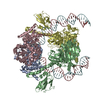 9o7oC  9o8pC  9o8zC  9o9mC M: map data used to model this data C: citing same article ( |
|---|---|
| Similar structure data |


-Links
 PDBj
PDBj


- Assembly
| Deposited unit | 
|
|---|---|
| 1 |
|
-Components
-Type 2 DNA topoisomerase 6 subunit ... , 2 types, 4 molecules ACBD
| #1: Protein | Mass: 42178.309 Da / Num. of mol.: 2 / Mutation: E342Q Source method: isolated from a genetically manipulated source Source: (gene. exp.) Methanosarcina mazei Go1 (archaea)Strain: ATCC BAA-159 / DSM 3647 / Goe1 / Go1 / JCM 11833 / OCM 88 Gene: top6A, MM_2418 / Plasmid: pST39 / Production host:  References: UniProt: Q8PUB7, DNA topoisomerase (ATP-hydrolysing) #2: Protein | Mass: 68853.367 Da / Num. of mol.: 2 Source method: isolated from a genetically manipulated source Source: (gene. exp.) Methanosarcina mazei Go1 (archaea)Strain: ATCC BAA-159 / DSM 3647 / Goe1 / Go1 / JCM 11833 / OCM 88 Gene: top6B, MM_2417 / Plasmid: pST39 / Production host: References: UniProt: Q8PUB8, DNA topoisomerase (ATP-hydrolysing) |
|---|
-Minicircle DNA ... , 4 types, 4 molecules FGHI
| #3: DNA chain | Mass: 10781.896 Da / Num. of mol.: 1 Source method: isolated from a genetically manipulated source Source: (gene. exp.) unidentified (others) / Production host: |
|---|---|
| #4: DNA chain | Mass: 12011.686 Da / Num. of mol.: 1 Source method: isolated from a genetically manipulated source Source: (gene. exp.) unidentified (others) / Plasmid: pUC / Production host: |
| #5: DNA chain | Mass: 11360.259 Da / Num. of mol.: 1 Source method: isolated from a genetically manipulated source Source: (gene. exp.) unidentified (others) / Production host: |
| #6: DNA chain | Mass: 11402.311 Da / Num. of mol.: 1 Source method: isolated from a genetically manipulated source Source: (gene. exp.) unidentified (others) / Production host: |
-Non-polymers , 3 types, 8 molecules 


| #7: Chemical | ChemComp-CA /  Mass: 40.078 Da / Num. of mol.: 4 / Source method: obtained synthetically / Formula: Ca Mass: 40.078 Da / Num. of mol.: 4 / Source method: obtained synthetically / Formula: Ca#8: Chemical |  Mass: 506.196 Da / Num. of mol.: 2 / Source method: obtained synthetically / Formula: C10H17N6O12P3 / Comment: AMP-PNP, energy-carrying molecule analogue*YM Mass: 506.196 Da / Num. of mol.: 2 / Source method: obtained synthetically / Formula: C10H17N6O12P3 / Comment: AMP-PNP, energy-carrying molecule analogue*YM#9: Chemical |  Mass: 39.098 Da / Num. of mol.: 2 / Source method: obtained synthetically / Formula: K Mass: 39.098 Da / Num. of mol.: 2 / Source method: obtained synthetically / Formula: K |
|---|
-Details
| Has ligand of interest | N |
|---|---|
| Has protein modification | N |
-Experimental details
-Experiment
| Experiment | Method: ELECTRON MICROSCOPY |
|---|---|
| EM experiment | Aggregation state: PARTICLE / 3D reconstruction method: single particle reconstruction |
- Sample preparation
| Component |
| ||||||||||||||||||||||||||||||||||||||||
|---|---|---|---|---|---|---|---|---|---|---|---|---|---|---|---|---|---|---|---|---|---|---|---|---|---|---|---|---|---|---|---|---|---|---|---|---|---|---|---|---|---|
| Molecular weight |
| ||||||||||||||||||||||||||||||||||||||||
| Source (natural) |
| ||||||||||||||||||||||||||||||||||||||||
| Source (recombinant) |
| ||||||||||||||||||||||||||||||||||||||||
| Buffer solution | pH: 7.5 / Details: Etoposide was not observed in the structure | ||||||||||||||||||||||||||||||||||||||||
| Buffer component |
| ||||||||||||||||||||||||||||||||||||||||
| Specimen | Conc.: 0.1 mg/ml / Embedding applied: NO / Shadowing applied: NO / Staining applied: NO / Vitrification applied: YES Details: Summary of order of assembly: minicircle DNA, Top6, then additional buffer components, ending with ADPNP. Sample was soluble and monodisperse. | ||||||||||||||||||||||||||||||||||||||||
| Specimen support | Details: Current set to 15 milliamp / Grid material: GOLD / Grid mesh size: 300 divisions/in. / Grid type: Au-flat 1.2/1.3 | ||||||||||||||||||||||||||||||||||||||||
| Vitrification | Instrument: FEI VITROBOT MARK IV / Cryogen name: ETHANE / Humidity: 100 % / Chamber temperature: 283 K / Details: Vitrification carried out in air |
- Electron microscopy imaging
| Experimental equipment |  Model: Titan Krios / Image courtesy: FEI Company |
|---|---|
| Microscopy | Model: TFS KRIOS |
| Electron gun | Electron source:  FIELD EMISSION GUN / Accelerating voltage: 300 kV / Illumination mode: FLOOD BEAM FIELD EMISSION GUN / Accelerating voltage: 300 kV / Illumination mode: FLOOD BEAM |
| Electron lens | Mode: BRIGHT FIELD / Nominal magnification: 130000 X / Nominal defocus max: 1800 nm / Nominal defocus min: 800 nm / Calibrated defocus min: 800 nm / Calibrated defocus max: 1800 nm / Cs: 2.7 mm / C2 aperture diameter: 50 µm / Alignment procedure: ZEMLIN TABLEAU |
| Specimen holder | Cryogen: NITROGEN / Specimen holder model: FEI TITAN KRIOS AUTOGRID HOLDER |
| Image recording | Average exposure time: 4 sec. / Electron dose: 30 e/Å2 / Film or detector model: TFS FALCON 4i (4k x 4k) / Num. of grids imaged: 1 / Num. of real images: 26728 |
| EM imaging optics | Energyfilter name: TFS Selectris / Energyfilter slit width: 10 eV |
| Image scans | Width: 4096 / Height: 4096 |
- Processing
| EM software |
| ||||||||||||||||||||||||||||||||||||||||||||
|---|---|---|---|---|---|---|---|---|---|---|---|---|---|---|---|---|---|---|---|---|---|---|---|---|---|---|---|---|---|---|---|---|---|---|---|---|---|---|---|---|---|---|---|---|---|
| CTF correction | Type: PHASE FLIPPING AND AMPLITUDE CORRECTION | ||||||||||||||||||||||||||||||||||||||||||||
| Particle selection | Num. of particles selected: 2479118 | ||||||||||||||||||||||||||||||||||||||||||||
| 3D reconstruction | Resolution: 2.5 Å / Resolution method: FSC 0.143 CUT-OFF / Num. of particles: 329731 / Algorithm: FOURIER SPACE / Symmetry type: POINT | ||||||||||||||||||||||||||||||||||||||||||||
| Atomic model building | Protocol: FLEXIBLE FIT / Space: REAL | ||||||||||||||||||||||||||||||||||||||||||||
| Atomic model building | 3D fitting-ID: 1
| ||||||||||||||||||||||||||||||||||||||||||||
| Refinement | Cross valid method: NONE Stereochemistry target values: GeoStd + Monomer Library + CDL v1.2 | ||||||||||||||||||||||||||||||||||||||||||||
| Displacement parameters | Biso mean: 74.27 Å2 | ||||||||||||||||||||||||||||||||||||||||||||
| Refine LS restraints |
|

