Movie
Movie Controller

[English] 日本語
 Yorodumi
- EMDB-48184: Antibody fragments from mAb824 and mAb926 bound to the adhesin pr... -
+ Open data

- Basic information
| Entry |  | |||||||||||||||
|---|---|---|---|---|---|---|---|---|---|---|---|---|---|---|---|---|
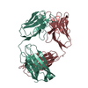
| Title | Antibody fragments from mAb824 and mAb926 bound to the adhesin protein FimH | |||||||||||||||
 Map data Map data | Antibody fragments from mAb824 and mAb926 bound to the E. coli adhesin protein FimH | |||||||||||||||
 Sample Sample |
| |||||||||||||||
 Keywords Keywords | Fimbrial tip / Lectin domain / Antibody fragments / Antibody-target complex / CELL ADHESION / CELL ADHESION-IMMUNE SYSTEM complex | |||||||||||||||
| Function / homology |  Function and homology information Function and homology informationpilus tip / mechanosensory behavior / Attachment of bacteria to epithelial cells / cell adhesion involved in single-species biofilm formation / pilus / cell-substrate adhesion / D-mannose binding / host cell membrane / cell adhesion Similarity search - Function | |||||||||||||||
| Biological species |   | |||||||||||||||
| Method | single particle reconstruction / cryo EM / Resolution: 3.1 Å | |||||||||||||||
 Authors Authors | Hvorecny KL / Magala P / Klevit RE / Kollman JM | |||||||||||||||
| Funding support |  United States, 4 items United States, 4 items
| |||||||||||||||
 Citation Citation | Journal: Nat Commun / Year: 2025 Title: Antibodies disrupt bacterial adhesion by ligand mimicry and allosteric interference. Authors: Kelli L Hvorecny / Gianluca Interlandi / Tim S Veth / Pavel Aprikian / Anna Manchenko / Veronika L Tchesnokova / Miles S Dickinson / Joel D Quispe / Nicholas M Riley / Rachel E Klevit / ...Authors: Kelli L Hvorecny / Gianluca Interlandi / Tim S Veth / Pavel Aprikian / Anna Manchenko / Veronika L Tchesnokova / Miles S Dickinson / Joel D Quispe / Nicholas M Riley / Rachel E Klevit / Pearl Magala / Evgeni V Sokurenko / Justin M Kollman / Abstract: A critical step in infections is the attachment of microorganisms to host cells using lectins that bind glycans, making lectins promising antimicrobial targets. Upon binding mannosylated glycans, ...A critical step in infections is the attachment of microorganisms to host cells using lectins that bind glycans, making lectins promising antimicrobial targets. Upon binding mannosylated glycans, FimH, an adhesin in E. coli, undergoes an allosteric transition from an inactive to an active conformation that can act as a catch-bond. Distinct monoclonal antibodies that alter FimH glycan binding are available, but the mechanisms of action remain unclear. Here, we use cryo-electron microscopy, mass spectrometry, adhesion assays, and molecular dynamics simulations to determine the structure-function relationships underlying antibody-FimH binding. Our study demonstrates four mechanisms of action: ligand mimicry by an N-linked, high-mannose glycan; stabilization of the ligand pocket in the inactive state; conformational trapping of the active and inactive states; and locking of the ligand pocket through long-range allosteric effects. These structures reveal multiple mechanisms of antibody responses to an allosteric protein and provide blueprints for antimicrobials that target adhesins. | |||||||||||||||
| History |
|
- Structure visualization
| Supplemental images |
|---|

- Downloads & links
-EMDB archive
| Map data | emd_48184.map.gz | 12.3 MB | EMDB map data format | |
|---|---|---|---|---|
| Header (meta data) | emd-48184-v30.xmlemd-48184.xml | 32.3 KB 32.3 KB | Display Display | EMDB header |
| Images |  emd_48184.png emd_48184.png | 60.8 KB | ||
| Filedesc metadata | emd-48184.cif.gz | 7.3 KB | ||
| Others | emd_48184_additional_1.map.gzemd_48184_additional_2.map.gzemd_48184_additional_3.map.gzemd_48184_half_map_1.map.gzemd_48184_half_map_2.map.gz | 3.3 MB 391 MB 391 MB 392 MB 392 MB | ||
| Archive directory |  http://ftp.pdbj.org/pub/emdb/structures/EMD-48184ftp://ftp.pdbj.org/pub/emdb/structures/EMD-48184 http://ftp.pdbj.org/pub/emdb/structures/EMD-48184ftp://ftp.pdbj.org/pub/emdb/structures/EMD-48184 | HTTPS FTP |
-Related structure data
| Related structure data |  9me5MC  9me4C  9me6C  9me7C  9ptmC M: atomic model generated by this map C: citing same article ( |
|---|---|
| Similar structure data |


-Links
| EMDB pages | EMDB (EBI/PDBe) / EMDataResource |
|---|
-Map
| File | Download / File: emd_48184.map.gz / Format: CCP4 / Size: 421.9 MB / Type: IMAGE STORED AS FLOATING POINT NUMBER (4 BYTES) | ||||||||||||||||||||||||||||||||||||
|---|---|---|---|---|---|---|---|---|---|---|---|---|---|---|---|---|---|---|---|---|---|---|---|---|---|---|---|---|---|---|---|---|---|---|---|---|---|
| Annotation | Antibody fragments from mAb824 and mAb926 bound to the E. coli adhesin protein FimH | ||||||||||||||||||||||||||||||||||||
| Projections & slices | Image control
Images are generated by Spider. | ||||||||||||||||||||||||||||||||||||
| Voxel size | X=Y=Z: 1.124 Å | ||||||||||||||||||||||||||||||||||||
| Density |
| ||||||||||||||||||||||||||||||||||||
| Symmetry | Space group: 1 | ||||||||||||||||||||||||||||||||||||
| Details | EMDB XML:
|
 X (Sec.)
 Y (Row.)
 Z (Col.)


-Supplemental data
-Additional map: Antibody fragments from mAb824 and mAb926 bound to...
| File | emd_48184_additional_1.map | ||||||||||||
|---|---|---|---|---|---|---|---|---|---|---|---|---|---|
| Annotation | Antibody fragments from mAb824 and mAb926 bound to the E. coli adhesin protein FimH masked around variable regions and FimH lectin domain | ||||||||||||
| Projections & Slices |
| ||||||||||||
| Density Histograms |


-Additional map: Half map of antibody fragments from mAb824 and...
| File | emd_48184_additional_2.map | ||||||||||||
|---|---|---|---|---|---|---|---|---|---|---|---|---|---|
| Annotation | Half map of antibody fragments from mAb824 and mAb926 bound to the E. coli adhesin protein FimH masked around variable regions and FimH lectin domain | ||||||||||||
| Projections & Slices |
| ||||||||||||
| Density Histograms |


-Additional map: Half map of antibody fragments from mAb824 and...
| File | emd_48184_additional_3.map | ||||||||||||
|---|---|---|---|---|---|---|---|---|---|---|---|---|---|
| Annotation | Half map of antibody fragments from mAb824 and mAb926 bound to the E. coli adhesin protein FimH masked around variable regions and FimH lectin domain | ||||||||||||
| Projections & Slices |
| ||||||||||||
| Density Histograms |


-Half map: Half map of antibody fragments from mAb824 and...
| File | emd_48184_half_map_1.map | ||||||||||||
|---|---|---|---|---|---|---|---|---|---|---|---|---|---|
| Annotation | Half map of antibody fragments from mAb824 and mAb926 bound to the E. coli adhesin protein FimH | ||||||||||||
| Projections & Slices |
| ||||||||||||
| Density Histograms |


-Half map: Half map of antibody fragments from mAb824 and...
| File | emd_48184_half_map_2.map | ||||||||||||
|---|---|---|---|---|---|---|---|---|---|---|---|---|---|
| Annotation | Half map of antibody fragments from mAb824 and mAb926 bound to the E. coli adhesin protein FimH | ||||||||||||
| Projections & Slices |
| ||||||||||||
| Density Histograms |


- Sample components
-Entire : Antibody fragments from mAb824 and mAb926 bound to the E. coli ad...
| Entire | Name: Antibody fragments from mAb824 and mAb926 bound to the E. coli adhesin protein FimH |
|---|---|
| Components |
|
-Supramolecule #1: Antibody fragments from mAb824 and mAb926 bound to the E. coli ad...
| Supramolecule | Name: Antibody fragments from mAb824 and mAb926 bound to the E. coli adhesin protein FimH type: complex / ID: 1 / Parent: 0 / Macromolecule list: all |
|---|
-Supramolecule #2: Fimbrial tip (FimH and FimG)
| Supramolecule | Name: Fimbrial tip (FimH and FimG) / type: complex / ID: 2 / Parent: 1 / Macromolecule list: #1-#2 |
|---|---|
| Source (natural) | Organism: |
-Supramolecule #3: Antibody Fragments, Heavy and Light Chains
| Supramolecule | Name: Antibody Fragments, Heavy and Light Chains / type: complex / ID: 3 / Parent: 1 / Macromolecule list: #3-#6 |
|---|---|
| Source (natural) | Organism: |
-Macromolecule #1: Type 1 fimbrin D-mannose specific adhesin
| Macromolecule | Name: Type 1 fimbrin D-mannose specific adhesin / type: protein_or_peptide / ID: 1 / Number of copies: 1 / Enantiomer: LEVO |
|---|---|
| Source (natural) | Organism: |
| Molecular weight | Theoretical: 31.48826 KDa |
| Recombinant expression | Organism: |
| Sequence | String: MKRVITLFAV LLMGWSVNAW SFACKTANGT AIPIGGGSAN VYVNLAPVVN VGQNLVVDLS TQIFCHNDYP ETITDYVTLQ RGSAYGGVL SNFSGTVKYS GSSYPFPTTS ETPRVVYNSR TDKPWPVALY LTPVSSAGGV AIKAGSLIAV LILRQTNNYN S DDFQFVWN ...String: MKRVITLFAV LLMGWSVNAW SFACKTANGT AIPIGGGSAN VYVNLAPVVN VGQNLVVDLS TQIFCHNDYP ETITDYVTLQ RGSAYGGVL SNFSGTVKYS GSSYPFPTTS ETPRVVYNSR TDKPWPVALY LTPVSSAGGV AIKAGSLIAV LILRQTNNYN S DDFQFVWN IYANNDVVVP TGGCDVSARD VTVTLPDYPG SVPIPLTVYC AKSQNLGYYL SGTTADAGNS IFTNTASFSP AQ GVGVQLT RNGTIIPANN TVSLGAVGTS AVSLGLTANY ARTGGQVTAG NVQSIIGVTF VYQ UniProtKB: Type 1 fimbrin D-mannose specific adhesin |
-Macromolecule #2: Protein FimG
| Macromolecule | Name: Protein FimG / type: protein_or_peptide / ID: 2 / Number of copies: 1 / Enantiomer: LEVO |
|---|---|
| Source (natural) | Organism: |
| Molecular weight | Theoretical: 17.328258 KDa |
| Recombinant expression | Organism: |
| Sequence | String: MKWCKRGYVL AAILALASAT IQAADVTITV NGKVVAKPCT VSTTNATVDL GDLYSFSLMS AGAASAWHDV ALELTNCPVG TSRVTASFS GAADSTGYYK NQGTAQNIQL ELQDDSGNTL NTGATKTVQV DDSSQSAHFP LQVRALTVNG GATQGTIQAV I SITYTYS UniProtKB: Protein FimG |
-Macromolecule #3: mAb926 Heavy Chain Fragment
| Macromolecule | Name: mAb926 Heavy Chain Fragment / type: protein_or_peptide / ID: 3 / Number of copies: 1 / Enantiomer: LEVO |
|---|---|
| Source (natural) | Organism: |
| Molecular weight | Theoretical: 22.703312 KDa |
| Sequence | String: QVQLQQSGAE LATPGASVKM SCKASGYTST NYWIHWVKQR PGQGLEWIGY INPTSGYTEY NQNFKDKATL TADKSSSTAY MQLTSLTSE DSAVYYCARG VIRDFWGQGT TLTVSSAKTT PPSVYPLAPG SAAQTNSMVT LGCLVKGYFP EPVTVTWNSG S LSSGVHTF ...String: QVQLQQSGAE LATPGASVKM SCKASGYTST NYWIHWVKQR PGQGLEWIGY INPTSGYTEY NQNFKDKATL TADKSSSTAY MQLTSLTSE DSAVYYCARG VIRDFWGQGT TLTVSSAKTT PPSVYPLAPG SAAQTNSMVT LGCLVKGYFP EPVTVTWNSG S LSSGVHTF PAVLQSDLYT LSSSVTVPSS PRPSETVTCN VAHPASSTKV DKKI |
-Macromolecule #4: mAb824 Heavy Chain Fragment
| Macromolecule | Name: mAb824 Heavy Chain Fragment / type: protein_or_peptide / ID: 4 / Number of copies: 1 / Enantiomer: LEVO |
|---|---|
| Source (natural) | Organism: |
| Molecular weight | Theoretical: 22.526971 KDa |
| Sequence | String: EIQLQQSGPE RMKPGASVKI SCKASGYSFT TYYIHWVKQS HGRSLEWIGY IDPFNDDTNY NQKFKGKATL TVDKSSSTAY MHLSSLTSE DSAVYYCARS YYGSLDYWGQ GTTLTVSSAK TTPPSVYPLA PGSAAQTNSM VTLGCLVKGY FPEPVTVTWN S GSLSSGVH ...String: EIQLQQSGPE RMKPGASVKI SCKASGYSFT TYYIHWVKQS HGRSLEWIGY IDPFNDDTNY NQKFKGKATL TVDKSSSTAY MHLSSLTSE DSAVYYCARS YYGSLDYWGQ GTTLTVSSAK TTPPSVYPLA PGSAAQTNSM VTLGCLVKGY FPEPVTVTWN S GSLSSGVH TFPAVLQSDL YTLSSSVTVP SSTWPSETVT CNVAHPASST |
-Macromolecule #5: mAb926 Light Chain Fragment
| Macromolecule | Name: mAb926 Light Chain Fragment / type: protein_or_peptide / ID: 5 / Number of copies: 1 / Enantiomer: LEVO |
|---|---|
| Source (natural) | Organism: |
| Molecular weight | Theoretical: 24.222803 KDa |
| Sequence | String: ELVMTQTPLS LPVSLGDQAS ISCRSSQNIV HNNGNTYLEW YLQSPGQSPK LLIYKVSNRF SGVPDRFSGS GSGTDFTLKI SRVEAEDLG VYYCFQGSHV PFTFGSGTKL EIKRADAAPT VSIFPPSSEQ LTSGGASVVC FLNNFYPKDI NVKWKIDGSE R QNGVLNSW ...String: ELVMTQTPLS LPVSLGDQAS ISCRSSQNIV HNNGNTYLEW YLQSPGQSPK LLIYKVSNRF SGVPDRFSGS GSGTDFTLKI SRVEAEDLG VYYCFQGSHV PFTFGSGTKL EIKRADAAPT VSIFPPSSEQ LTSGGASVVC FLNNFYPKDI NVKWKIDGSE R QNGVLNSW TDQDSKDSTY SMSSTLTLTK DEYERHNSYT CEATHKTSTS PIVKSFNRNE C |
-Macromolecule #6: mAb824 Light Chain Fragment
| Macromolecule | Name: mAb824 Light Chain Fragment / type: protein_or_peptide / ID: 6 / Number of copies: 1 / Enantiomer: LEVO |
|---|---|
| Source (natural) | Organism: |
| Molecular weight | Theoretical: 21.757852 KDa |
| Sequence | String: DIQMTQTTSS LSASLGDRVT ISCRASQGVN NYLNWYQQKP DGSVKLLIYY TSNLHSGAPS RFSGSGSGTD YSLTISNLEQ EDIATYFCQ QANMVPWTFG GGTKLEIKRA DAAPTVSIFP PSSEQLTSGG ASVVCFLNNF YPKDINVKWK IDGSERQNGV L NSWTDQDS ...String: DIQMTQTTSS LSASLGDRVT ISCRASQGVN NYLNWYQQKP DGSVKLLIYY TSNLHSGAPS RFSGSGSGTD YSLTISNLEQ EDIATYFCQ QANMVPWTFG GGTKLEIKRA DAAPTVSIFP PSSEQLTSGG ASVVCFLNNF YPKDINVKWK IDGSERQNGV L NSWTDQDS KDSTYSMSST LTLTKDEYER HNSYTCEAR |
-Experimental details
-Structure determination
| Method | cryo EM |
|---|---|
 Processing Processing | single particle reconstruction |
| Aggregation state | particle |
-Sample preparation
| Buffer | pH: 7.4 |
|---|---|
| Vitrification | Cryogen name: ETHANE / Instrument: HOMEMADE PLUNGER |
- Electron microscopy
| Microscope | TFS KRIOS |
|---|---|
| Image recording | #0 - Image recording ID: 1 / #0 - Film or detector model: GATAN K3 BIOQUANTUM (6k x 4k) / #0 - Number grids imaged: 1 / #0 - Number real images: 15349 / #0 - Average electron dose: 42.0 e/Å2 / #0 - Details: SerialEM / #1 - Image recording ID: 2 / #1 - Film or detector model: GATAN K3 BIOQUANTUM (6k x 4k) / #1 - Number grids imaged: 1 / #1 - Number real images: 1651 / #1 - Average electron dose: 55.0 e/Å2 / #1 - Details: Leginon |
| Electron beam | Acceleration voltage: 300 kV / Electron source:  FIELD EMISSION GUN FIELD EMISSION GUN |
| Electron optics | Illumination mode: FLOOD BEAM / Imaging mode: BRIGHT FIELD / Nominal defocus max: 1.5 µm / Nominal defocus min: 0.75 µm / Nominal magnification: 105000 |
| Sample stage | Specimen holder model: FEI TITAN KRIOS AUTOGRID HOLDER / Cooling holder cryogen: NITROGEN |
| Experimental equipment |  Model: Titan Krios / Image courtesy: FEI Company |