 Movie
Movie Controller


+ Open data

- Basic information
| Entry | 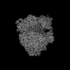 | ||||||||||||||||||
|---|---|---|---|---|---|---|---|---|---|---|---|---|---|---|---|---|---|---|---|
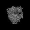
| Title | 70S initiation complex (tRNA-fMet M1 + AUG start codon) | ||||||||||||||||||
 Map data Map data | Masked, sharpened map from homogeneous 3D refinement (sharpening B factor of -61.5 A^2), used for modeling | ||||||||||||||||||
 Sample Sample |
| ||||||||||||||||||
 Keywords Keywords | translation initiation / tRNA-fMet M1 / frameshifting / ribosome | ||||||||||||||||||
| Function / homology |  Function and homology information Function and homology informationDnaA-L2 complex / negative regulation of translational initiation / negative regulation of DNA-templated DNA replication initiation / mRNA regulatory element binding translation repressor activity / cytosolic ribosome assembly / ribosome assembly / assembly of large subunit precursor of preribosome / transcription antitermination / DNA-templated transcription termination / response to radiation ...DnaA-L2 complex / negative regulation of translational initiation / negative regulation of DNA-templated DNA replication initiation / mRNA regulatory element binding translation repressor activity / cytosolic ribosome assembly / ribosome assembly / assembly of large subunit precursor of preribosome / transcription antitermination / DNA-templated transcription termination / response to radiation / maintenance of translational fidelity / mRNA 5'-UTR binding / large ribosomal subunit / transferase activity / ribosomal small subunit assembly / ribosome binding / ribosomal small subunit biogenesis / 5S rRNA binding / small ribosomal subunit / ribosomal large subunit assembly / small ribosomal subunit rRNA binding / cytosolic small ribosomal subunit / large ribosomal subunit rRNA binding / cytosolic large ribosomal subunit / cytoplasmic translation / tRNA binding / negative regulation of translation / rRNA binding / structural constituent of ribosome / ribosome / translation / ribonucleoprotein complex / response to antibiotic / mRNA binding / RNA binding / zinc ion binding / membrane / metal ion binding / cytoplasm / cytosol Similarity search - Function | ||||||||||||||||||
| Biological species |  | ||||||||||||||||||
| Method | single particle reconstruction / cryo EM / Resolution: 2.75 Å | ||||||||||||||||||
 Authors Authors | Mattingly JM / Nguyen HA / Dunham CM | ||||||||||||||||||
| Funding support |  United States, 5 items United States, 5 items
| ||||||||||||||||||
 Citation Citation | Journal: J Biol Chem / Year: 2024 Title: Structural analysis of noncanonical translation initiation complexes. Authors: Jacob M Mattingly / Ha An Nguyen / Bappaditya Roy / Kurt Fredrick / Christine M Dunham / Abstract: Translation initiation is a highly regulated, multi-step process that is critical for efficient and accurate protein synthesis. In bacteria, initiation begins when mRNA, initiation factors, and a ...Translation initiation is a highly regulated, multi-step process that is critical for efficient and accurate protein synthesis. In bacteria, initiation begins when mRNA, initiation factors, and a dedicated initiator fMet-tRNA bind the small (30S) ribosomal subunit. Specific binding of fMet-tRNA in the peptidyl (P) site is mediated by the inspection of the fMet moiety by initiation factor IF2 and of three conserved G-C base pairs in the tRNA anticodon stem by the 30S head domain. Tandem A-minor interactions form between 16S ribosomal RNA nucleotides A1339 and G1338 and tRNA base pairs G30-C40 and G29-C41, respectively. Swapping the G30-C40 pair of tRNA with C-G (called tRNA M1) reduces discrimination against the noncanonical start codon CUG in vitro, suggesting crosstalk between the gripping of the anticodon stem and recognition of the start codon. Here, we solved electron cryomicroscopy structures of Escherichia coli 70S initiation complexes containing the fMet-tRNA M1 variant paired to the noncanonical CUG start codon, in the presence or absence of IF2 and the non-hydrolyzable GTP analog GDPCP, alongside structures of 70S initiation complexes containing this tRNA variant paired to the canonical bacterial start codons AUG, GUG, and UUG. We find that the M1 mutation weakens A-minor interactions between tRNA and 16S nucleotides A1339 and G1338, with IF2 strengthening the interaction of G1338 with the tRNA minor groove. These structures suggest how even slight changes to the recognition of the fMet-tRNA anticodon stem by the ribosome can impact the start codon selection. | ||||||||||||||||||
| History |
|
- Structure visualization
| Supplemental images |
|---|

- Downloads & links
-EMDB archive
| Map data | emd_45573.map.gz | 230.3 MB | EMDB map data format | |
|---|---|---|---|---|
| Header (meta data) | emd-45573-v30.xmlemd-45573.xml | 80.6 KB 80.6 KB | Display Display | EMDB header |
| FSC (resolution estimation) | emd_45573_fsc.xml | 13.1 KB | Display | FSC data file |
| Images |  emd_45573.png emd_45573.png | 224.5 KB | ||
| Masks | emd_45573_msk_1.map | 244.1 MB | Mask map | |
| Filedesc metadata | emd-45573.cif.gz | 15.9 KB | ||
| Others | emd_45573_additional_1.map.gzemd_45573_half_map_1.map.gzemd_45573_half_map_2.map.gz | 183.8 MB 226.8 MB 226.8 MB | ||
| Archive directory |  http://ftp.pdbj.org/pub/emdb/structures/EMD-45573ftp://ftp.pdbj.org/pub/emdb/structures/EMD-45573 http://ftp.pdbj.org/pub/emdb/structures/EMD-45573ftp://ftp.pdbj.org/pub/emdb/structures/EMD-45573 | HTTPS FTP |
-Related structure data
| Related structure data |  9cg7MC  9ax7C 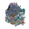 9ax8C  9cg5C 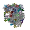 9cg6C C: citing same article ( M: atomic model generated by this map |
|---|---|
| Similar structure data |


-Links
| EMDB pages | EMDB (EBI/PDBe) / EMDataResource |
|---|---|
| Related items in Molecule of the Month |


-Map
| File | Download / File: emd_45573.map.gz / Format: CCP4 / Size: 244.1 MB / Type: IMAGE STORED AS FLOATING POINT NUMBER (4 BYTES) | ||||||||||||||||||||||||||||||||||||
|---|---|---|---|---|---|---|---|---|---|---|---|---|---|---|---|---|---|---|---|---|---|---|---|---|---|---|---|---|---|---|---|---|---|---|---|---|---|
| Annotation | Masked, sharpened map from homogeneous 3D refinement (sharpening B factor of -61.5 A^2), used for modeling | ||||||||||||||||||||||||||||||||||||
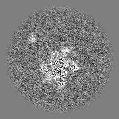
| Projections & slices | Image control
Images are generated by Spider. | ||||||||||||||||||||||||||||||||||||
| Voxel size | X=Y=Z: 1.045 Å | ||||||||||||||||||||||||||||||||||||
| Density |
| ||||||||||||||||||||||||||||||||||||
| Symmetry | Space group: 1 | ||||||||||||||||||||||||||||||||||||
| Details | EMDB XML:
|
 Z (Sec.)
 Y (Row.)
 X (Col.)


-Supplemental data
-Mask #1
| File | emd_45573_msk_1.map | ||||||||||||
|---|---|---|---|---|---|---|---|---|---|---|---|---|---|
| Projections & Slices |
| ||||||||||||
| Density Histograms |


- Sample components
+Entire : 70S initiation complex (tRNA-fMet M1 + AUG start codon)
+Supramolecule #1: 70S initiation complex (tRNA-fMet M1 + AUG start codon)
+Macromolecule #1: 50S ribosomal protein L33
+Macromolecule #2: 50S ribosomal protein L34
+Macromolecule #3: 50S ribosomal protein L35
+Macromolecule #4: 50S ribosomal protein L36
+Macromolecule #5: 50S ribosomal protein L31
+Macromolecule #7: 30S ribosomal protein S2
+Macromolecule #8: 30S ribosomal protein S3
+Macromolecule #9: 30S ribosomal protein S4
+Macromolecule #10: 30S ribosomal protein S5
+Macromolecule #11: 30S ribosomal protein S6
+Macromolecule #12: 30S ribosomal protein S7
+Macromolecule #13: 30S ribosomal protein S8
+Macromolecule #14: 30S ribosomal protein S9
+Macromolecule #15: 30S ribosomal protein S10
+Macromolecule #16: 30S ribosomal protein S11
+Macromolecule #17: 30S ribosomal protein S12
+Macromolecule #18: 30S ribosomal protein S13
+Macromolecule #19: 30S ribosomal protein S14
+Macromolecule #20: 30S ribosomal protein S15
+Macromolecule #21: 30S ribosomal protein S16
+Macromolecule #22: 30S ribosomal protein S17
+Macromolecule #23: 30S ribosomal protein S18
+Macromolecule #24: 30S ribosomal protein S19
+Macromolecule #25: 30S ribosomal protein S20
+Macromolecule #26: 30S ribosomal protein S21
+Macromolecule #31: 50S ribosomal protein L2
+Macromolecule #32: 50S ribosomal protein L3
+Macromolecule #33: 50S ribosomal protein L4
+Macromolecule #34: 50S ribosomal protein L5
+Macromolecule #35: 50S ribosomal protein L6
+Macromolecule #36: 50S ribosomal protein L9
+Macromolecule #37: 50S ribosomal protein L13
+Macromolecule #38: 50S ribosomal protein L14
+Macromolecule #39: 50S ribosomal protein L15
+Macromolecule #40: 50S ribosomal protein L16
+Macromolecule #41: 50S ribosomal protein L17
+Macromolecule #42: 50S ribosomal protein L18
+Macromolecule #43: 50S ribosomal protein L19
+Macromolecule #44: 50S ribosomal protein L20
+Macromolecule #45: 50S ribosomal protein L21
+Macromolecule #46: 50S ribosomal protein L22
+Macromolecule #47: 50S ribosomal protein L23
+Macromolecule #48: 50S ribosomal protein L24
+Macromolecule #49: 50S ribosomal protein L25
+Macromolecule #50: 50S ribosomal protein L27
+Macromolecule #51: 50S ribosomal protein L28
+Macromolecule #52: 50S ribosomal protein L29
+Macromolecule #53: 50S ribosomal protein L30
+Macromolecule #54: 50S ribosomal protein L32
+Macromolecule #6: 16S ribosomal RNA
+Macromolecule #27: mRNA
+Macromolecule #28: P-site tRNA-fMet M1
+Macromolecule #29: 23S ribosomal RNA
+Macromolecule #30: 5S ribosomal RNA
+Macromolecule #55: ZINC ION
+Macromolecule #56: MAGNESIUM ION
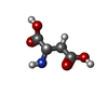
+Macromolecule #57: BETA-L-ASPARTIC ACID
-Experimental details
-Structure determination
| Method | cryo EM |
|---|---|
 Processing Processing | single particle reconstruction |
| Aggregation state | particle |
-Sample preparation
| Buffer | pH: 7.5 |
|---|---|
| Grid | Model: C-flat-1.2/1.3 / Material: GOLD / Mesh: 300 / Support film - Material: CARBON / Support film - topology: HOLEY / Pretreatment - Type: GLOW DISCHARGE / Pretreatment - Time: 30 sec. / Pretreatment - Atmosphere: AIR |
| Vitrification | Cryogen name: ETHANE / Chamber humidity: 100 % / Chamber temperature: 277 K / Instrument: FEI VITROBOT MARK IV |
- Electron microscopy
| Microscope | FEI TALOS ARCTICA |
|---|---|
| Specialist optics | Energy filter - Name: GIF Bioquantum / Energy filter - Slit width: 20 eV |
| Image recording | Film or detector model: GATAN K3 BIOQUANTUM (6k x 4k) / Digitization - Dimensions - Width: 5760 pixel / Digitization - Dimensions - Height: 4092 pixel / Number grids imaged: 1 / Number real images: 2265 / Average electron dose: 58.4 e/Å2 |
| Electron beam | Acceleration voltage: 200 kV / Electron source:  FIELD EMISSION GUN FIELD EMISSION GUN |
| Electron optics | Illumination mode: FLOOD BEAM / Imaging mode: BRIGHT FIELD / Cs: 2.7 mm / Nominal defocus max: 1.8 µm / Nominal defocus min: 0.6 µm / Nominal magnification: 79000 |
| Sample stage | Specimen holder model: FEI TITAN KRIOS AUTOGRID HOLDER / Cooling holder cryogen: NITROGEN |
| Experimental equipment |  Model: Talos Arctica / Image courtesy: FEI Company |
+Image processing

-Atomic model buiding 1
| Initial model | PDB ID: Chain - Source name: PDB / Chain - Initial model type: experimental model |
|---|---|
| Details | Starting model was fit into the unsharpened 3D reconstruction in UCSF ChimeraX before performing real-space refinement with the sharpened map in PHENIX |
| Refinement | Space: REAL / Protocol: FLEXIBLE FIT / Overall B value: 64.3 |
| Output model | PDB-9cg7: |

