[English] 日本語
Yorodumi- EMDB-45178: Cryo-EM structure of Danio rerio voltage-sensing phosphatase (VSP... -
+Open data
-Basic information
Entry | Database: EMDB / ID: EMD-45178 | |||||||||
---|---|---|---|---|---|---|---|---|---|---|
Title | Cryo-EM structure of Danio rerio voltage-sensing phosphatase (VSP) phosphatase domain | |||||||||
Map data | Sharpened map | |||||||||
Sample |
| |||||||||
Keywords | phosphatase / voltage-sensing / MEMBRANE PROTEIN | |||||||||
Function / homology | Function and homology information regulation of phosphatidylinositol 3-kinase/protein kinase B signal transduction / phosphatidylinositol-3,4,5-trisphosphate 3-phosphatase activity / phosphatidylinositol dephosphorylation / dephosphorylation / regulation of endocytosis / protein-tyrosine-phosphatase / protein tyrosine phosphatase activity / cell projection / cell motility / monoatomic ion channel activity ...regulation of phosphatidylinositol 3-kinase/protein kinase B signal transduction / phosphatidylinositol-3,4,5-trisphosphate 3-phosphatase activity / phosphatidylinositol dephosphorylation / dephosphorylation / regulation of endocytosis / protein-tyrosine-phosphatase / protein tyrosine phosphatase activity / cell projection / cell motility / monoatomic ion channel activity / negative regulation of cell population proliferation / nucleus / plasma membrane / cytosol Similarity search - Function | |||||||||
Biological species | Danio rerio (zebrafish) | |||||||||
Method | single particle reconstruction / cryo EM / Resolution: 2.97 Å | |||||||||
Authors | Zhang L / Brohawn SG | |||||||||
Funding support | United States, 1 items
| |||||||||
Citation | Journal: Nat Commun / Year: 2024 Title: Coupling sensor to enzyme in the voltage sensing phosphatase. Authors: Yawei Yu / Lin Zhang / Baobin Li / Zhu Fu / Stephen G Brohawn / Ehud Y Isacoff / Abstract: Voltage-sensing phosphatases (VSPs) dephosphorylate phosphoinositide (PIP) signaling lipids in response to membrane depolarization. VSPs possess an S4-containing voltage sensor domain (VSD), ...Voltage-sensing phosphatases (VSPs) dephosphorylate phosphoinositide (PIP) signaling lipids in response to membrane depolarization. VSPs possess an S4-containing voltage sensor domain (VSD), resembling that of voltage-gated cation channels, and a lipid phosphatase domain (PD). The mechanism by which voltage turns on enzyme activity is unclear. Structural analysis and modeling suggest several sites of VSD-PD interaction that could couple voltage sensing to catalysis. Voltage clamp fluorometry reveals voltage-driven rearrangements in three sites implicated earlier in enzyme activation-the VSD-PD linker, gating loop and R loop-as well as the N-terminal domain, which has not yet been explored. N-terminus mutations perturb both rearrangements in the other segments and enzyme activity. Our results provide a model for a dynamic assembly by which S4 controls the catalytic site. | |||||||||
History |
|
-Structure visualization
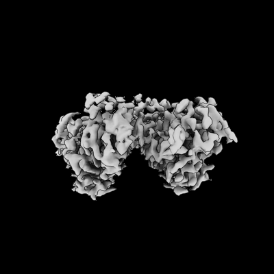
Supplemental images |
---|
-Downloads & links
-EMDB archive
Map data | emd_45178.map.gz | 56.2 MB | EMDB map data format | |
---|---|---|---|---|
Header (meta data) | emd-45178-v30.xml emd-45178.xml | 17.9 KB 17.9 KB | Display Display | EMDB header |
Images | emd_45178.png | 49 KB | ||
Filedesc metadata | emd-45178.cif.gz | 5.9 KB | ||
Others | emd_45178_additional_1.map.gz emd_45178_half_map_1.map.gz emd_45178_half_map_2.map.gz | 30 MB 55.4 MB 55.4 MB | ||
Archive directory | http://ftp.pdbj.org/pub/emdb/structures/EMD-45178
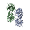
 ftp://ftp.pdbj.org/pub/emdb/structures/EMD-45178 | HTTPS FTP |
-Validation report
Summary document | emd_45178_validation.pdf.gz | 726.3 KB | Display | EMDB validaton report |
---|---|---|---|---|
Full document | emd_45178_full_validation.pdf.gz | 725.9 KB | Display | |
Data in XML | emd_45178_validation.xml.gz | 12.2 KB | Display | |
Data in CIF | emd_45178_validation.cif.gz | 14.3 KB | Display | |
Arichive directory | https://ftp.pdbj.org/pub/emdb/validation_reports/EMD-45178 ftp://ftp.pdbj.org/pub/emdb/validation_reports/EMD-45178 | HTTPS FTP |
-Related structure data
Related structure data | 9c49MC M: atomic model generated by this map C: citing same article (ref.) |
---|---|
Similar structure data | Similarity search - Function & homologyF&H Search |
-Links
EMDB pages | EMDB (EBI/PDBe) / EMDataResource |
---|---|
Related items in Molecule of the Month |
-Map
File | Download / File: emd_45178.map.gz / Format: CCP4 / Size: 59.6 MB / Type: IMAGE STORED AS FLOATING POINT NUMBER (4 BYTES) | ||||||||||||||||||||
---|---|---|---|---|---|---|---|---|---|---|---|---|---|---|---|---|---|---|---|---|---|
Annotation | Sharpened map | ||||||||||||||||||||
Voxel size | X=Y=Z: 1.115 Å | ||||||||||||||||||||
Density |
| ||||||||||||||||||||
Symmetry | Space group: 1 | ||||||||||||||||||||
Details | EMDB XML:
|
-Supplemental data
-Additional map: Unsharpened map
File | emd_45178_additional_1.map | ||||||||||||
---|---|---|---|---|---|---|---|---|---|---|---|---|---|
Annotation | Unsharpened map | ||||||||||||
Projections & Slices |
| ||||||||||||
Density Histograms |
-Half map: #1
File | emd_45178_half_map_1.map | ||||||||||||
---|---|---|---|---|---|---|---|---|---|---|---|---|---|
Projections & Slices |
| ||||||||||||
Density Histograms |
-Half map: #2
File | emd_45178_half_map_2.map | ||||||||||||
---|---|---|---|---|---|---|---|---|---|---|---|---|---|
Projections & Slices |
| ||||||||||||
Density Histograms |
-Sample components
-Entire : Voltage-sensing phosphatase (Phosphatidylinositol-3,4,5-trisphosp...
Entire | Name: Voltage-sensing phosphatase (Phosphatidylinositol-3,4,5-trisphosphate 3-phosphatase TPTE2) in lipid nanodisc |
---|---|
Components |
|
-Supramolecule #1: Voltage-sensing phosphatase (Phosphatidylinositol-3,4,5-trisphosp...
Supramolecule | Name: Voltage-sensing phosphatase (Phosphatidylinositol-3,4,5-trisphosphate 3-phosphatase TPTE2) in lipid nanodisc type: complex / ID: 1 / Parent: 0 / Macromolecule list: all |
---|---|
Source (natural) | Organism: Danio rerio (zebrafish) |
Molecular weight | Theoretical: 119 KDa |
-Macromolecule #1: Phosphatidylinositol-3,4,5-trisphosphate 3-phosphatase TPTE2
Macromolecule | Name: Phosphatidylinositol-3,4,5-trisphosphate 3-phosphatase TPTE2 type: protein_or_peptide / ID: 1 / Number of copies: 2 / Enantiomer: LEVO / EC number: protein-tyrosine-phosphatase |
---|---|
Source (natural) | Organism: Danio rerio (zebrafish) |
Molecular weight | Theoretical: 59.64052 KDa |
Recombinant expression | Organism: Komagataella pastoris (fungus) |
Sequence | String: MTSVHFNPGL DSKEVNGNSV KEEAEVQIGD GKEETKDPDT MYHQVRKKIT PFVMSFGFRV FGLVLIILDI IMVIVDLSLS EKSRDVGGA LETVSLVISF FFLIDVLLRV YVEGFKVYFS SKLNIVDACI VVITLVVTMI YAFSDFSGAS LIPRVVTFLR S LRILILVR ...String: MTSVHFNPGL DSKEVNGNSV KEEAEVQIGD GKEETKDPDT MYHQVRKKIT PFVMSFGFRV FGLVLIILDI IMVIVDLSLS EKSRDVGGA LETVSLVISF FFLIDVLLRV YVEGFKVYFS SKLNIVDACI VVITLVVTMI YAFSDFSGAS LIPRVVTFLR S LRILILVR IFRLASQKRE LEKVTRRMVS ENKRRYQKDG FDLDLTYVTE RVIAMSFPSS GKQALYRNPI REVVRFLDTK HM DHYKVFN LCSEKGYDPK FFHYRVERVM IDDHNVPSLD DMLRYTACVR DWMAADSRNV IAIHSKGGKG RTGTMVCTWL IDS DQFESA QESLDYFGER RTDKSMSSKF QGVETPSQSR YVGYYEIMKN QYNRQLPPRK SLKIKSIRIH SIAGVGKGNG SDLK LKIIV KHELVFQCVC AKQHNCTVFP DTGSNAVVIS LQDGPIVTGD VKVMFESSAG LPKGYEDCPF YFWFNTSFVE NYRLF LSRE ELDNPHKPKT WDIYKEDFGV TLSFTEPSNS LEVLFQ UniProtKB: Phosphatidylinositol-3,4,5-trisphosphate 3-phosphatase TPTE2 |
-Experimental details
-Structure determination
Method | cryo EM |
---|---|
Processing | single particle reconstruction |
Aggregation state | particle |
-Sample preparation
Concentration | 5.6 mg/mL | ||||||||||||
---|---|---|---|---|---|---|---|---|---|---|---|---|---|
Buffer | pH: 8 Component:
| ||||||||||||
Grid | Model: UltrAuFoil R1.2/1.3 / Material: GOLD / Mesh: 300 / Support film - Material: GOLD / Support film - topology: HOLEY / Pretreatment - Type: GLOW DISCHARGE / Pretreatment - Time: 25 sec. | ||||||||||||
Vitrification | Cryogen name: ETHANE / Chamber humidity: 100 % / Chamber temperature: 4 K / Instrument: FEI VITROBOT MARK IV / Details: 3s blot, blot force 1. | ||||||||||||
Details | VSP reconstituted in MSP2N2 lipid nanodiscs with 2:1:1:0.2 DOPE:POPS:POPC:Brain PI(3,4)P2 |
-Electron microscopy
Microscope | FEI TALOS ARCTICA |
---|---|
Image recording | Film or detector model: GATAN K3 (6k x 4k) / Average electron dose: 50.0 e/Å2 |
Electron beam | Acceleration voltage: 200 kV / Electron source: FIELD EMISSION GUN |
Electron optics | Illumination mode: FLOOD BEAM / Imaging mode: BRIGHT FIELD / Nominal defocus max: 1.8 µm / Nominal defocus min: 0.6 µm |
Experimental equipment | Model: Talos Arctica / Image courtesy: FEI Company |
-Image processing
Startup model | Type of model: NONE |
---|---|
Final reconstruction | Resolution.type: BY AUTHOR / Resolution: 2.97 Å / Resolution method: FSC 0.143 CUT-OFF / Software - Name: cryoSPARC / Number images used: 208690 |
Initial angle assignment | Type: MAXIMUM LIKELIHOOD |
Final angle assignment | Type: MAXIMUM LIKELIHOOD |