 Movie
Movie Controller

[English] 日本語
 Yorodumi
- PDB-9c49: Cryo-EM structure of Danio rerio voltage-sensing phosphatase (VSP... -
+ Open data

- Basic information
| Entry | Database: PDB / ID: 9c49 | ||||||
|---|---|---|---|---|---|---|---|
| Title | Cryo-EM structure of Danio rerio voltage-sensing phosphatase (VSP) phosphatase domain | ||||||
 Components Components | Phosphatidylinositol-3,4,5-trisphosphate 3-phosphatase TPTE2 | ||||||
 Keywords Keywords | MEMBRANE PROTEIN / phosphatase / voltage-sensing | ||||||
| Function / homology |  Function and homology information Function and homology informationphosphatidylinositol-3,4,5-trisphosphate 3-phosphatase activity / phosphatidylinositol dephosphorylation / regulation of endocytosis / monoatomic ion channel activity / cell projection / protein-tyrosine-phosphatase / protein tyrosine phosphatase activity / membrane Similarity search - Function | ||||||
| Biological species |  | ||||||
| Method | ELECTRON MICROSCOPY / single particle reconstruction / cryo EM / Resolution: 2.97 Å | ||||||
 Authors Authors | Zhang, L. / Brohawn, S.G. | ||||||
| Funding support |  United States, 1items United States, 1items
| ||||||
 Citation Citation | Journal: Nat Commun / Year: 2024 Title: Coupling sensor to enzyme in the voltage sensing phosphatase. Authors: Yawei Yu / Lin Zhang / Baobin Li / Zhu Fu / Stephen G Brohawn / Ehud Y Isacoff /  Abstract: Voltage-sensing phosphatases (VSPs) dephosphorylate phosphoinositide (PIP) signaling lipids in response to membrane depolarization. VSPs possess an S4-containing voltage sensor domain (VSD), ...Voltage-sensing phosphatases (VSPs) dephosphorylate phosphoinositide (PIP) signaling lipids in response to membrane depolarization. VSPs possess an S4-containing voltage sensor domain (VSD), resembling that of voltage-gated cation channels, and a lipid phosphatase domain (PD). The mechanism by which voltage turns on enzyme activity is unclear. Structural analysis and modeling suggest several sites of VSD-PD interaction that could couple voltage sensing to catalysis. Voltage clamp fluorometry reveals voltage-driven rearrangements in three sites implicated earlier in enzyme activation-the VSD-PD linker, gating loop and R loop-as well as the N-terminal domain, which has not yet been explored. N-terminus mutations perturb both rearrangements in the other segments and enzyme activity. Our results provide a model for a dynamic assembly by which S4 controls the catalytic site. | ||||||
| History |
|
- Structure visualization
| Structure viewer | Molecule: MolmilJmol/JSmol |
|---|
- Downloads & links
-Download
| PDBx/mmCIF format | 9c49.cif.gz | 162.3 KB | Display | PDBx/mmCIF format |
|---|---|---|---|---|
| PDB format | pdb9c49.ent.gz | 100.9 KB | Display | PDB format |
| PDBx/mmJSON format | 9c49.json.gz | Tree view | PDBx/mmJSON format | |
| Others |  Other downloads Other downloads |
-Validation report
| Arichive directory | https://data.pdbj.org/pub/pdb/validation_reports/c4/9c49ftp://data.pdbj.org/pub/pdb/validation_reports/c4/9c49 | HTTPS FTP |
|---|
-Related structure data
| Related structure data |  45178MC M: map data used to model this data C: citing same article ( |
|---|---|
| Similar structure data |
-Links
 PDBj
PDBj


- Assembly
| Deposited unit | 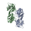
|
|---|---|
| 1 |
|
-Components
| #1: Protein | Mass: 59640.520 Da / Num. of mol.: 2 / Mutation: C302S Source method: isolated from a genetically manipulated source Source: (gene. exp.)  Komagataella pastoris (fungus) / References: UniProt: Q4V9E4, protein-tyrosine-phosphatase Komagataella pastoris (fungus) / References: UniProt: Q4V9E4, protein-tyrosine-phosphatase |
|---|
-Experimental details
-Experiment
| Experiment | Method: ELECTRON MICROSCOPY |
|---|---|
| EM experiment | Aggregation state: PARTICLE / 3D reconstruction method: single particle reconstruction |
- Sample preparation
| Component | Name: Voltage-sensing phosphatase (Phosphatidylinositol-3,4,5-trisphosphate 3-phosphatase TPTE2) in lipid nanodisc Type: COMPLEX / Entity ID: all / Source: RECOMBINANT | ||||||||||||||||||||
|---|---|---|---|---|---|---|---|---|---|---|---|---|---|---|---|---|---|---|---|---|---|
| Molecular weight | Value: 0.119 MDa / Experimental value: NO | ||||||||||||||||||||
| Source (natural) | Organism: | ||||||||||||||||||||
| Source (recombinant) | Organism: Komagataella pastoris (fungus) | ||||||||||||||||||||
| Buffer solution | pH: 8 | ||||||||||||||||||||
| Buffer component |
| ||||||||||||||||||||
| Specimen | Conc.: 5.6 mg/ml / Embedding applied: NO / Shadowing applied: NO / Staining applied: NO / Vitrification applied: YES Details: VSP reconstituted in MSP2N2 lipid nanodiscs with 2:1:1:0.2 DOPE:POPS:POPC:Brain PI(3,4)P2 | ||||||||||||||||||||
| Specimen support | Grid material: GOLD / Grid mesh size: 300 divisions/in. / Grid type: UltrAuFoil R1.2/1.3 | ||||||||||||||||||||
| Vitrification | Instrument: FEI VITROBOT MARK IV / Cryogen name: ETHANE / Humidity: 100 % / Chamber temperature: 4 K / Details: 3s blot, blot force 1 |
- Electron microscopy imaging
| Experimental equipment |  Model: Talos Arctica / Image courtesy: FEI Company |
|---|---|
| Microscopy | Model: FEI TALOS ARCTICA |
| Electron gun | Electron source:  FIELD EMISSION GUN / Accelerating voltage: 200 kV / Illumination mode: FLOOD BEAM FIELD EMISSION GUN / Accelerating voltage: 200 kV / Illumination mode: FLOOD BEAM |
| Electron lens | Mode: BRIGHT FIELD / Nominal defocus max: 1800 nm / Nominal defocus min: 600 nm |
| Image recording | Electron dose: 50 e/Å2 / Film or detector model: GATAN K3 (6k x 4k) |
- Processing
| EM software |
| ||||||||||||||||||||||||
|---|---|---|---|---|---|---|---|---|---|---|---|---|---|---|---|---|---|---|---|---|---|---|---|---|---|
| CTF correction | Type: PHASE FLIPPING AND AMPLITUDE CORRECTION | ||||||||||||||||||||||||
| 3D reconstruction | Resolution: 2.97 Å / Resolution method: FSC 0.143 CUT-OFF / Num. of particles: 208690 / Symmetry type: POINT | ||||||||||||||||||||||||
| Refinement | Cross valid method: NONE Stereochemistry target values: GeoStd + Monomer Library + CDL v1.2 | ||||||||||||||||||||||||
| Displacement parameters | Biso mean: 49.17 Å2 | ||||||||||||||||||||||||
| Refine LS restraints |
|
