- EMDB-43973: Non-translating S. pombe ribosome large subunit -
+
データを開く
IDまたはキーワード:
読み込み中...
-
基本情報
登録情報
データベース: EMDB / ID: EMD-43973
タイトル
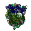
Non-translating S. pombe ribosome large subunit
マップデータ
試料
複合体: Non-translating S. pombe ribosome large subunit
タンパク質・ペプチド: x 40種
RNA: x 3種
リガンド: x 2種
キーワード
Non-translating / 60S / Large subunit / S. pombe / Schizosasccharomyces pombe / RIBOSOME
機能・相同性
機能・相同性情報
cytosolic large ribosomal subunit assembly / ribonuclease MRP complex / L13a-mediated translational silencing of Ceruloplasmin expression / Formation of a pool of free 40S subunits / GTP hydrolysis and joining of the 60S ribosomal subunit / SRP-dependent cotranslational protein targeting to membrane / Nonsense Mediated Decay (NMD) independent of the Exon Junction Complex (EJC) / Nonsense Mediated Decay (NMD) enhanced by the Exon Junction Complex (EJC) / preribosome / tRNA processing ...cytosolic large ribosomal subunit assembly / ribonuclease MRP complex / L13a-mediated translational silencing of Ceruloplasmin expression / Formation of a pool of free 40S subunits / GTP hydrolysis and joining of the 60S ribosomal subunit / SRP-dependent cotranslational protein targeting to membrane / Nonsense Mediated Decay (NMD) independent of the Exon Junction Complex (EJC) / Nonsense Mediated Decay (NMD) enhanced by the Exon Junction Complex (EJC) / preribosome / tRNA processing / pre-mRNA intronic binding / protein-RNA complex assembly / endonucleolytic cleavage in ITS1 to separate SSU-rRNA from 5.8S rRNA and LSU-rRNA from tricistronic rRNA transcript (SSU-rRNA, 5.8S rRNA, LSU-rRNA) / maturation of LSU-rRNA / maturation of LSU-rRNA from tricistronic rRNA transcript (SSU-rRNA, 5.8S rRNA, LSU-rRNA) / ribosomal large subunit biogenesis / rRNA processing / ribosome biogenesis / 5S rRNA binding / ribosomal large subunit assembly / cytosolic small ribosomal subunit / large ribosomal subunit rRNA binding / cytosolic large ribosomal subunit / cytoplasmic translation / negative regulation of translation / rRNA binding / ribosome / structural constituent of ribosome / translation / negative regulation of gene expression / mRNA binding / nucleolus / RNA binding / zinc ion binding / nucleus / cytosol / cytoplasm 類似検索 - 分子機能
Ribosomal protein L28e / : / Ribosomal L28e/Mak16 / Ribosomal L28e protein family / Ribosomal protein L29e / Ribosomal L29e protein family / Ribosomal protein L13e, conserved site / Ribosomal protein L13e signature. / Ribosomal protein L22e / Ribosomal protein L22e superfamily ...Ribosomal protein L28e / : / Ribosomal L28e/Mak16 / Ribosomal L28e protein family / Ribosomal protein L29e / Ribosomal L29e protein family / Ribosomal protein L13e, conserved site / Ribosomal protein L13e signature. / Ribosomal protein L22e / Ribosomal protein L22e superfamily / Ribosomal L22e protein family / Ribosomal protein L10e, conserved site / Ribosomal protein L10e signature. / Ribosomal protein L27e, conserved site / Ribosomal protein L27e signature. / Ribosomal protein L10e / Ribosomal protein L38e / Ribosomal protein L38e superfamily / Ribosomal L38e protein family / Ribosomal protein L44e signature. / Ribosomal protein L24e, conserved site / Ribosomal protein L24e signature. / : / Ribosomal protein L19, eukaryotic / Ribosomal protein L6e signature. / Ribosomal protein L13e / Ribosomal protein L13e / 60S ribosomal protein L18a/ L20, eukaryotes / : / Ribosomal protein L44e / Ribosomal protein L44 / Ribosomal protein L34e, conserved site / Ribosomal protein L34e signature. / Ribosomal protein L5 eukaryotic, C-terminal / Ribosomal L18 C-terminal region / Ribosomal protein L30e signature 1. / 50S ribosomal protein L18Ae/60S ribosomal protein L20 and L18a / Ribosomal protein 50S-L18Ae/60S-L20/60S-L18A / Ribosomal proteins 50S-L18Ae/60S-L20/60S-L18A / Ribosomal protein L23/L25, N-terminal / : / Ribosomal protein L23, N-terminal domain / Eukaryotic Ribosomal Protein L27, KOW domain / Ribosomal protein 60S L18 and 50S L18e / Ribosomal Protein L6, KOW domain / Ribosomal protein L18/L18-A/B/e, conserved site / Ribosomal protein L18e signature. / Ribosomal protein L30e signature 2. / Ribosomal protein L27e / Ribosomal protein L27e superfamily / Ribosomal L27e protein family / Ribosomal protein L36e signature. / Ribosomal protein L35Ae, conserved site / Ribosomal protein L30e, conserved site / Ribosomal protein L35Ae signature. / Ribosomal protein L39e, conserved site / Ribosomal protein L39e signature. / : / Ribosomal protein L6e / Ribosomal protein L34Ae / Ribosomal protein L34e / 60S ribosomal protein L19 / Ribosomal protein L30/YlxQ / Ribosomal protein L7A/L8 / 60S ribosomal protein L6E / 60S ribosomal protein L35 / Ribosomal protein L13, eukaryotic/archaeal / Ribosomal protein L18e / 60S ribosomal protein L4, C-terminal domain / 60S ribosomal protein L4 C-terminal domain / Ribosomal protein L7, eukaryotic / Ribosomal protein L30, N-terminal / Ribosomal protein L31e, conserved site / Ribosomal L30 N-terminal domain / Ribosomal protein L31e signature. / Ribosomal protein L37ae / Ribosomal L37ae protein family / Ribosomal protein L36e / Ribosomal protein L36e domain superfamily / Ribosomal protein L36e / Ribosomal_L19e / Ribosomal protein L19/L19e / Ribosomal protein L19/L19e, domain 1 / Ribosomal protein L19/L19e superfamily / Ribosomal protein L19e, N-terminal domain / Ribosomal protein L14e domain / Ribosomal protein L14 / Ribosomal protein L35A / Ribosomal protein L35Ae / Ribosomal protein L39e / Ribosomal protein L39e domain superfamily / Ribosomal protein L35A superfamily / Ribosomal L39 protein / Ribosomal protein L32e, conserved site / Ribosomal protein L32e signature. / Ribosomal protein L5 eukaryotic/L18 archaeal / Ribosomal large subunit proteins 60S L5, and 50S L18 / Ribosomal protein L6, conserved site-2 / Ribosomal protein L6 signature 2. / Ribosomal protein L14 類似検索 - ドメイン・相同性
Large ribosomal subunit protein eL8 / Large ribosomal subunit protein eL28 / Large ribosomal subunit protein uL22A / Large ribosomal subunit protein eL27A / Large ribosomal subunit protein eL19B / Large ribosomal subunit protein eL21B / Large ribosomal subunit protein uL13A / Large ribosomal subunit protein uL30C / Large ribosomal subunit protein eL13 / Large ribosomal subunit protein uL18B ...Large ribosomal subunit protein eL8 / Large ribosomal subunit protein eL28 / Large ribosomal subunit protein uL22A / Large ribosomal subunit protein eL27A / Large ribosomal subunit protein eL19B / Large ribosomal subunit protein eL21B / Large ribosomal subunit protein uL13A / Large ribosomal subunit protein uL30C / Large ribosomal subunit protein eL13 / Large ribosomal subunit protein uL18B / Large ribosomal subunit protein eL24B / Large ribosomal subunit protein uL29 / Large ribosomal subunit protein uL6B / Large ribosomal subunit protein eL14 / Large ribosomal subunit protein eL36B / Large ribosomal subunit protein eL37B / Large ribosomal subunit protein eL39 / Large ribosomal subunit protein uL14B / Large ribosomal subunit protein eL20A / Large ribosomal subunit protein uL2C / Large ribosomal subunit protein uL5A / Large ribosomal subunit protein uL4A / Large ribosomal subunit protein uL3A / Large ribosomal subunit protein eL30A / Large ribosomal subunit protein uL15B / Large ribosomal subunit protein uL24 / Large ribosomal subunit protein eL32A / Large ribosomal subunit protein eL6 / Large ribosomal subunit protein uL16A / Large ribosomal subunit protein eL22 / Large ribosomal subunit protein uL23A / Large ribosomal subunit protein eL18B / Large ribosomal subunit protein eL29 / Large ribosomal subunit protein eL43A / Large ribosomal subunit protein eL34B / Large ribosomal subunit protein eL31 / Large ribosomal subunit protein eL15B / Large ribosomal subunit protein eL38A / Large ribosomal subunit protein eL33A / Large ribosomal subunit protein eL42 類似検索 - 構成要素
EIPOD fellowship under Marie Sklodowska-Curie Actions COFUND
ドイツ
引用
ジャーナル: Nat Commun / 年: 2024 タイトル: Ribosomes hibernate on mitochondria during cellular stress. 著者: Olivier Gemin / Maciej Gluc / Higor Rosa / Michael Purdy / Moritz Niemann / Yelena Peskova / Simone Mattei / Ahmad Jomaa / 要旨: Cell survival under nutrient-deprived conditions relies on cells' ability to adapt their organelles and rewire their metabolic pathways. In yeast, glucose depletion induces a stress response mediated ...Cell survival under nutrient-deprived conditions relies on cells' ability to adapt their organelles and rewire their metabolic pathways. In yeast, glucose depletion induces a stress response mediated by mitochondrial fragmentation and sequestration of cytosolic ribosomes on mitochondria. This cellular adaptation promotes survival under harsh environmental conditions; however, the underlying mechanism of this response remains unknown. Here, we demonstrate that upon glucose depletion protein synthesis is halted. Cryo-electron microscopy structure of the ribosomes show that they are devoid of both tRNA and mRNA, and a subset of the particles depicted a conformational change in rRNA H69 that could prevent tRNA binding. Our in situ structural analyses reveal that the hibernating ribosomes tether to fragmented mitochondria and establish eukaryotic-specific, higher-order storage structures by assembling into oligomeric arrays on the mitochondrial surface. Notably, we show that hibernating ribosomes exclusively bind to the outer mitochondrial membrane via the small ribosomal subunit during cellular stress. We identify the ribosomal protein Cpc2/RACK1 as the molecule mediating ribosomal tethering to mitochondria. This study unveils the molecular mechanism connecting mitochondrial stress with the shutdown of protein synthesis and broadens our understanding of cellular responses to nutrient scarcity and cell quiescence.
 ムービー
ムービー コントローラー


 データを開く
 基本情報
基本情報
 マップデータ
 試料
試料 キーワード
キーワード 機能・相同性情報
 データ登録者
 米国,
米国,  ドイツ, 2件
  引用
引用 構造の表示
構造の表示
 ダウンロードとリンク
 emd_43973.png
 http://ftp.pdbj.org/pub/emdb/structures/EMD-43973


 Z (Sec.)
 Y (Row.)
 X (Col.)


 試料の構成要素
 解析
解析 電子顕微鏡法
 FIELD EMISSION GUN

