Protein or peptide: Isoform ATE1-2 of Arginyl-tRNA--protein transferase 1
Ligand: ZINC ION
Keywords
arginylation / ATE1 / apo / TRANSFERASE
Function / homology
Function and homology information
protein arginylation / arginyltransferase / arginyl-tRNA--protein transferase activity / ubiquitin-dependent protein catabolic process via the N-end rule pathway / proteasomal protein catabolic process / response to oxidative stress / ubiquitin-dependent protein catabolic process / nucleus / cytoplasm Similarity search - Function
National Institutes of Health/National Cancer Institute (NIH/NCI)
CA241301
United States
National Institutes of Health/National Cancer Institute (NIH/NCI)
CA240993
United States
National Institutes of Health/National Institute of General Medical Sciences (NIH/NIGMS)
GM133841
United States
National Institutes of Health/National Institute of General Medical Sciences (NIH/NIGMS)
GM142002
United States
Citation
Journal: Nat Commun / Year: 2024 Title: Oligomerization and a distinct tRNA-binding loop are important regulators of human arginyl-transferase function. Authors: Xin Lan / Wei Huang / Su Bin Kim / Dechen Fu / Thilini Abeywansha / Jiemin Lou / Udayakumaran Balamurugan / Yong Tae Kwon / Chang Hoon Ji / Derek J Taylor / Yi Zhang / Abstract: The arginyl-transferase ATE1 is a tRNA-dependent enzyme that covalently attaches an arginine molecule to a protein substrate. Conserved from yeast to humans, ATE1 deficiency in mice correlates with ...The arginyl-transferase ATE1 is a tRNA-dependent enzyme that covalently attaches an arginine molecule to a protein substrate. Conserved from yeast to humans, ATE1 deficiency in mice correlates with defects in cardiovascular development and angiogenesis and results in embryonic lethality, while conditional knockouts exhibit reproductive, developmental, and neurological deficiencies. Despite the recent revelation of the tRNA binding mechanism and the catalytic cycle of yeast ATE1, the structure-function relationship of ATE1 in higher organisms is not well understood. In this study, we present the three-dimensional structure of human ATE1 in an apo-state and in complex with its tRNA cofactor and a peptide substrate. In contrast to its yeast counterpart, human ATE1 forms a symmetric homodimer, which dissociates upon binding of a substrate. Furthermore, human ATE1 includes a unique and extended loop that wraps around tRNA, creating extensive contacts with the T-arm of the tRNA cofactor. Substituting key residues identified in the substrate binding site of ATE1 abolishes enzymatic activity and results in the accumulation of ATE1 substrates in cells.
In the structure databanks used in Yorodumi, some data are registered as the other names, "COVID-19 virus" and "2019-nCoV". Here are the details of the virus and the list of structure data.
Jan 31, 2019. EMDB accession codes are about to change! (news from PDBe EMDB page)
EMDB accession codes are about to change! (news from PDBe EMDB page)
The allocation of 4 digits for EMDB accession codes will soon come to an end. Whilst these codes will remain in use, new EMDB accession codes will include an additional digit and will expand incrementally as the available range of codes is exhausted. The current 4-digit format prefixed with “EMD-” (i.e. EMD-XXXX) will advance to a 5-digit format (i.e. EMD-XXXXX), and so on. It is currently estimated that the 4-digit codes will be depleted around Spring 2019, at which point the 5-digit format will come into force.
The EM Navigator/Yorodumi systems omit the EMD- prefix.
Related info.:Q: What is EMD? / ID/Accession-code notation in Yorodumi/EM Navigator
Yorodumi is a browser for structure data from EMDB, PDB, SASBDB, etc.
This page is also the successor to EM Navigator detail page, and also detail information page/front-end page for Omokage search.
The word "yorodu" (or yorozu) is an old Japanese word meaning "ten thousand". "mi" (miru) is to see.
Related info.:EMDB / PDB / SASBDB / Comparison of 3 databanks / Yorodumi Search / Aug 31, 2016. New EM Navigator & Yorodumi / Yorodumi Papers / Jmol/JSmol / Function and homology information / Changes in new EM Navigator and Yorodumi
 Movie
Movie Controller


 Open data
 Basic information
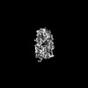
 Map data
 Sample
 Keywords
 Function and homology information
 Homo sapiens (human)
 Authors
 United States, 4 items
  Citation
 Structure visualization
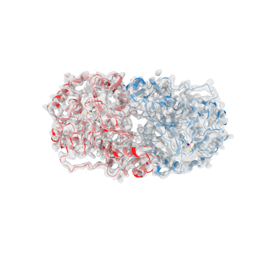
 Downloads & links
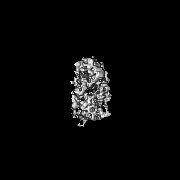
 emd_41770.png
 http://ftp.pdbj.org/pub/emdb/structures/EMD-41770
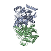


 Z (Sec.)
 Y (Row.)
 X (Col.)


 Sample components
 Processing
 Electron microscopy
 FIELD EMISSION GUN
