+ Open data
- Basic information
| Entry |  | |||||||||
|---|---|---|---|---|---|---|---|---|---|---|
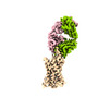
| Title | Structure of a class A GPCR/Fab complex | |||||||||
 Map data Map data | ||||||||||
 Sample Sample |
| |||||||||
 Keywords Keywords | GPCR Fab / STRUCTURAL PROTEIN | |||||||||
| Function / homology |  Function and homology information Function and homology informationchemokine receptor activity / C-C chemokine receptor activity / C-C chemokine binding / Chemokine receptors bind chemokines / coreceptor activity / bioluminescence / cell chemotaxis / generation of precursor metabolites and energy / calcium-mediated signaling / chemotaxis ...chemokine receptor activity / C-C chemokine receptor activity / C-C chemokine binding / Chemokine receptors bind chemokines / coreceptor activity / bioluminescence / cell chemotaxis / generation of precursor metabolites and energy / calcium-mediated signaling / chemotaxis / positive regulation of cytosolic calcium ion concentration / G alpha (i) signalling events / cell adhesion / immune response / G protein-coupled receptor signaling pathway / external side of plasma membrane / plasma membrane Similarity search - Function | |||||||||
| Biological species |  Homo sapiens (human) / Homo sapiens (human) /   | |||||||||
| Method | single particle reconstruction / cryo EM / Resolution: 2.9 Å | |||||||||
 Authors Authors | Sun D / Johnson M / Masureel M | |||||||||
| Funding support | 1 items
| |||||||||
 Citation Citation |  Journal: Nat Commun / Year: 2023 Journal: Nat Commun / Year: 2023Title: Structural basis of antibody inhibition and chemokine activation of the human CC chemokine receptor 8. Authors: Dawei Sun / Yonglian Sun / Eric Janezic / Tricia Zhou / Matthew Johnson / Caleigh Azumaya / Sigrid Noreng / Cecilia Chiu / Akiko Seki / Teresita L Arenzana / John M Nicoludis / Yongchang Shi ...Authors: Dawei Sun / Yonglian Sun / Eric Janezic / Tricia Zhou / Matthew Johnson / Caleigh Azumaya / Sigrid Noreng / Cecilia Chiu / Akiko Seki / Teresita L Arenzana / John M Nicoludis / Yongchang Shi / Baomei Wang / Hoangdung Ho / Prajakta Joshi / Christine Tam / Jian Payandeh / Laëtitia Comps-Agrar / Jianyong Wang / Sascha Rutz / James T Koerber / Matthieu Masureel /  Abstract: The C-C motif chemokine receptor 8 (CCR8) is a class A G-protein coupled receptor that has emerged as a promising therapeutic target in cancer. Targeting CCR8 with an antibody has appeared to be an ...The C-C motif chemokine receptor 8 (CCR8) is a class A G-protein coupled receptor that has emerged as a promising therapeutic target in cancer. Targeting CCR8 with an antibody has appeared to be an attractive therapeutic approach, but the molecular basis for chemokine-mediated activation and antibody-mediated inhibition of CCR8 are not fully elucidated. Here, we obtain an antagonist antibody against human CCR8 and determine structures of CCR8 in complex with either the antibody or the endogenous agonist ligand CCL1. Our studies reveal characteristic antibody features allowing recognition of the CCR8 extracellular loops and CCL1-CCR8 interaction modes that are distinct from other chemokine receptor - ligand pairs. Informed by these structural insights, we demonstrate that CCL1 follows a two-step, two-site binding sequence to CCR8 and that antibody-mediated inhibition of CCL1 signaling can occur by preventing the second binding event. Together, our results provide a detailed structural and mechanistic framework of CCR8 activation and inhibition that expands our molecular understanding of chemokine - receptor interactions and offers insight into the development of therapeutic antibodies targeting chemokine GPCRs. | |||||||||
| History |
|
- Structure visualization
| Supplemental images |
|---|
- Downloads & links
-EMDB archive
| Map data |  emd_41370.map.gz emd_41370.map.gz | 43.3 MB |  EMDB map data format EMDB map data format | |
|---|---|---|---|---|
| Header (meta data) |  emd-41370-v30.xml emd-41370-v30.xml emd-41370.xml emd-41370.xml | 17 KB 17 KB | Display Display |  EMDB header EMDB header |
| FSC (resolution estimation) |  emd_41370_fsc.xml emd_41370_fsc.xml | 9.2 KB | Display |  FSC data file FSC data file |
| Images |  emd_41370.png emd_41370.png | 59.1 KB | ||
| Masks |  emd_41370_msk_1.map emd_41370_msk_1.map | 83.7 MB |  Mask map Mask map | |
| Filedesc metadata |  emd-41370.cif.gz emd-41370.cif.gz | 6.3 KB | ||
| Others |  emd_41370_half_map_1.map.gz emd_41370_half_map_1.map.gz emd_41370_half_map_2.map.gz emd_41370_half_map_2.map.gz | 77.7 MB 77.7 MB | ||
| Archive directory |  http://ftp.pdbj.org/pub/emdb/structures/EMD-41370 http://ftp.pdbj.org/pub/emdb/structures/EMD-41370 ftp://ftp.pdbj.org/pub/emdb/structures/EMD-41370 ftp://ftp.pdbj.org/pub/emdb/structures/EMD-41370 | HTTPS FTP |
-Validation report
| Summary document |  emd_41370_validation.pdf.gz emd_41370_validation.pdf.gz | 925.7 KB | Display |  EMDB validaton report EMDB validaton report |
|---|---|---|---|---|
| Full document |  emd_41370_full_validation.pdf.gz emd_41370_full_validation.pdf.gz | 925.3 KB | Display | |
| Data in XML |  emd_41370_validation.xml.gz emd_41370_validation.xml.gz | 17.3 KB | Display | |
| Data in CIF |  emd_41370_validation.cif.gz emd_41370_validation.cif.gz | 22.2 KB | Display | |
| Arichive directory |  https://ftp.pdbj.org/pub/emdb/validation_reports/EMD-41370 https://ftp.pdbj.org/pub/emdb/validation_reports/EMD-41370 ftp://ftp.pdbj.org/pub/emdb/validation_reports/EMD-41370 ftp://ftp.pdbj.org/pub/emdb/validation_reports/EMD-41370 | HTTPS FTP |
-Related structure data
| Related structure data |  8tlmMC  8u1uC M: atomic model generated by this map C: citing same article ( |
|---|---|
| Similar structure data | Similarity search - Function & homology  F&H Search F&H Search |
- Links
Links
| EMDB pages |  EMDB (EBI/PDBe) / EMDB (EBI/PDBe) /  EMDataResource EMDataResource |
|---|---|
| Related items in Molecule of the Month |
- Map
Map
| File |  Download / File: emd_41370.map.gz / Format: CCP4 / Size: 83.7 MB / Type: IMAGE STORED AS FLOATING POINT NUMBER (4 BYTES) Download / File: emd_41370.map.gz / Format: CCP4 / Size: 83.7 MB / Type: IMAGE STORED AS FLOATING POINT NUMBER (4 BYTES) | ||||||||||||||||||||||||||||||||||||
|---|---|---|---|---|---|---|---|---|---|---|---|---|---|---|---|---|---|---|---|---|---|---|---|---|---|---|---|---|---|---|---|---|---|---|---|---|---|
| Projections & slices | Image control
Images are generated by Spider. | ||||||||||||||||||||||||||||||||||||
| Voxel size | X=Y=Z: 0.97 Å | ||||||||||||||||||||||||||||||||||||
| Density |
| ||||||||||||||||||||||||||||||||||||
| Symmetry | Space group: 1 | ||||||||||||||||||||||||||||||||||||
| Details | EMDB XML:
|
-Supplemental data
-Mask #1
| File |  emd_41370_msk_1.map emd_41370_msk_1.map | ||||||||||||
|---|---|---|---|---|---|---|---|---|---|---|---|---|---|
| Projections & Slices |
| ||||||||||||
| Density Histograms |
-Half map: #2
| File | emd_41370_half_map_1.map | ||||||||||||
|---|---|---|---|---|---|---|---|---|---|---|---|---|---|
| Projections & Slices |
| ||||||||||||
| Density Histograms |
-Half map: #1
| File | emd_41370_half_map_2.map | ||||||||||||
|---|---|---|---|---|---|---|---|---|---|---|---|---|---|
| Projections & Slices |
| ||||||||||||
| Density Histograms |
- Sample components
-Entire : CCR8_Fab complex
| Entire | Name: CCR8_Fab complex |
|---|---|
| Components |
|
-Supramolecule #1: CCR8_Fab complex
| Supramolecule | Name: CCR8_Fab complex / type: complex / ID: 1 / Parent: 0 / Macromolecule list: all |
|---|---|
| Source (natural) | Organism:  Homo sapiens (human) Homo sapiens (human) |
-Macromolecule #1: Fab heavy chain
| Macromolecule | Name: Fab heavy chain / type: protein_or_peptide / ID: 1 / Number of copies: 1 / Enantiomer: LEVO |
|---|---|
| Source (natural) | Organism:  |
| Molecular weight | Theoretical: 23.775549 KDa |
| Recombinant expression | Organism:  |
| Sequence | String: QSVEESGGRL VTPGTPLTLT CTVSGFSLSN YAMIWVRQAP GEGLEWVGTI SLGGYTYYAN WAKGRFTISK TSTTVDLKIS SPTTEDTAT YFCARARWST DSAIYTYAFD PWGPGTLVTV SSASTKGPSV FPLAPSSKST SGGTAALGCL VKDYFPEPVT V SWNSGALT ...String: QSVEESGGRL VTPGTPLTLT CTVSGFSLSN YAMIWVRQAP GEGLEWVGTI SLGGYTYYAN WAKGRFTISK TSTTVDLKIS SPTTEDTAT YFCARARWST DSAIYTYAFD PWGPGTLVTV SSASTKGPSV FPLAPSSKST SGGTAALGCL VKDYFPEPVT V SWNSGALT SGVHTFPAVL QSSGLYSLSS VVTVPSSSLG TQTYICNVNH KPSNTKVDKK VEPKSCD |
-Macromolecule #2: Fab light chain
| Macromolecule | Name: Fab light chain / type: protein_or_peptide / ID: 2 / Number of copies: 1 / Enantiomer: LEVO |
|---|---|
| Source (natural) | Organism:  |
| Molecular weight | Theoretical: 23.402865 KDa |
| Recombinant expression | Organism:  |
| Sequence | String: AYDMTQTPAS VEVAVGGTVT IKCQASQSIS SYLSWYQQKP GQRPELLIYK ASTLASGVSS RFKGSGSGTQ FTLTISDLEA ADAATYYCQ QGYTSSNIDN IFGGGTEVVV KRTVAAPSVF IFPPSDEQLK SGTASVVCLL NNFYPREAKV QWKVDNALQS G NSQESVTE ...String: AYDMTQTPAS VEVAVGGTVT IKCQASQSIS SYLSWYQQKP GQRPELLIYK ASTLASGVSS RFKGSGSGTQ FTLTISDLEA ADAATYYCQ QGYTSSNIDN IFGGGTEVVV KRTVAAPSVF IFPPSDEQLK SGTASVVCLL NNFYPREAKV QWKVDNALQS G NSQESVTE QDSKDSTYSL SSTLTLSKAD YEKHKVYACE VTHQGLSSPV TKSFNRGEC |
-Macromolecule #3: C-C chemokine receptor type 8, Green fluorescent protein fusion
| Macromolecule | Name: C-C chemokine receptor type 8, Green fluorescent protein fusion type: protein_or_peptide / ID: 3 / Number of copies: 1 / Enantiomer: LEVO |
|---|---|
| Source (natural) | Organism:  |
| Molecular weight | Theoretical: 76.003508 KDa |
| Recombinant expression | Organism: Mammalian expression vector Flag-MCS-pcDNA3.1 (others) |
| Sequence | String: MKTIIALSYI FCLVFADYKD DDDKGSENLY FQSGSMDYTL DLSVTTVTDY YYPDIFSSPC DAELIQTNGK LLLAVFYCLL FVFSLLGNS LVILVLVVCK KLRSITDVYL LNLALSDLLF VFSFPFQTYY LLDQWVFGTV MCKVVSGFYY IGFYSSMFFI T LMSVDRYL ...String: MKTIIALSYI FCLVFADYKD DDDKGSENLY FQSGSMDYTL DLSVTTVTDY YYPDIFSSPC DAELIQTNGK LLLAVFYCLL FVFSLLGNS LVILVLVVCK KLRSITDVYL LNLALSDLLF VFSFPFQTYY LLDQWVFGTV MCKVVSGFYY IGFYSSMFFI T LMSVDRYL AVVHAVYALK VRTIRMGTTL CLAVWLTAIM ATIPLLVFYQ VASEDGVLQC YSFYNQQTLK WKIFTNFKMN IL GLLIPFT IFMFCYIKIL HQLKRCQNHN KTKAIRLVLI VVIASLLFWV PFNVVLFLTS LHSMHILDGC SISQQLTYAT HVT EIISFT HCCVNPVIYA FVGEKFKKHL SEIFQKSCSQ IFNYLGRQMP RESCEKSSSC QQHSSRSSSV DYILGNSLEV LFQG PMVSK GEELFTGVVP ILVELDGDVN GHKFSVSGEG EGDATYGKLT LKLICTTGKL PVPWPTLVTT LGYGLQCFAR YPDHM KQHD FFKSAMPEGY VQERTIFFKD DGNYKTRAEV KFEGDTLVNR IELKGIDFKE DGNILGHKLE YNYNSHNVYI TADKQK NGI KANFKIRHNI EDGGVQLADH YQQNTPIGDG PVLLPDNHYL SYQSKLSKDP NEKRDHMVLL EFVTAAGITL GMDELYK GS AWSHPQFEKG GGSGGGSGGS AWSHPQFEK UniProtKB: C-C chemokine receptor type 8, Green fluorescent protein |
-Experimental details
-Structure determination
| Method | cryo EM |
|---|---|
 Processing Processing | single particle reconstruction |
| Aggregation state | particle |
- Sample preparation
| Buffer | pH: 7.5 |
|---|---|
| Vitrification | Cryogen name: ETHANE |
- Electron microscopy
| Microscope | FEI TITAN KRIOS |
|---|---|
| Image recording | Film or detector model: FEI FALCON III (4k x 4k) / Average electron dose: 1.067 e/Å2 |
| Electron beam | Acceleration voltage: 300 kV / Electron source:  FIELD EMISSION GUN FIELD EMISSION GUN |
| Electron optics | Illumination mode: SPOT SCAN / Imaging mode: OTHER / Nominal defocus max: 1.8 µm / Nominal defocus min: 0.8 µm |
| Experimental equipment |  Model: Titan Krios / Image courtesy: FEI Company |
 Movie
Movie Controller


 Z (Sec.)
 Y (Row.)
 X (Col.)


