 Movie
Movie Controller


+ Open data

- Basic information
| Entry |  | |||||||||
|---|---|---|---|---|---|---|---|---|---|---|
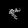
| Title | Structure of a class A GPCR/agonist complex | |||||||||
 Map data Map data | ||||||||||
 Sample Sample |
| |||||||||
 Keywords Keywords | GPCR / agonist / STRUCTURAL PROTEIN | |||||||||
| Function / homology |  Function and homology information Function and homology informationchemokine receptor activity / CCR chemokine receptor binding / C-C chemokine binding / C-C chemokine receptor activity / eosinophil chemotaxis / positive regulation of monocyte chemotaxis / chemokine activity / Chemokine receptors bind chemokines / positive regulation of interleukin-17 production / viral process ...chemokine receptor activity / CCR chemokine receptor binding / C-C chemokine binding / C-C chemokine receptor activity / eosinophil chemotaxis / positive regulation of monocyte chemotaxis / chemokine activity / Chemokine receptors bind chemokines / positive regulation of interleukin-17 production / viral process / coreceptor activity / adenylate cyclase inhibitor activity / positive regulation of protein localization to cell cortex / T cell migration / positive regulation of relaxation of smooth muscle / Adenylate cyclase inhibitory pathway / D2 dopamine receptor binding / adenylate cyclase-inhibiting serotonin receptor signaling pathway / G protein-coupled serotonin receptor binding / cellular response to forskolin / bioluminescence / regulation of mitotic spindle organization / chemokine-mediated signaling pathway / cell chemotaxis / generation of precursor metabolites and energy / calcium-mediated signaling / Regulation of insulin secretion / neuropeptide signaling pathway / response to prostaglandin E / positive regulation of cholesterol biosynthetic process / negative regulation of insulin secretion / G protein-coupled receptor binding / response to peptide hormone / G protein-coupled receptor activity / chemotaxis / intracellular calcium ion homeostasis / centriolar satellite / G-protein beta/gamma-subunit complex binding / adenylate cyclase-modulating G protein-coupled receptor signaling pathway / adenylate cyclase-inhibiting G protein-coupled receptor signaling pathway / Olfactory Signaling Pathway / positive regulation of inflammatory response / Activation of the phototransduction cascade / G protein-coupled acetylcholine receptor signaling pathway / G beta:gamma signalling through PLC beta / Presynaptic function of Kainate receptors / Thromboxane signalling through TP receptor / Activation of G protein gated Potassium channels / Inhibition of voltage gated Ca2+ channels via Gbeta/gamma subunits / G-protein activation / Glucagon signaling in metabolic regulation / G beta:gamma signalling through CDC42 / Prostacyclin signalling through prostacyclin receptor / Synthesis, secretion, and inactivation of Glucagon-like Peptide-1 (GLP-1) / G beta:gamma signalling through BTK / photoreceptor disc membrane / ADP signalling through P2Y purinoceptor 12 / GDP binding / Glucagon-type ligand receptors / Sensory perception of sweet, bitter, and umami (glutamate) taste / Adrenaline,noradrenaline inhibits insulin secretion / Vasopressin regulates renal water homeostasis via Aquaporins / Glucagon-like Peptide-1 (GLP1) regulates insulin secretion / G alpha (z) signalling events / cellular response to catecholamine stimulus / ADP signalling through P2Y purinoceptor 1 / antimicrobial humoral immune response mediated by antimicrobial peptide / G beta:gamma signalling through PI3Kgamma / ADORA2B mediated anti-inflammatory cytokines production / adenylate cyclase-activating dopamine receptor signaling pathway / Cooperation of PDCL (PhLP1) and TRiC/CCT in G-protein beta folding / GPER1 signaling / cellular response to prostaglandin E stimulus / heterotrimeric G-protein complex / G alpha (12/13) signalling events / Inactivation, recovery and regulation of the phototransduction cascade / G-protein beta-subunit binding / extracellular vesicle / sensory perception of taste / Thrombin signalling through proteinase activated receptors (PARs) / sperm principal piece / adenylate cyclase-activating G protein-coupled receptor signaling pathway / signaling receptor complex adaptor activity / positive regulation of cytosolic calcium ion concentration / retina development in camera-type eye / fibroblast proliferation / GTPase binding / G protein activity / midbody / Ca2+ pathway / cell cortex / High laminar flow shear stress activates signaling by PIEZO1 and PECAM1:CDH5:KDR in endothelial cells / G alpha (i) signalling events / G alpha (s) signalling events / G alpha (q) signalling events / phospholipase C-activating G protein-coupled receptor signaling pathway / Hydrolases; Acting on acid anhydrides; Acting on GTP to facilitate cellular and subcellular movement / Ras protein signal transduction / cell adhesion / Extra-nuclear estrogen signaling Similarity search - Function | |||||||||
| Biological species |  Homo sapiens (human) / Homo sapiens (human) /   Human adenovirus C serotype 2 / Human adenovirus C serotype 2 /  | |||||||||
| Method | single particle reconstruction / cryo EM / Resolution: 3.1 Å | |||||||||
 Authors Authors | Sun D / Johnson M / Masureel M | |||||||||
| Funding support | 1 items
| |||||||||
 Citation Citation | Journal: Nat Commun / Year: 2023 Title: Structural basis of antibody inhibition and chemokine activation of the human CC chemokine receptor 8. Authors: Dawei Sun / Yonglian Sun / Eric Janezic / Tricia Zhou / Matthew Johnson / Caleigh Azumaya / Sigrid Noreng / Cecilia Chiu / Akiko Seki / Teresita L Arenzana / John M Nicoludis / Yongchang Shi ...Authors: Dawei Sun / Yonglian Sun / Eric Janezic / Tricia Zhou / Matthew Johnson / Caleigh Azumaya / Sigrid Noreng / Cecilia Chiu / Akiko Seki / Teresita L Arenzana / John M Nicoludis / Yongchang Shi / Baomei Wang / Hoangdung Ho / Prajakta Joshi / Christine Tam / Jian Payandeh / Laëtitia Comps-Agrar / Jianyong Wang / Sascha Rutz / James T Koerber / Matthieu Masureel /  Abstract: The C-C motif chemokine receptor 8 (CCR8) is a class A G-protein coupled receptor that has emerged as a promising therapeutic target in cancer. Targeting CCR8 with an antibody has appeared to be an ...The C-C motif chemokine receptor 8 (CCR8) is a class A G-protein coupled receptor that has emerged as a promising therapeutic target in cancer. Targeting CCR8 with an antibody has appeared to be an attractive therapeutic approach, but the molecular basis for chemokine-mediated activation and antibody-mediated inhibition of CCR8 are not fully elucidated. Here, we obtain an antagonist antibody against human CCR8 and determine structures of CCR8 in complex with either the antibody or the endogenous agonist ligand CCL1. Our studies reveal characteristic antibody features allowing recognition of the CCR8 extracellular loops and CCL1-CCR8 interaction modes that are distinct from other chemokine receptor - ligand pairs. Informed by these structural insights, we demonstrate that CCL1 follows a two-step, two-site binding sequence to CCR8 and that antibody-mediated inhibition of CCL1 signaling can occur by preventing the second binding event. Together, our results provide a detailed structural and mechanistic framework of CCR8 activation and inhibition that expands our molecular understanding of chemokine - receptor interactions and offers insight into the development of therapeutic antibodies targeting chemokine GPCRs. | |||||||||
| History |
|
- Structure visualization
| Supplemental images |
|---|

- Downloads & links
-EMDB archive
| Map data | emd_41829.map.gz | 43.5 MB | EMDB map data format | |
|---|---|---|---|---|
| Header (meta data) | emd-41829-v30.xmlemd-41829.xml | 17.2 KB 17.2 KB | Display Display | EMDB header |
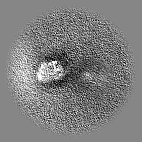
| Images | 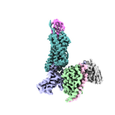 emd_41829.png emd_41829.png | 76.7 KB | ||
| Filedesc metadata | emd-41829.cif.gz | 6.9 KB | ||
| Archive directory |  http://ftp.pdbj.org/pub/emdb/structures/EMD-41829ftp://ftp.pdbj.org/pub/emdb/structures/EMD-41829 http://ftp.pdbj.org/pub/emdb/structures/EMD-41829ftp://ftp.pdbj.org/pub/emdb/structures/EMD-41829 | HTTPS FTP |
-Related structure data
| Related structure data |  8u1uMC  8tlmC C: citing same article ( M: atomic model generated by this map |
|---|---|
| Similar structure data |

-Links
| EMDB pages | EMDB (EBI/PDBe) / EMDataResource |
|---|---|
| Related items in Molecule of the Month |


-Map
| File | Download / File: emd_41829.map.gz / Format: CCP4 / Size: 91.1 MB / Type: IMAGE STORED AS FLOATING POINT NUMBER (4 BYTES) | ||||||||||||||||||||||||||||||||||||
|---|---|---|---|---|---|---|---|---|---|---|---|---|---|---|---|---|---|---|---|---|---|---|---|---|---|---|---|---|---|---|---|---|---|---|---|---|---|
| Projections & slices | Image control
Images are generated by Spider. | ||||||||||||||||||||||||||||||||||||
| Voxel size | X=Y=Z: 1.0153 Å | ||||||||||||||||||||||||||||||||||||
| Density |
| ||||||||||||||||||||||||||||||||||||
| Symmetry | Space group: 1 | ||||||||||||||||||||||||||||||||||||
| Details | EMDB XML:
|
 X (Sec.)
 Y (Row.)
 Z (Col.)


-Supplemental data
- Sample components
-Entire : CCR8 in complex with CCL1 and Gi
| Entire | Name: CCR8 in complex with CCL1 and Gi |
|---|---|
| Components |
|
-Supramolecule #1: CCR8 in complex with CCL1 and Gi
| Supramolecule | Name: CCR8 in complex with CCL1 and Gi / type: complex / ID: 1 / Parent: 0 / Macromolecule list: #1-#5 |
|---|---|
| Source (natural) | Organism: Homo sapiens (human) |
-Macromolecule #1: C-C motif chemokine 1,C-C chemokine receptor type 8,EGFP fusion p...
| Macromolecule | Name: C-C motif chemokine 1,C-C chemokine receptor type 8,EGFP fusion protein type: protein_or_peptide / ID: 1 / Number of copies: 1 / Enantiomer: LEVO |
|---|---|
| Source (natural) | Organism: Human adenovirus C serotype 2 |
| Molecular weight | Theoretical: 83.23668 KDa |
| Recombinant expression | Organism: Mammalian expression vector Flag-MCS-pcDNA3.1 (others) |
| Sequence | String: KSMQVPFSRC CFSFCEQEIP LRAILCYRNT SSICSNEGLI FKLKRGKEAC ALDTVGWVQR HRKMLRHCPS KRKGSGSGSG SGSGSGSGS GSGSGSGSDY TLDLSVTTVT DYYYPDICSS PCDAELIQTN GKLLLAVFYC LLFVFSLLGN SLVILVLVVC K KLRSITDV ...String: KSMQVPFSRC CFSFCEQEIP LRAILCYRNT SSICSNEGLI FKLKRGKEAC ALDTVGWVQR HRKMLRHCPS KRKGSGSGSG SGSGSGSGS GSGSGSGSDY TLDLSVTTVT DYYYPDICSS PCDAELIQTN GKLLLAVFYC LLFVFSLLGN SLVILVLVVC K KLRSITDV YLLNLALSDL LFVFSFPFQT YYLLDQWVFG TVMCKVVSGF YYIGFYSSMF FITLMSVDRY LAVVHAVYAL KV RTIRMGT TLCLAVWLTA IMATIPLLVF YQVASEDGVL QCYSFYNQQT LKWKIFTNFK MNILGLLIPF TIFMFCYIKI LHQ LKRCQN HNKTKAIRLV LIVVIASLLF WVPFNVVLFL TSLHSMHILD GCSISQQLTY ATHVTEIISF THCCVNPVIY AFVG EKFKK HLSEIFQKSC SQIFNYLGRQ MPRESCEKSS SCQQHSSRSS SVDYILGGSD YKDDDDKGGS LEVLFQGPMV SKGEE LFTG VVPILVELDG DVNGHKFSVS GEGEGDATYG KLTLKLICTT GKLPVPWPTL VTTLGYGLQC FARYPDHMKQ HDFFKS AMP EGYVQERTIF FKDDGNYKTR AEVKFEGDTL VNRIELKGID FKEDGNILGH KLEYNYNSHN VYITADKQKN GIKANFK IR HNIEDGGVQL ADHYQQNTPI GDGPVLLPDN HYLSYQSKLS KDPNEKRDHM VLLEFVTAAG ITLGMDELYK GSAWSHPQ F EKGGGSGGGS GGSAWSHPQF EK UniProtKB: C-C motif chemokine 1, C-C chemokine receptor type 8, EGFP |
-Macromolecule #2: Guanine nucleotide-binding protein G(i) subunit alpha-1
| Macromolecule | Name: Guanine nucleotide-binding protein G(i) subunit alpha-1 type: protein_or_peptide / ID: 2 / Number of copies: 1 / Enantiomer: LEVO |
|---|---|
| Source (natural) | Organism: Homo sapiens (human) |
| Molecular weight | Theoretical: 43.182078 KDa |
| Recombinant expression | Organism:  |
| Sequence | String: MKKHHHHHHH HHHENLYFQG GSMGCTLSAE DKAAVERSKM IDRNLREDGE KAAREVKLLL LGAGESGKST IVKQMKIIHE AGYSEEECK QYKAVVYSNT IQSIIAIIRA MGRLKIDFGD SARADDARQL FVLAGAAEEG FMTAELAGVI KRLWKDSGVQ A CFNRSREY ...String: MKKHHHHHHH HHHENLYFQG GSMGCTLSAE DKAAVERSKM IDRNLREDGE KAAREVKLLL LGAGESGKST IVKQMKIIHE AGYSEEECK QYKAVVYSNT IQSIIAIIRA MGRLKIDFGD SARADDARQL FVLAGAAEEG FMTAELAGVI KRLWKDSGVQ A CFNRSREY QLNDSAAYYL NDLDRIAQPN YIPTQQDVLR TRVKTTGIVE THFTFKDLHF KMFDVGGQRS ERKKWIHCFE GV TAIIFCV ALSDYDLVLA EDEEMNRMHE SMKLFDSICN NKWFTDTSII LFLNKKDLFE EKIKKSPLTI CYPEYAGSNT YEE AAAYIQ CQFEDLNKRK DTKEIYTHFT CATDTKNVQF VFDAVTDVII KNNLKDCGLF UniProtKB: Guanine nucleotide-binding protein G(i) subunit alpha-1 |
-Macromolecule #3: Guanine nucleotide-binding protein G(I)/G(S)/G(T) subunit beta-1
| Macromolecule | Name: Guanine nucleotide-binding protein G(I)/G(S)/G(T) subunit beta-1 type: protein_or_peptide / ID: 3 / Number of copies: 1 / Enantiomer: LEVO |
|---|---|
| Source (natural) | Organism: Homo sapiens (human) |
| Molecular weight | Theoretical: 39.518121 KDa |
| Recombinant expression | Organism:   Spodoptera frugiperda (fall armyworm) Spodoptera frugiperda (fall armyworm) |
| Sequence | String: MHHHHHHHHG ENLYFQGSSE LDQLRQEAEQ LKNQIRDARK ACADATLSQI TNNIDPVGRI QMRTRRTLRG HLAKIYAMHW GTDSRLLVS ASQDGKLIIW DSYTTNKVHA IPLRSSWVMT CAYAPSGNYV ACGGLDNICS IYNLKTREGN VRVSRELAGH T GYLSCCRF ...String: MHHHHHHHHG ENLYFQGSSE LDQLRQEAEQ LKNQIRDARK ACADATLSQI TNNIDPVGRI QMRTRRTLRG HLAKIYAMHW GTDSRLLVS ASQDGKLIIW DSYTTNKVHA IPLRSSWVMT CAYAPSGNYV ACGGLDNICS IYNLKTREGN VRVSRELAGH T GYLSCCRF LDDNQIVTSS GDTTCALWDI ETGQQTTTFT GHTGDVMSLS LAPDTRLFVS GACDASAKLW DVREGMCRQT FT GHESDIN AICFFPNGNA FATGSDDATC RLFDLRADQE LMTYSHDNII CGITSVSFSK SGRLLLAGYD DFNCNVWDAL KAD RAGVLA GHDNRVSCLG VTDDGMAVAT GSWDSFLKIW N UniProtKB: Guanine nucleotide-binding protein G(I)/G(S)/G(T) subunit beta-1 |
-Macromolecule #4: Guanine nucleotide-binding protein G(I)/G(S)/G(O) subunit gamma-2
| Macromolecule | Name: Guanine nucleotide-binding protein G(I)/G(S)/G(O) subunit gamma-2 type: protein_or_peptide / ID: 4 / Number of copies: 1 / Enantiomer: LEVO |
|---|---|
| Source (natural) | Organism: Homo sapiens (human) |
| Molecular weight | Theoretical: 7.861143 KDa |
| Recombinant expression | Organism: Spodoptera frugiperda (fall armyworm) |
| Sequence | String: MASNNTASIA QARKLVEQLK MEANIDRIKV SKAAADLMAY CEAHAKEDPL LTPVPASENP FREKKFFCAI L UniProtKB: Guanine nucleotide-binding protein G(I)/G(S)/G(O) subunit gamma-2 |
-Macromolecule #5: scFv fragment
| Macromolecule | Name: scFv fragment / type: protein_or_peptide / ID: 5 / Number of copies: 1 / Enantiomer: LEVO |
|---|---|
| Source (natural) | Organism: |
| Molecular weight | Theoretical: 28.124387 KDa |
| Recombinant expression | Organism: Spodoptera frugiperda (fall armyworm) |
| Sequence | String: AGSDVQLVES GGGLVQPGGS RKLSCSASGF AFSSFGMHWV RQAPEKGLEW VAYISSGSGT IYYADTVKGR FTISRDDPKN TLFLQMTSL RSEDTAMYYC VRSIYYYGSS PFDFWGQGTT LTVSSGGSDI VMTQATSSVP VTPGESVSIS CRSSKSLLHS N GNTYLYWF ...String: AGSDVQLVES GGGLVQPGGS RKLSCSASGF AFSSFGMHWV RQAPEKGLEW VAYISSGSGT IYYADTVKGR FTISRDDPKN TLFLQMTSL RSEDTAMYYC VRSIYYYGSS PFDFWGQGTT LTVSSGGSDI VMTQATSSVP VTPGESVSIS CRSSKSLLHS N GNTYLYWF LQRPGQSPQL LIYRMSNLAS GVPDRFSGSG SGTAFTLTIS RLEAEDVGVY YCMQHLEYPL TFGAGTKLEL KA AAGNSLV PRGSHHHHHH HH |
-Macromolecule #6: 2-acetamido-2-deoxy-beta-D-glucopyranose
| Macromolecule | Name: 2-acetamido-2-deoxy-beta-D-glucopyranose / type: ligand / ID: 6 / Number of copies: 1 / Formula: NAG |
|---|---|
| Molecular weight | Theoretical: 221.208 Da |
| Chemical component information |  ChemComp-NAG: |
-Experimental details
-Structure determination
| Method | cryo EM |
|---|---|
 Processing Processing | single particle reconstruction |
| Aggregation state | particle |
-Sample preparation
| Buffer | pH: 7.5 |
|---|---|
| Vitrification | Cryogen name: ETHANE |
- Electron microscopy
| Microscope | FEI TITAN KRIOS |
|---|---|
| Image recording | Film or detector model: FEI FALCON IV (4k x 4k) / Average electron dose: 40.814 e/Å2 |
| Electron beam | Acceleration voltage: 300 kV / Electron source:  FIELD EMISSION GUN FIELD EMISSION GUN |
| Electron optics | Illumination mode: FLOOD BEAM / Imaging mode: BRIGHT FIELD / Nominal defocus max: 1.8 µm / Nominal defocus min: 0.8 µm |
| Experimental equipment |  Model: Titan Krios / Image courtesy: FEI Company |
-Image processing
| Startup model | Type of model: OTHER |
|---|---|
| Final reconstruction | Resolution.type: BY AUTHOR / Resolution: 3.1 Å / Resolution method: OTHER / Number images used: 201761 |
| Initial angle assignment | Type: OTHER / Details: Ab initio in cryoSPARC |
| Final angle assignment | Type: MAXIMUM LIKELIHOOD |
