 Movie
Movie Controller

[English] 日本語
 Yorodumi
- EMDB-3883: High-resolution cryo-EM structure of the human 80S ribosome - (co... -
+ Open data

- Basic information
| Entry | Database: EMDB / ID: EMD-3883 | |||||||||
|---|---|---|---|---|---|---|---|---|---|---|
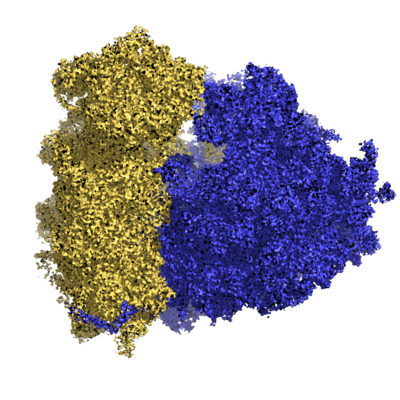
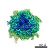
| Title | High-resolution cryo-EM structure of the human 80S ribosome - (composite structure) | |||||||||
 Map data Map data | Human 80S ribosome high-resolution cryo-EM map | |||||||||
 Sample Sample |
| |||||||||
| Function / homology |  Function and homology information Function and homology informationtranslation at presynapse / exit from mitosis / optic nerve development / response to insecticide / regulation of translation involved in cellular response to UV / eukaryotic 80S initiation complex / ribosomal protein import into nucleus / negative regulation of formation of translation preinitiation complex / negative regulation of endoplasmic reticulum unfolded protein response / axial mesoderm development ...translation at presynapse / exit from mitosis / optic nerve development / response to insecticide / regulation of translation involved in cellular response to UV / eukaryotic 80S initiation complex / ribosomal protein import into nucleus / negative regulation of formation of translation preinitiation complex / negative regulation of endoplasmic reticulum unfolded protein response / axial mesoderm development / regulation of G1 to G0 transition / retinal ganglion cell axon guidance / oxidized pyrimidine DNA binding / response to TNF agonist / positive regulation of base-excision repair / protein-DNA complex disassembly / positive regulation of intrinsic apoptotic signaling pathway in response to DNA damage by p53 class mediator / positive regulation of respiratory burst involved in inflammatory response / positive regulation of gastrulation / 90S preribosome assembly / positive regulation of ubiquitin-protein transferase activity / protein tyrosine kinase inhibitor activity / positive regulation of DNA-templated transcription initiation / positive regulation of intrinsic apoptotic signaling pathway in response to DNA damage / IRE1-RACK1-PP2A complex / positive regulation of Golgi to plasma membrane protein transport / alpha-beta T cell differentiation / nucleolus organization / TNFR1-mediated ceramide production / positive regulation of DNA damage response, signal transduction by p53 class mediator / GAIT complex / negative regulation of RNA splicing / TORC2 complex binding / neural crest cell differentiation / supercoiled DNA binding / G1 to G0 transition / cytoplasmic translational initiation / NF-kappaB complex / negative regulation of DNA repair / middle ear morphogenesis / oxidized purine DNA binding / cysteine-type endopeptidase activator activity involved in apoptotic process / rRNA modification in the nucleus and cytosol / negative regulation of intrinsic apoptotic signaling pathway in response to hydrogen peroxide / negative regulation of bicellular tight junction assembly / ubiquitin-like protein conjugating enzyme binding / regulation of establishment of cell polarity / negative regulation of phagocytosis / erythrocyte homeostasis / cytoplasmic side of rough endoplasmic reticulum membrane / Formation of the ternary complex, and subsequently, the 43S complex / ion channel inhibitor activity / protein kinase A binding / laminin receptor activity / homeostatic process / pigmentation / Ribosomal scanning and start codon recognition / positive regulation of mitochondrial depolarization / macrophage chemotaxis / Translation initiation complex formation / lung morphogenesis / positive regulation of natural killer cell proliferation / negative regulation of Wnt signaling pathway / fibroblast growth factor binding / male meiosis I / Protein hydroxylation / monocyte chemotaxis / BH3 domain binding / negative regulation of translational frameshifting / regulation of adenylate cyclase-activating G protein-coupled receptor signaling pathway / positive regulation of GTPase activity / TOR signaling / mTORC1-mediated signalling / SARS-CoV-1 modulates host translation machinery / iron-sulfur cluster binding / regulation of cell division / Peptide chain elongation / cellular response to ethanol / Selenocysteine synthesis / Formation of a pool of free 40S subunits / negative regulation of protein binding / blastocyst development / protein serine/threonine kinase inhibitor activity / Eukaryotic Translation Termination / positive regulation of intrinsic apoptotic signaling pathway by p53 class mediator / ubiquitin ligase inhibitor activity / SRP-dependent cotranslational protein targeting to membrane / Response of EIF2AK4 (GCN2) to amino acid deficiency / negative regulation of respiratory burst involved in inflammatory response / Viral mRNA Translation / endonucleolytic cleavage to generate mature 3'-end of SSU-rRNA from (SSU-rRNA, 5.8S rRNA, LSU-rRNA) / positive regulation of signal transduction by p53 class mediator / protein localization to nucleus / negative regulation of ubiquitin-dependent protein catabolic process / Nonsense Mediated Decay (NMD) independent of the Exon Junction Complex (EJC) / GTP hydrolysis and joining of the 60S ribosomal subunit / L13a-mediated translational silencing of Ceruloplasmin expression / Major pathway of rRNA processing in the nucleolus and cytosol / protein targeting / regulation of translational fidelity Similarity search - Function | |||||||||
| Biological species |  Homo sapiens (human) / Human (human) Homo sapiens (human) / Human (human) | |||||||||
| Method | single particle reconstruction / cryo EM / Resolution: 2.9 Å | |||||||||
 Authors Authors | Natchiar SK / Myasnikov AG / Kratzat H / Hazemann I / Klaholz BP | |||||||||
 Citation Citation | Journal: Nature / Year: 2017 Title: Visualization of chemical modifications in the human 80S ribosome structure. Authors: S Kundhavai Natchiar / Alexander G Myasnikov / Hanna Kratzat / Isabelle Hazemann / Bruno P Klaholz /  Abstract: Chemical modifications of human ribosomal RNA (rRNA) are introduced during biogenesis and have been implicated in the dysregulation of protein synthesis, as is found in cancer and other diseases. ...Chemical modifications of human ribosomal RNA (rRNA) are introduced during biogenesis and have been implicated in the dysregulation of protein synthesis, as is found in cancer and other diseases. However, their role in this phenomenon is unknown. Here we visualize more than 130 individual rRNA modifications in the three-dimensional structure of the human ribosome, explaining their structural and functional roles. In addition to a small number of universally conserved sites, we identify many eukaryote- or human-specific modifications and unique sites that form an extended shell in comparison to bacterial ribosomes, and which stabilize the RNA. Several of the modifications are associated with the binding sites of three ribosome-targeting antibiotics, or are associated with degenerate states in cancer, such as keto alkylations on nucleotide bases reminiscent of specialized ribosomes. This high-resolution structure of the human 80S ribosome paves the way towards understanding the role of epigenetic rRNA modifications in human diseases and suggests new possibilities for designing selective inhibitors and therapeutic drugs. | |||||||||
| History |
|
- Structure visualization
| Movie |
Movie viewer |
|---|---|
| Structure viewer | EM map: SurfViewMolmilJmol/JSmol |
| Supplemental images |
- Downloads & links
-EMDB archive
| Map data | emd_3883.map.gz | 52.1 MB | EMDB map data format | |
|---|---|---|---|---|
| Header (meta data) | emd-3883-v30.xmlemd-3883.xml | 94 KB 94 KB | Display Display | EMDB header |
| FSC (resolution estimation) | emd_3883_fsc.xmlemd_3883_fsc_1.xmlemd_3883_fsc_2.xmlemd_3883_fsc_3.xml | 22.4 KB 22.4 KB 22.4 KB 22.4 KB | Display Display Display Display | FSC data file |
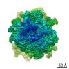
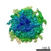
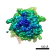
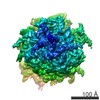
| Images |  emd_3883.png emd_3883.png | 222.4 KB | ||
| Archive directory |  http://ftp.pdbj.org/pub/emdb/structures/EMD-3883ftp://ftp.pdbj.org/pub/emdb/structures/EMD-3883 http://ftp.pdbj.org/pub/emdb/structures/EMD-3883ftp://ftp.pdbj.org/pub/emdb/structures/EMD-3883 | HTTPS FTP |
-Related structure data
| Related structure data |  6qzpMC  4243C 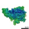 4244C 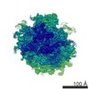 4245C 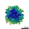 4263C  6ek0 C: citing same article ( M: atomic model generated by this map |
|---|---|
| Similar structure data |

-Links
| EMDB pages | EMDB (EBI/PDBe) / EMDataResource |
|---|---|
| Related items in Molecule of the Month |


-Map
| File | Download / File: emd_3883.map.gz / Format: CCP4 / Size: 1000 MB / Type: IMAGE STORED AS FLOATING POINT NUMBER (4 BYTES) | ||||||||||||||||||||||||||||||||||||||||||||||||||||||||||||
|---|---|---|---|---|---|---|---|---|---|---|---|---|---|---|---|---|---|---|---|---|---|---|---|---|---|---|---|---|---|---|---|---|---|---|---|---|---|---|---|---|---|---|---|---|---|---|---|---|---|---|---|---|---|---|---|---|---|---|---|---|---|
| Annotation | Human 80S ribosome high-resolution cryo-EM map | ||||||||||||||||||||||||||||||||||||||||||||||||||||||||||||
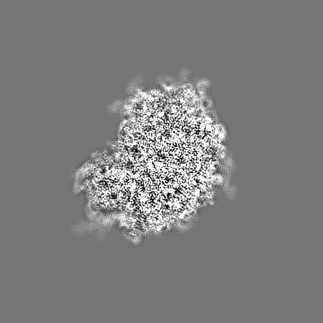
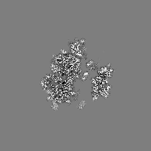
| Projections & slices | Image control
Images are generated by Spider. | ||||||||||||||||||||||||||||||||||||||||||||||||||||||||||||
| Voxel size | X=Y=Z: 0.85 Å | ||||||||||||||||||||||||||||||||||||||||||||||||||||||||||||
| Density |
| ||||||||||||||||||||||||||||||||||||||||||||||||||||||||||||
| Symmetry | Space group: 1 | ||||||||||||||||||||||||||||||||||||||||||||||||||||||||||||
| Details | EMDB XML:
CCP4 map header:
| ||||||||||||||||||||||||||||||||||||||||||||||||||||||||||||
 Z (Sec.)
 Y (Row.)
 X (Col.)


-Supplemental data
- Sample components
+Entire : 80S ribosome with ligand HHT and HYG
+Supramolecule #1: 80S ribosome with ligand HHT and HYG
+Macromolecule #1: 28S ribosomal RNA
+Macromolecule #2: 5S ribosomal RNA
+Macromolecule #3: 5.8S ribosomal RNA
+Macromolecule #47: 18S ribosomal RNA
+Macromolecule #48: Human initiator Met-tRNA-i
+Macromolecule #4: 60S ribosomal protein L8
+Macromolecule #5: 60S ribosomal protein L3
+Macromolecule #6: 60S ribosomal protein L4
+Macromolecule #7: 60S ribosomal protein L5
+Macromolecule #8: 60S ribosomal protein L6
+Macromolecule #9: 60S ribosomal protein L7
+Macromolecule #10: 60S ribosomal protein L7a
+Macromolecule #11: 60S ribosomal protein L9
+Macromolecule #12: 60S ribosomal protein L10-like
+Macromolecule #13: 60S ribosomal protein L11
+Macromolecule #14: 60S ribosomal protein L13
+Macromolecule #15: 60S ribosomal protein L14
+Macromolecule #16: 60S ribosomal protein L15
+Macromolecule #17: 60S ribosomal protein L13a
+Macromolecule #18: 60S ribosomal protein L17
+Macromolecule #19: 60S ribosomal protein L18
+Macromolecule #20: 60S ribosomal protein L19
+Macromolecule #21: 60S ribosomal protein L18a
+Macromolecule #22: 60S ribosomal protein L21
+Macromolecule #23: 60S ribosomal protein L22
+Macromolecule #24: 60S ribosomal protein L23
+Macromolecule #25: 60S ribosomal protein L24
+Macromolecule #26: 60S ribosomal protein L23a
+Macromolecule #27: 60S ribosomal protein L26
+Macromolecule #28: 60S ribosomal protein L27
+Macromolecule #29: 60S ribosomal protein L27a
+Macromolecule #30: 60S ribosomal protein L29
+Macromolecule #31: 60S ribosomal protein L30
+Macromolecule #32: 60S ribosomal protein L31
+Macromolecule #33: 60S ribosomal protein L32
+Macromolecule #34: 60S ribosomal protein L35a
+Macromolecule #35: 60S ribosomal protein L34
+Macromolecule #36: 60S ribosomal protein L35
+Macromolecule #37: 60S ribosomal protein L36
+Macromolecule #38: 60S ribosomal protein L37
+Macromolecule #39: 60S ribosomal protein L38
+Macromolecule #40: 60S ribosomal protein L39
+Macromolecule #41: Ubiquitin-60S ribosomal protein L40
+Macromolecule #42: 60S ribosomal protein L41
+Macromolecule #43: 60S ribosomal protein L36a
+Macromolecule #44: 60S ribosomal protein L37a
+Macromolecule #45: 60S ribosomal protein L28
+Macromolecule #46: 60S ribosomal protein L10a
+Macromolecule #49: 40S ribosomal protein SA
+Macromolecule #50: 40S ribosomal protein S3a
+Macromolecule #51: 40S ribosomal protein S3
+Macromolecule #52: 40S ribosomal protein S4, X isoform
+Macromolecule #53: 40S ribosomal protein S5
+Macromolecule #54: 40S ribosomal protein S7
+Macromolecule #55: 40S ribosomal protein S8
+Macromolecule #56: 40S ribosomal protein S10
+Macromolecule #57: 40S ribosomal protein S11
+Macromolecule #58: 40S ribosomal protein S15
+Macromolecule #59: 40S ribosomal protein S16
+Macromolecule #60: 40S ribosomal protein S17
+Macromolecule #61: 40S ribosomal protein S18
+Macromolecule #62: 40S ribosomal protein S19
+Macromolecule #63: 40S ribosomal protein S20
+Macromolecule #64: 40S ribosomal protein S21
+Macromolecule #65: 40S ribosomal protein S23
+Macromolecule #66: 40S ribosomal protein S26
+Macromolecule #67: 40S ribosomal protein S28
+Macromolecule #68: 40S ribosomal protein S29
+Macromolecule #69: Receptor of activated protein C kinase 1
+Macromolecule #70: 40S ribosomal protein S2
+Macromolecule #71: 40S ribosomal protein S6
+Macromolecule #72: 40S ribosomal protein S9
+Macromolecule #73: 40S ribosomal protein S12
+Macromolecule #74: 40S ribosomal protein S13
+Macromolecule #75: 40S ribosomal protein S14
+Macromolecule #76: 40S ribosomal protein S15a
+Macromolecule #77: 40S ribosomal protein S24
+Macromolecule #78: 40S ribosomal protein S25
+Macromolecule #79: 40S ribosomal protein S27
+Macromolecule #80: 40S ribosomal protein S30
+Macromolecule #81: Ubiquitin-40S ribosomal protein S27a
+Macromolecule #82: MAGNESIUM ION
+Macromolecule #83: (3beta)-O~3~-[(2R)-2,6-dihydroxy-2-(2-methoxy-2-oxoethyl)-6-methy...

+Macromolecule #84: ZINC ION
+Macromolecule #85: HYGROMYCIN B

+Macromolecule #86: water

-Experimental details
-Structure determination
| Method | cryo EM |
|---|---|
 Processing Processing | single particle reconstruction |
| Aggregation state | particle |
-Sample preparation
| Concentration | 0.5 mg/mL |
|---|---|
| Buffer | pH: 7.5 / Details: 100mM KCl, 5mM MgAc2, 20mM Hepes, 10mM NH4Cl |
| Vitrification | Cryogen name: ETHANE / Chamber humidity: 100 % / Chamber temperature: 283 K / Instrument: FEI VITROBOT MARK IV |
- Electron microscopy
| Microscope | FEI TITAN KRIOS |
|---|---|
| Image recording | Film or detector model: FEI FALCON II (4k x 4k) / Average electron dose: 3.5 e/Å2 |
| Electron beam | Acceleration voltage: 300 kV / Electron source:  FIELD EMISSION GUN FIELD EMISSION GUN |
| Electron optics | Illumination mode: SPOT SCAN / Imaging mode: BRIGHT FIELD |
| Sample stage | Specimen holder model: FEI TITAN KRIOS AUTOGRID HOLDER |
| Experimental equipment |  Model: Titan Krios / Image courtesy: FEI Company |
-Image processing
| Final reconstruction | Resolution.type: BY AUTHOR / Resolution: 2.9 Å / Resolution method: FSC 0.143 CUT-OFF / Number images used: 138234 |
|---|---|
| Initial angle assignment | Type: NOT APPLICABLE |
| Final angle assignment | Type: NOT APPLICABLE |
| FSC plot (resolution estimation) |  |
-Atomic model buiding 1
| Refinement | Protocol: OTHER |
|---|---|
| Output model | PDB-6qzp: |
