 Movie
Movie Controller


+ Open data

- Basic information
| Entry | 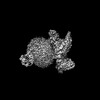 | |||||||||
|---|---|---|---|---|---|---|---|---|---|---|
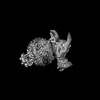
| Title | Structure of CXCR2 bound to CXCL3 (CXCR2-CXCL3-Go Full map) | |||||||||
 Map data Map data | ||||||||||
 Sample Sample |
| |||||||||
 Keywords Keywords | GPCR / Arrestin / SIGNALING PROTEIN-IMMUNE SYSTEM complex | |||||||||
| Function / homology |  Function and homology information Function and homology informationinterleukin-8-mediated signaling pathway / interleukin-8 receptor activity / mast cell granule / interleukin-8 binding / C-X-C chemokine receptor activity / neutrophil activation / mu-type opioid receptor binding / C-C chemokine receptor activity / : / corticotropin-releasing hormone receptor 1 binding ...interleukin-8-mediated signaling pathway / interleukin-8 receptor activity / mast cell granule / interleukin-8 binding / C-X-C chemokine receptor activity / neutrophil activation / mu-type opioid receptor binding / C-C chemokine receptor activity / : / corticotropin-releasing hormone receptor 1 binding / C-C chemokine binding / chemokine activity / G protein-coupled dopamine receptor signaling pathway / Chemokine receptors bind chemokines / dendritic cell chemotaxis / regulation of heart contraction / parallel fiber to Purkinje cell synapse / postsynaptic modulation of chemical synaptic transmission / cellular defense response / neutrophil chemotaxis / adenylate cyclase-inhibiting serotonin receptor signaling pathway / G protein-coupled serotonin receptor binding / secretory granule membrane / muscle contraction / calcium-mediated signaling / locomotory behavior / negative regulation of insulin secretion / receptor internalization / GABA-ergic synapse / G protein-coupled receptor activity / chemotaxis / G-protein beta/gamma-subunit complex binding / adenylate cyclase-modulating G protein-coupled receptor signaling pathway / adenylate cyclase-inhibiting G protein-coupled receptor signaling pathway / Olfactory Signaling Pathway / Activation of the phototransduction cascade / G protein-coupled acetylcholine receptor signaling pathway / G beta:gamma signalling through PLC beta / Presynaptic function of Kainate receptors / Thromboxane signalling through TP receptor / Activation of G protein gated Potassium channels / Inhibition of voltage gated Ca2+ channels via Gbeta/gamma subunits / G-protein activation / Glucagon signaling in metabolic regulation / mitotic spindle / Prostacyclin signalling through prostacyclin receptor / G beta:gamma signalling through CDC42 / Synthesis, secretion, and inactivation of Glucagon-like Peptide-1 (GLP-1) / G beta:gamma signalling through BTK / photoreceptor disc membrane / ADP signalling through P2Y purinoceptor 12 / Sensory perception of sweet, bitter, and umami (glutamate) taste / Glucagon-type ligand receptors / Adrenaline,noradrenaline inhibits insulin secretion / Vasopressin regulates renal water homeostasis via Aquaporins / Glucagon-like Peptide-1 (GLP1) regulates insulin secretion / G alpha (z) signalling events / cellular response to catecholamine stimulus / ADP signalling through P2Y purinoceptor 1 / ADORA2B mediated anti-inflammatory cytokines production / G beta:gamma signalling through PI3Kgamma / adenylate cyclase-activating dopamine receptor signaling pathway / Cooperation of PDCL (PhLP1) and TRiC/CCT in G-protein beta folding / GPER1 signaling / cellular response to prostaglandin E stimulus / heterotrimeric G-protein complex / G alpha (12/13) signalling events / Inactivation, recovery and regulation of the phototransduction cascade / G-protein beta-subunit binding / extracellular vesicle / sensory perception of taste / Thrombin signalling through proteinase activated receptors (PARs) / signaling receptor complex adaptor activity / positive regulation of cytosolic calcium ion concentration / microtubule cytoskeleton / retina development in camera-type eye / cell body / GTPase binding / fibroblast proliferation / G protein activity / presynaptic membrane / Ca2+ pathway / High laminar flow shear stress activates signaling by PIEZO1 and PECAM1:CDH5:KDR in endothelial cells / G alpha (i) signalling events / G alpha (s) signalling events / phospholipase C-activating G protein-coupled receptor signaling pathway / G alpha (q) signalling events / Hydrolases; Acting on acid anhydrides; Acting on GTP to facilitate cellular and subcellular movement / Ras protein signal transduction / postsynaptic membrane / cell surface receptor signaling pathway / Extra-nuclear estrogen signaling / cell population proliferation / immune response / G protein-coupled receptor signaling pathway / inflammatory response / external side of plasma membrane / lysosomal membrane / GTPase activity / positive regulation of cell population proliferation Similarity search - Function | |||||||||
| Biological species |  Homo sapiens (human) / Homo sapiens (human) /  | |||||||||
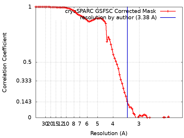
| Method | single particle reconstruction / cryo EM / Resolution: 3.38 Å | |||||||||
 Authors Authors | Sano FK / Saha S / Sharma S / Ganguly M / Shihoya W / Nureki O / Shukla AK / Banerjee R | |||||||||
| Funding support |  India, 1 items India, 1 items
| |||||||||
 Citation Citation | Journal: Mol Cell / Year: 2025 Title: Molecular basis of promiscuous chemokine binding and structural mimicry at the C-X-C chemokine receptor, CXCR2. Authors: Shirsha Saha / Fumiya K Sano / Saloni Sharma / Manisankar Ganguly / Sudha Mishra / Annu Dalal / Hiroaki Akasaka / Takaaki A Kobayashi / Nashrah Zaidi / Divyanshu Tiwari / Nabarun Roy / ...Authors: Shirsha Saha / Fumiya K Sano / Saloni Sharma / Manisankar Ganguly / Sudha Mishra / Annu Dalal / Hiroaki Akasaka / Takaaki A Kobayashi / Nashrah Zaidi / Divyanshu Tiwari / Nabarun Roy / Manish K Yadav / Nilanjana Banerjee / Sayantan Saha / Samanwita Mohapatra / Yuzuru Itoh / Andy Chevigné / Ramanuj Banerjee / Wataru Shihoya / Osamu Nureki / Arun K Shukla /  Abstract: Selectivity of natural agonists for their cognate receptors is a hallmark of G-protein-coupled receptors (GPCRs); however, this selectivity often breaks down at the chemokine receptors. Chemokines ...Selectivity of natural agonists for their cognate receptors is a hallmark of G-protein-coupled receptors (GPCRs); however, this selectivity often breaks down at the chemokine receptors. Chemokines often display promiscuous binding to chemokine receptors, but the underlying molecular determinants remain mostly elusive. Here, we perform a comprehensive transducer-coupling analysis, testing all known C-X-C chemokines on every C-X-C type chemokine receptor to generate a global fingerprint of the selectivity and promiscuity encoded within this system. Taking lead from this, we determine cryoelectron microscopy (cryo-EM) structures of the most promiscuous receptor, C-X-C chemokine receptor 2 (CXCR2), in complex with several chemokines. These structural snapshots elucidate the details of ligand-receptor interactions, including structural motifs, which are validated using mutagenesis and functional experiments. We also observe that most chemokines position themselves on CXCR2 as a dimer while CXCL6 exhibits a monomeric binding pose. Taken together, our findings provide the molecular basis of chemokine promiscuity at CXCR2 with potential implications for developing therapeutic molecules. | |||||||||
| History |
|
- Structure visualization
| Supplemental images |
|---|

- Downloads & links
-EMDB archive
| Map data | emd_38744.map.gz | 48.5 MB | EMDB map data format | |
|---|---|---|---|---|
| Header (meta data) | emd-38744-v30.xmlemd-38744.xml | 24 KB 24 KB | Display Display | EMDB header |
| FSC (resolution estimation) | emd_38744_fsc.xml | 7.9 KB | Display | FSC data file |
| Images |  emd_38744.png emd_38744.png | 71.8 KB | ||
| Filedesc metadata | emd-38744.cif.gz | 7.3 KB | ||
| Others | emd_38744_half_map_1.map.gzemd_38744_half_map_2.map.gz | 48.9 MB 48.9 MB | ||
| Archive directory |  http://ftp.pdbj.org/pub/emdb/structures/EMD-38744ftp://ftp.pdbj.org/pub/emdb/structures/EMD-38744 http://ftp.pdbj.org/pub/emdb/structures/EMD-38744ftp://ftp.pdbj.org/pub/emdb/structures/EMD-38744 | HTTPS FTP |
-Related structure data
| Related structure data |  8xx3MC  8xvuC  8xwaC  8xwfC  8xwmC  8xwnC  8xwsC 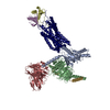 8xwvC  8xx6C  8xx7C  8xxhC  8xxrC 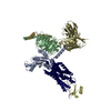 8xxxC M: atomic model generated by this map C: citing same article ( |
|---|---|
| Similar structure data |
-Links
| EMDB pages | EMDB (EBI/PDBe) / EMDataResource |
|---|---|
| Related items in Molecule of the Month |


-Map
| File | Download / File: emd_38744.map.gz / Format: CCP4 / Size: 52.7 MB / Type: IMAGE STORED AS FLOATING POINT NUMBER (4 BYTES) | ||||||||||||||||||||||||||||||||||||
|---|---|---|---|---|---|---|---|---|---|---|---|---|---|---|---|---|---|---|---|---|---|---|---|---|---|---|---|---|---|---|---|---|---|---|---|---|---|
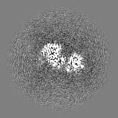
| Projections & slices | Image control
Images are generated by Spider. | ||||||||||||||||||||||||||||||||||||
| Voxel size | X=Y=Z: 1.0375 Å | ||||||||||||||||||||||||||||||||||||
| Density |
| ||||||||||||||||||||||||||||||||||||
| Symmetry | Space group: 1 | ||||||||||||||||||||||||||||||||||||
| Details | EMDB XML:
|
 X (Sec.)
 Y (Row.)
 Z (Col.)


-Supplemental data
-Half map: #1
| File | emd_38744_half_map_1.map | ||||||||||||
|---|---|---|---|---|---|---|---|---|---|---|---|---|---|
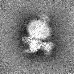
| Projections & Slices |
| ||||||||||||
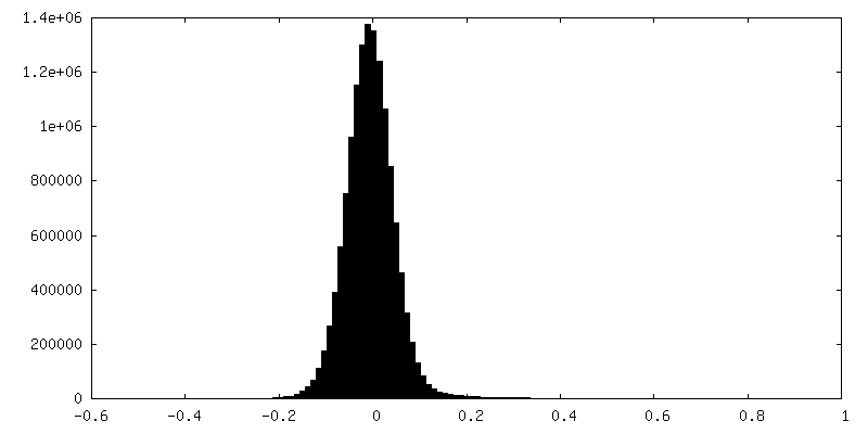
| Density Histograms |


-Half map: #2
| File | emd_38744_half_map_2.map | ||||||||||||
|---|---|---|---|---|---|---|---|---|---|---|---|---|---|
| Projections & Slices |
| ||||||||||||
| Density Histograms |


- Sample components
-Entire : C-X-C chemokine receptor type 2 in complex with C-X-C motif chemo...
| Entire | Name: C-X-C chemokine receptor type 2 in complex with C-X-C motif chemokine 3 and Go |
|---|---|
| Components |
|
-Supramolecule #1: C-X-C chemokine receptor type 2 in complex with C-X-C motif chemo...
| Supramolecule | Name: C-X-C chemokine receptor type 2 in complex with C-X-C motif chemokine 3 and Go type: complex / ID: 1 / Parent: 0 / Macromolecule list: all |
|---|---|
| Source (natural) | Organism: Homo sapiens (human) |
-Macromolecule #1: C-X-C motif chemokine 3
| Macromolecule | Name: C-X-C motif chemokine 3 / type: protein_or_peptide / ID: 1 / Number of copies: 2 / Enantiomer: LEVO |
|---|---|
| Source (natural) | Organism: Homo sapiens (human) |
| Molecular weight | Theoretical: 7.876251 KDa |
| Recombinant expression | Organism:  |
| Sequence | String: ASVVTELRCQ CLQTLQGIHL KNIQSVNVRS PGPHCAQTEV IATLKNGKKA CLNPASPMVQ KIIEKILNKG STN UniProtKB: C-X-C motif chemokine 3 |
-Macromolecule #2: C-X-C chemokine receptor type 2
| Macromolecule | Name: C-X-C chemokine receptor type 2 / type: protein_or_peptide / ID: 2 / Number of copies: 1 / Enantiomer: LEVO |
|---|---|
| Source (natural) | Organism: Homo sapiens (human) |
| Molecular weight | Theoretical: 46.699609 KDa |
| Recombinant expression | Organism:   Spodoptera frugiperda (fall armyworm) Spodoptera frugiperda (fall armyworm) |
| Sequence | String: MGKTIIALSY IFCLVFADYK DDDDAANFTP VNGSSGNQSV RLVTSSSLEV LFQGPGSEDF NMESDSFEDF WKGEDLSNYS YSSTLPPFL LDAAPCEPES LEINKYFVVI IYALVFLLSL LGNSLVMLVI LYSRVGRSVT DVYLLNLALA DLLFALTLPI W AASKVNGW ...String: MGKTIIALSY IFCLVFADYK DDDDAANFTP VNGSSGNQSV RLVTSSSLEV LFQGPGSEDF NMESDSFEDF WKGEDLSNYS YSSTLPPFL LDAAPCEPES LEINKYFVVI IYALVFLLSL LGNSLVMLVI LYSRVGRSVT DVYLLNLALA DLLFALTLPI W AASKVNGW IFGTFLCKVV SLLKEVNFYS GILLLACISV DRYLAIVHAT RTLTQKRYLV KFICLSIWGL SLLLALPVLL FR RTVYSSN VSPACYEDMG NNTANWRMLL RILPQSFGFI VPLLIMLFCY GFTLRTLFKA HMGQKHRAMR VIFAVVLIFL LCW LPYNLV LLADTLMRTQ VIQETCERRN HIDRALDATE ILGILHSCLN PLIYAFIGQK FRHGLLKILA IHGLISKDSL PKDS RPSFV GSSSGHTSTT L UniProtKB: C-X-C chemokine receptor type 2 |
-Macromolecule #3: Guanine nucleotide-binding protein G(o) subunit alpha
| Macromolecule | Name: Guanine nucleotide-binding protein G(o) subunit alpha / type: protein_or_peptide / ID: 3 / Number of copies: 1 / Enantiomer: LEVO |
|---|---|
| Source (natural) | Organism: Homo sapiens (human) |
| Molecular weight | Theoretical: 27.024762 KDa |
| Recombinant expression | Organism: |
| Sequence | String: MGHHHHHHEN LYFQGTLSAE ERAALERSKA IEKNLKEDGI SAAKDVKLLL LGADNSGKST IVKQMKIIHG GSGGSGGTTG IVETHFTFK NLHFRLFDVG GQRSERKKWI HCFEDVTAII FCVDLSDYNR MHESLMLFDS ICNNKFFIDT SIILFLNKKD L FGEKIKKS ...String: MGHHHHHHEN LYFQGTLSAE ERAALERSKA IEKNLKEDGI SAAKDVKLLL LGADNSGKST IVKQMKIIHG GSGGSGGTTG IVETHFTFK NLHFRLFDVG GQRSERKKWI HCFEDVTAII FCVDLSDYNR MHESLMLFDS ICNNKFFIDT SIILFLNKKD L FGEKIKKS PLTICFPEYT GPNTYEDAAA YIQAQFESKN RSPNKEIYCH MTCATDTNNA QVIFDAVTDI IIANNLRGCG LY UniProtKB: Guanine nucleotide-binding protein G(o) subunit alpha, Guanine nucleotide-binding protein G(o) subunit alpha |
-Macromolecule #4: Guanine nucleotide-binding protein G(I)/G(S)/G(T) subunit beta-1
| Macromolecule | Name: Guanine nucleotide-binding protein G(I)/G(S)/G(T) subunit beta-1 type: protein_or_peptide / ID: 4 / Number of copies: 1 / Enantiomer: LEVO |
|---|---|
| Source (natural) | Organism: Homo sapiens (human) |
| Molecular weight | Theoretical: 38.534062 KDa |
| Recombinant expression | Organism: Spodoptera frugiperda (fall armyworm) |
| Sequence | String: MHHHHHHGSS GSELDQLRQE AEQLKNQIRD ARKACADATL SQITNNIDPV GRIQMRTRRT LRGHLAKIYA MHWGTDSRLL VSASQDGKL IIWDSYTTNK VHAIPLRSSW VMTCAYAPSG NYVACGGLDN ICSIYNLKTR EGNVRVSREL AGHTGYLSCC R FLDDNQIV ...String: MHHHHHHGSS GSELDQLRQE AEQLKNQIRD ARKACADATL SQITNNIDPV GRIQMRTRRT LRGHLAKIYA MHWGTDSRLL VSASQDGKL IIWDSYTTNK VHAIPLRSSW VMTCAYAPSG NYVACGGLDN ICSIYNLKTR EGNVRVSREL AGHTGYLSCC R FLDDNQIV TSSGDTTCAL WDIETGQQTT TFTGHTGDVM SLSLAPDTRL FVSGACDASA KLWDVREGMC RQTFTGHESD IN AICFFPN GNAFATGSDD ATCRLFDLRA DQELMTYSHD NIICGITSVS FSKSGRLLLA GYDDFNCNVW DALKADRAGV LAG HDNRVS CLGVTDDGMA VATGSWDSFL KIWN UniProtKB: Guanine nucleotide-binding protein G(I)/G(S)/G(T) subunit beta-1 |
-Macromolecule #5: Guanine nucleotide-binding protein G(I)/G(S)/G(O) subunit gamma-2
| Macromolecule | Name: Guanine nucleotide-binding protein G(I)/G(S)/G(O) subunit gamma-2 type: protein_or_peptide / ID: 5 / Number of copies: 1 / Enantiomer: LEVO |
|---|---|
| Source (natural) | Organism: Homo sapiens (human) |
| Molecular weight | Theoretical: 7.861143 KDa |
| Recombinant expression | Organism: Spodoptera frugiperda (fall armyworm) |
| Sequence | String: MASNNTASIA QARKLVEQLK MEANIDRIKV SKAAADLMAY CEAHAKEDPL LTPVPASENP FREKKFFCAI L UniProtKB: Guanine nucleotide-binding protein G(I)/G(S)/G(O) subunit gamma-2 |
-Macromolecule #6: Antibody fragment ScFv16
| Macromolecule | Name: Antibody fragment ScFv16 / type: protein_or_peptide / ID: 6 / Number of copies: 1 / Enantiomer: LEVO |
|---|---|
| Source (natural) | Organism: |
| Molecular weight | Theoretical: 26.466486 KDa |
| Recombinant expression | Organism: |
| Sequence | String: DVQLVESGGG LVQPGGSRKL SCSASGFAFS SFGMHWVRQA PEKGLEWVAY ISSGSGTIYY ADTVKGRFTI SRDDPKNTLF LQMTSLRSE DTAMYYCVRS IYYYGSSPFD FWGQGTTLTV SSGGGGSGGG GSGGGGSDIV MTQATSSVPV TPGESVSISC R SSKSLLHS ...String: DVQLVESGGG LVQPGGSRKL SCSASGFAFS SFGMHWVRQA PEKGLEWVAY ISSGSGTIYY ADTVKGRFTI SRDDPKNTLF LQMTSLRSE DTAMYYCVRS IYYYGSSPFD FWGQGTTLTV SSGGGGSGGG GSGGGGSDIV MTQATSSVPV TPGESVSISC R SSKSLLHS NGNTYLYWFL QRPGQSPQLL IYRMSNLASG VPDRFSGSGS GTAFTLTISR LEAEDVGVYY CMQHLEYPLT FG AGTKLEL K |
-Experimental details
-Structure determination
| Method | cryo EM |
|---|---|
 Processing Processing | single particle reconstruction |
| Aggregation state | particle |
-Sample preparation
| Buffer | pH: 7.4 |
|---|---|
| Vitrification | Cryogen name: ETHANE |
- Electron microscopy
| Microscope | FEI TITAN KRIOS |
|---|---|
| Image recording | Film or detector model: GATAN K3 (6k x 4k) / Detector mode: COUNTING / Average electron dose: 50.2 e/Å2 |
| Electron beam | Acceleration voltage: 300 kV / Electron source:  FIELD EMISSION GUN FIELD EMISSION GUN |
| Electron optics | Illumination mode: FLOOD BEAM / Imaging mode: BRIGHT FIELD / Cs: 2.7 mm / Nominal defocus max: 1.6 µm / Nominal defocus min: 0.8 µm |
| Sample stage | Specimen holder model: FEI TITAN KRIOS AUTOGRID HOLDER / Cooling holder cryogen: NITROGEN |
| Experimental equipment |  Model: Titan Krios / Image courtesy: FEI Company |
+Image processing
-Atomic model buiding 1
| Initial model | Chain - Source name: SwissModel / Chain - Initial model type: in silico model |
|---|---|
| Refinement | Space: REAL / Protocol: FLEXIBLE FIT |
| Output model | PDB-8xx3: |
