 Movie
Movie Controller


+ Open data

- Basic information
| Entry | Database: PDB / ID: 8xvu | ||||||
|---|---|---|---|---|---|---|---|
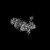
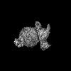
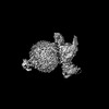
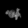
| Title | Structure of CXCR2 bound to CXCL2 (Ligand-receptor focused map) | ||||||
 Components Components |
| ||||||
 Keywords Keywords | SIGNALING PROTEIN/IMMUNE SYSTEM / GPCR / Arrestin / SIGNALING PROTEIN-IMMUNE SYSTEM complex | ||||||
| Function / homology |  Function and homology information Function and homology informationinterleukin-8-mediated signaling pathway / interleukin-8 receptor activity / mast cell granule / interleukin-8 binding / C-X-C chemokine receptor activity / neutrophil activation / regulation of chemotaxis / C-C chemokine binding / C-C chemokine receptor activity / chemokine activity ...interleukin-8-mediated signaling pathway / interleukin-8 receptor activity / mast cell granule / interleukin-8 binding / C-X-C chemokine receptor activity / neutrophil activation / regulation of chemotaxis / C-C chemokine binding / C-C chemokine receptor activity / chemokine activity / Chemokine receptors bind chemokines / dendritic cell chemotaxis / Interleukin-10 signaling / cellular defense response / neutrophil chemotaxis / secretory granule membrane / calcium-mediated signaling / response to molecule of bacterial origin / receptor internalization / chemotaxis / G protein-coupled receptor activity / phospholipase C-activating G protein-coupled receptor signaling pathway / positive regulation of cytosolic calcium ion concentration / G alpha (i) signalling events / cell surface receptor signaling pathway / immune response / inflammatory response / G protein-coupled receptor signaling pathway / external side of plasma membrane / positive regulation of cell population proliferation / Neutrophil degranulation / cell surface / signal transduction / : / extracellular region / membrane / plasma membrane Similarity search - Function | ||||||
| Biological species |  Homo sapiens (human) Homo sapiens (human) | ||||||
| Method | ELECTRON MICROSCOPY / single particle reconstruction / cryo EM / Resolution: 3.09 Å | ||||||
 Authors Authors | Sano, F.K. / Saha, S. / Sharma, S. / Ganguly, M. / Shihoya, W. / Nureki, O. / Shukla, A.K. / Banerjee, R. | ||||||
| Funding support |  India, 1items India, 1items
| ||||||
 Citation Citation | Journal: Mol Cell / Year: 2025 Title: Molecular basis of promiscuous chemokine binding and structural mimicry at the C-X-C chemokine receptor, CXCR2. Authors: Shirsha Saha / Fumiya K Sano / Saloni Sharma / Manisankar Ganguly / Sudha Mishra / Annu Dalal / Hiroaki Akasaka / Takaaki A Kobayashi / Nashrah Zaidi / Divyanshu Tiwari / Nabarun Roy / ...Authors: Shirsha Saha / Fumiya K Sano / Saloni Sharma / Manisankar Ganguly / Sudha Mishra / Annu Dalal / Hiroaki Akasaka / Takaaki A Kobayashi / Nashrah Zaidi / Divyanshu Tiwari / Nabarun Roy / Manish K Yadav / Nilanjana Banerjee / Sayantan Saha / Samanwita Mohapatra / Yuzuru Itoh / Andy Chevigné / Ramanuj Banerjee / Wataru Shihoya / Osamu Nureki / Arun K Shukla /  Abstract: Selectivity of natural agonists for their cognate receptors is a hallmark of G-protein-coupled receptors (GPCRs); however, this selectivity often breaks down at the chemokine receptors. Chemokines ...Selectivity of natural agonists for their cognate receptors is a hallmark of G-protein-coupled receptors (GPCRs); however, this selectivity often breaks down at the chemokine receptors. Chemokines often display promiscuous binding to chemokine receptors, but the underlying molecular determinants remain mostly elusive. Here, we perform a comprehensive transducer-coupling analysis, testing all known C-X-C chemokines on every C-X-C type chemokine receptor to generate a global fingerprint of the selectivity and promiscuity encoded within this system. Taking lead from this, we determine cryoelectron microscopy (cryo-EM) structures of the most promiscuous receptor, C-X-C chemokine receptor 2 (CXCR2), in complex with several chemokines. These structural snapshots elucidate the details of ligand-receptor interactions, including structural motifs, which are validated using mutagenesis and functional experiments. We also observe that most chemokines position themselves on CXCR2 as a dimer while CXCL6 exhibits a monomeric binding pose. Taken together, our findings provide the molecular basis of chemokine promiscuity at CXCR2 with potential implications for developing therapeutic molecules. | ||||||
| History |
|
- Structure visualization
| Structure viewer | Molecule: MolmilJmol/JSmol |
|---|
- Downloads & links
-Download
| PDBx/mmCIF format | 8xvu.cif.gz | 88.8 KB | Display | PDBx/mmCIF format |
|---|---|---|---|---|
| PDB format | pdb8xvu.ent.gz | 62.8 KB | Display | PDB format |
| PDBx/mmJSON format | 8xvu.json.gz | Tree view | PDBx/mmJSON format | |
| Others |  Other downloads Other downloads |
-Validation report
| Arichive directory | https://data.pdbj.org/pub/pdb/validation_reports/xv/8xvuftp://data.pdbj.org/pub/pdb/validation_reports/xv/8xvu | HTTPS FTP |
|---|
-Related structure data
| Related structure data | 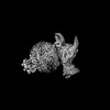 38719MC  8xwaC  8xwfC  8xwmC  8xwnC  8xwsC 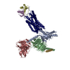 8xwvC  8xx3C  8xx6C  8xx7C  8xxhC  8xxrC 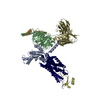 8xxxC M: map data used to model this data C: citing same article ( |
|---|---|
| Similar structure data |
-Links
 PDBj
PDBj


- Assembly
| Deposited unit | 
|
|---|---|
| 1 |
|
-Components
| #1: Protein | Mass: 46699.609 Da / Num. of mol.: 1 Source method: isolated from a genetically manipulated source Source: (gene. exp.) Homo sapiens (human) / Gene: CXCR2 / Production host:   Spodoptera frugiperda (fall armyworm) / References: UniProt: P25025 Spodoptera frugiperda (fall armyworm) / References: UniProt: P25025 | ||
|---|---|---|---|
| #2: Protein | Mass: 7905.440 Da / Num. of mol.: 2 Source method: isolated from a genetically manipulated source Source: (gene. exp.) Homo sapiens (human) / Gene: CXCL2 / Production host:  Has protein modification | Y | |
-Experimental details
-Experiment
| Experiment | Method: ELECTRON MICROSCOPY |
|---|---|
| EM experiment | Aggregation state: PARTICLE / 3D reconstruction method: single particle reconstruction |
- Sample preparation
| Component |
| ||||||||||||||||||||||||
|---|---|---|---|---|---|---|---|---|---|---|---|---|---|---|---|---|---|---|---|---|---|---|---|---|---|
| Molecular weight |
| ||||||||||||||||||||||||
| Source (natural) |
| ||||||||||||||||||||||||
| Source (recombinant) |
| ||||||||||||||||||||||||
| Buffer solution | pH: 7.4 | ||||||||||||||||||||||||
| Specimen | Embedding applied: NO / Shadowing applied: NO / Staining applied: NO / Vitrification applied: YES | ||||||||||||||||||||||||
| Vitrification | Cryogen name: ETHANE |
- Electron microscopy imaging
| Experimental equipment |  Model: Titan Krios / Image courtesy: FEI Company |
|---|---|
| Microscopy | Model: FEI TITAN KRIOS |
| Electron gun | Electron source:  FIELD EMISSION GUN / Accelerating voltage: 300 kV / Illumination mode: FLOOD BEAM FIELD EMISSION GUN / Accelerating voltage: 300 kV / Illumination mode: FLOOD BEAM |
| Electron lens | Mode: BRIGHT FIELD / Nominal defocus max: 1600 nm / Nominal defocus min: 800 nm / Cs: 2.7 mm |
| Specimen holder | Cryogen: NITROGEN / Specimen holder model: FEI TITAN KRIOS AUTOGRID HOLDER |
| Image recording | Electron dose: 50.2 e/Å2 / Detector mode: COUNTING / Film or detector model: GATAN K3 (6k x 4k) |
| Image scans | Movie frames/image: 40 |
- Processing
| EM software |
| ||||||||||||||||||||||||||||
|---|---|---|---|---|---|---|---|---|---|---|---|---|---|---|---|---|---|---|---|---|---|---|---|---|---|---|---|---|---|
| CTF correction | Type: NONE | ||||||||||||||||||||||||||||
| Particle selection | Num. of particles selected: 1927680 | ||||||||||||||||||||||||||||
| 3D reconstruction | Resolution: 3.09 Å / Resolution method: FSC 0.143 CUT-OFF / Num. of particles: 285884 / Symmetry type: POINT | ||||||||||||||||||||||||||||
| Atomic model building | Protocol: FLEXIBLE FIT / Space: REAL | ||||||||||||||||||||||||||||
| Atomic model building | Source name: SwissModel / Type: in silico model | ||||||||||||||||||||||||||||
| Refine LS restraints |
|
