 Movie
Movie Controller


+ Open data

- Basic information
| Entry |  | |||||||||
|---|---|---|---|---|---|---|---|---|---|---|
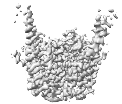
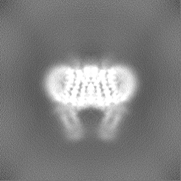
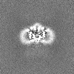
| Title | Cryo-EM structure of sheep VMAT2 dimer in an atypical fold | |||||||||
 Map data Map data | OaVMAT2 TM8/9 replaced by BRIL. | |||||||||
 Sample Sample |
| |||||||||
 Keywords Keywords | Vesicular monoamine transporter / SLC18A2 / MFS / Apo / Conformation / TRANSPORT PROTEIN | |||||||||
| Biological species |  | |||||||||
| Method | single particle reconstruction / cryo EM / Resolution: 3.2 Å | |||||||||
 Authors Authors | Lyu Y / Fu C / Ma H / Sun Z / Su Z / Zhou X | |||||||||
| Funding support |  China, 1 items China, 1 items
| |||||||||
 Citation Citation | Journal: Nat Commun / Year: 2024 Title: Engineering of a mammalian VMAT2 for cryo-EM analysis results in non-canonical protein folding. Authors: Ying Lyu / Chunting Fu / Haiyun Ma / Zhaoming Su / Ziyi Sun / Xiaoming Zhou / Abstract: Vesicular monoamine transporter 2 (VMAT2) belongs to the major facilitator superfamily (MFS), and mediates cytoplasmic monoamine packaging into presynaptic vesicles. Here, we present two cryo-EM ...Vesicular monoamine transporter 2 (VMAT2) belongs to the major facilitator superfamily (MFS), and mediates cytoplasmic monoamine packaging into presynaptic vesicles. Here, we present two cryo-EM structures of VMAT2, with a frog VMAT2 adopting a canonical MFS fold and an engineered sheep VMAT2 adopting a non-canonical fold. Both VMAT2 proteins mediate uptake of a selective fluorescent VMAT2 substrate into cells. Molecular docking, substrate binding and transport analysis reveal potential substrate binding mechanism in VMAT2. Meanwhile, caution is advised when interpreting engineered membrane protein structures. | |||||||||
| History |
|
- Structure visualization
| Supplemental images |
|---|
- Downloads & links
-EMDB archive
| Map data | emd_38389.map.gz | 59.7 MB |  EMDB map data format EMDB map data format | |
|---|---|---|---|---|
| Header (meta data) | emd-38389-v30.xmlemd-38389.xml | 15.8 KB 15.8 KB | Display Display | EMDB header |
| FSC (resolution estimation) | emd_38389_fsc.xml | 8.5 KB | Display | FSC data file |
| Images |  emd_38389.png emd_38389.png | 89.2 KB | ||
| Filedesc metadata | emd-38389.cif.gz | 5.9 KB | ||
| Others | emd_38389_half_map_1.map.gzemd_38389_half_map_2.map.gz | 59.2 MB 59.2 MB | ||
| Archive directory |  http://ftp.pdbj.org/pub/emdb/structures/EMD-38389ftp://ftp.pdbj.org/pub/emdb/structures/EMD-38389 http://ftp.pdbj.org/pub/emdb/structures/EMD-38389ftp://ftp.pdbj.org/pub/emdb/structures/EMD-38389 | HTTPS FTP |
-Related structure data
-Links
| EMDB pages | EMDB (EBI/PDBe) / EMDataResource |
|---|
-Map
| File | Download / File: emd_38389.map.gz / Format: CCP4 / Size: 64 MB / Type: IMAGE STORED AS FLOATING POINT NUMBER (4 BYTES) | ||||||||||||||||||||||||||||||||||||
|---|---|---|---|---|---|---|---|---|---|---|---|---|---|---|---|---|---|---|---|---|---|---|---|---|---|---|---|---|---|---|---|---|---|---|---|---|---|
| Annotation | OaVMAT2 TM8/9 replaced by BRIL. | ||||||||||||||||||||||||||||||||||||
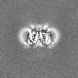
| Projections & slices | Image control
Images are generated by Spider. | ||||||||||||||||||||||||||||||||||||
| Voxel size | X=Y=Z: 0.85 Å | ||||||||||||||||||||||||||||||||||||
| Density |
| ||||||||||||||||||||||||||||||||||||
| Symmetry | Space group: 1 | ||||||||||||||||||||||||||||||||||||
| Details | EMDB XML:
|
 Z (Sec.)
 Y (Row.)
 X (Col.)


-Supplemental data
-Half map: Half map B.
| File | emd_38389_half_map_1.map | ||||||||||||
|---|---|---|---|---|---|---|---|---|---|---|---|---|---|
| Annotation | Half map B. | ||||||||||||
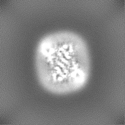
| Projections & Slices |
| ||||||||||||
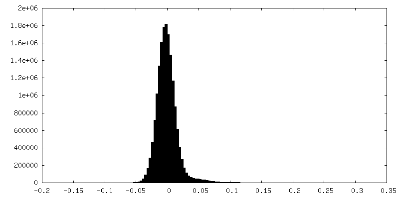
| Density Histograms |


-Half map: Half map A.
| File | emd_38389_half_map_2.map | ||||||||||||
|---|---|---|---|---|---|---|---|---|---|---|---|---|---|
| Annotation | Half map A. | ||||||||||||
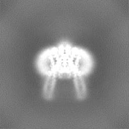
| Projections & Slices |
| ||||||||||||
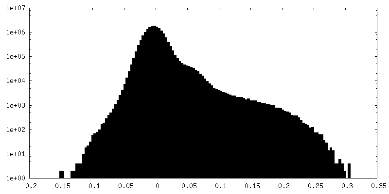
| Density Histograms |


- Sample components
-Entire : OaVMAT2 TM8/9 loop replaced by BRIL
| Entire | Name: OaVMAT2 TM8/9 loop replaced by BRIL |
|---|---|
| Components |
|
-Supramolecule #1: OaVMAT2 TM8/9 loop replaced by BRIL
| Supramolecule | Name: OaVMAT2 TM8/9 loop replaced by BRIL / type: complex / ID: 1 / Parent: 0 / Macromolecule list: all |
|---|---|
| Source (natural) | Organism: |
-Macromolecule #1: OaVMAT2-BRIL
| Macromolecule | Name: OaVMAT2-BRIL / type: protein_or_peptide / ID: 1 / Number of copies: 2 / Enantiomer: LEVO |
|---|---|
| Source (natural) | Organism: |
| Molecular weight | Theoretical: 68.592688 KDa |
| Recombinant expression | Organism:  Komagataella pastoris (fungus) Komagataella pastoris (fungus) |
| Sequence | String: MALSELALLR RLQESRHSRK LVLFIVFLAL LLDNMLLTVV VPIIPSYLYS IEHEKDALEI QTAKPGLTAS APGSFQNIFS YYDNSTMVT GNNTDHLQGP LVHEATTQRM VTNSSSAPSD CPSEDKDLLN ENVQVGLLFA SKATVQLLTN PFIGLLTNRI G YPIPMFTG ...String: MALSELALLR RLQESRHSRK LVLFIVFLAL LLDNMLLTVV VPIIPSYLYS IEHEKDALEI QTAKPGLTAS APGSFQNIFS YYDNSTMVT GNNTDHLQGP LVHEATTQRM VTNSSSAPSD CPSEDKDLLN ENVQVGLLFA SKATVQLLTN PFIGLLTNRI G YPIPMFTG FCIMFISTVM FAFSRSYAFL LFARSLQGIG SSCSSVAGMG MLASVYTDDE ERGNAMGIAL GGLAMGVLVG PP FGSVLYE FVGKTAPFLV LAALVLLDGA IQLFVLQPSR VQPESQKGTP LTTLLRDPYI LIAAGSICFA NMGIAMLEPA LPI WMMETM CSHKWQLGVA FLPASISYLI GTNVFGILAR RQLADLEDNW ETLNDNLKVI EKADNAAQVK DALTKMRAAA LDAQ KATPP KLEDKSPDSP EMKDFRHGFD ILVGQIDDAL KLANEGKVKE AQAAAEQLKT TRNAYIQKYL ERARSTLRWL CALLG MIIV GMSILCIPLA KNIYGLIAPN FGVGFAIGMV DSSMMPIMGY LVDLRHVSVY GSVYAIADVA FCMGYAIGPS AGGAIA KAI GFPWLMTIIG IIDILFAPLC FFLRSPPAKE EKMAILMDHN CPIKTKMYTQ NSSQSHPIGE DEDSESD |
-Experimental details
-Structure determination
| Method | cryo EM |
|---|---|
 Processing Processing | single particle reconstruction |
| Aggregation state | particle |
-Sample preparation
| Concentration | 8.0 mg/mL |
|---|---|
| Buffer | pH: 7.4 / Details: 150 mm NaCl, 20 mM HEPES-Na pH 7.4, 0.4 mM DDM |
| Vitrification | Cryogen name: ETHANE / Chamber humidity: 100 % / Chamber temperature: 277 K / Instrument: FEI VITROBOT MARK IV / Details: blot for 3 s before plunging. |
| Details | This sample was monodisperse. |
- Electron microscopy
| Microscope | FEI TITAN KRIOS |
|---|---|
| Image recording | Film or detector model: GATAN K2 SUMMIT (4k x 4k) / Detector mode: COUNTING / Average electron dose: 62.64 e/Å2 |
| Electron beam | Acceleration voltage: 300 kV / Electron source:  FIELD EMISSION GUN FIELD EMISSION GUN |
| Electron optics | Illumination mode: FLOOD BEAM / Imaging mode: BRIGHT FIELD / Nominal defocus max: 1.6 µm / Nominal defocus min: 1.0 µm |
| Sample stage | Specimen holder model: FEI TITAN KRIOS AUTOGRID HOLDER / Cooling holder cryogen: NITROGEN |
| Experimental equipment |  Model: Titan Krios / Image courtesy: FEI Company |
+Image processing

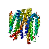
-Atomic model buiding 1
| Initial model | Chain - Source name: AlphaFold / Chain - Initial model type: in silico model |
|---|---|
| Output model | 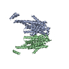 PDB-8xit: |
