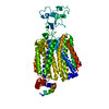
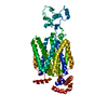
Journal: Cell Rep / Year: 2024 Title: Structural basis for the transport and substrate selection of human urate transporter 1. Authors: Jingjing He / Guoyun Liu / Fang Kong / Qiulong Tan / Zhenzhou Wang / Meng Yang / Yonglin He / Xiaoxiao Jia / Chuangye Yan / Chao Wang / Hongwu Qian / Abstract: High serum urate levels are the major risk factor for gout. URAT1, the primary transporter for urate absorption in the kidneys, is well known as an anti-hyperuricemia drug target. However, the ...High serum urate levels are the major risk factor for gout. URAT1, the primary transporter for urate absorption in the kidneys, is well known as an anti-hyperuricemia drug target. However, the clinical application of URAT1-targeted drugs is limited because of their low specificity and severe side effects. The lack of structural information impedes elucidation of the transport mechanism and the development of new drugs. Here, we present the cryoelectron microscopy (cryo-EM) structures of human URAT1(R477S), its complex with urate, and its closely related homolog OAT4. URAT1(R477S) and OAT4 exhibit major facilitator superfamily (MFS) folds with outward- and inward-open conformations, respectively. Structural comparison reveals a 30° rotation between the N-terminal and C-terminal domains, supporting an alternating access mechanism. A conserved arginine (OAT4-Arg473/URAT1-Arg477) is found to be essential for chloride-mediated inhibition. The URAT1(R477S)-urate complex reveals the specificity of urate recognition. Taken together, our study promotes our understanding of the transport mechanism and substrate selection of URAT1.
In the structure databanks used in Yorodumi, some data are registered as the other names, "COVID-19 virus" and "2019-nCoV". Here are the details of the virus and the list of structure data.
Jan 31, 2019. EMDB accession codes are about to change! (news from PDBe EMDB page)
EMDB accession codes are about to change! (news from PDBe EMDB page)
The allocation of 4 digits for EMDB accession codes will soon come to an end. Whilst these codes will remain in use, new EMDB accession codes will include an additional digit and will expand incrementally as the available range of codes is exhausted. The current 4-digit format prefixed with “EMD-” (i.e. EMD-XXXX) will advance to a 5-digit format (i.e. EMD-XXXXX), and so on. It is currently estimated that the 4-digit codes will be depleted around Spring 2019, at which point the 5-digit format will come into force.
The EM Navigator/Yorodumi systems omit the EMD- prefix.
Related info.:Q: What is EMD? / ID/Accession-code notation in Yorodumi/EM Navigator
Yorodumi is a browser for structure data from EMDB, PDB, SASBDB, etc.
This page is also the successor to EM Navigator detail page, and also detail information page/front-end page for Omokage search.
The word "yorodu" (or yorozu) is an old Japanese word meaning "ten thousand". "mi" (miru) is to see.
Related info.:EMDB / PDB / SASBDB / Comparison of 3 databanks / Yorodumi Search / Aug 31, 2016. New EM Navigator & Yorodumi / Yorodumi Papers / Jmol/JSmol / Function and homology information / Changes in new EM Navigator and Yorodumi
 Movie
Movie Controller


 Open data
 Basic information
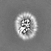
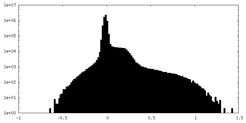
 Map data
 Sample
 Keywords
 Function and homology information
 Homo sapiens (human)
 Authors
 China, 1 items
  Citation
 Structure visualization
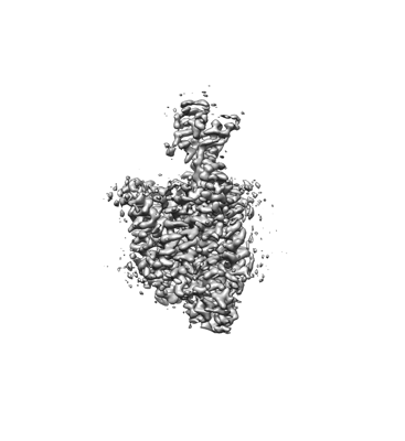
 Downloads & links
 emd_37579.png
 http://ftp.pdbj.org/pub/emdb/structures/EMD-37579
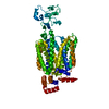


 Z (Sec.)
 Y (Row.)
 X (Col.)


 Sample components
 Processing
 Electron microscopy
 FIELD EMISSION GUN
