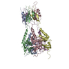
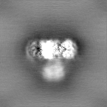
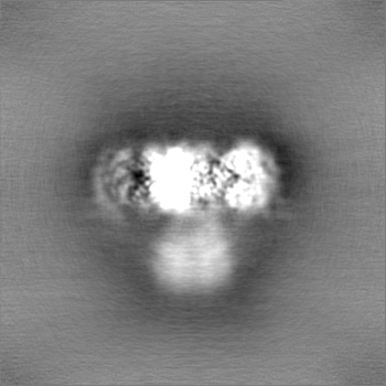
登録情報 データベース : EMDB / ID : EMD-36367タイトル Cryo-EM structure of KCTD5 in complex with Gbeta gamma subunits EM density map of KCTD5_Gng2/Gnb1 complex 複合体 : Cryo-EM structure of KCTD5 in complex with G protein betagamma subunitsタンパク質・ペプチド : Guanine nucleotide-binding protein G(I)/G(S)/G(T) subunit beta-1タンパク質・ペプチド : Guanine nucleotide-binding protein G(I)/G(S)/G(O) subunit gamma-2タンパク質・ペプチド : BTB/POZ domain-containing protein KCTD5 / / 機能・相同性 分子機能 ドメイン・相同性 構成要素
/ / / / / / / / / / / / / / / / / / / / / / / / / / / / / / / / / / / / / / / / / / / / / / / / / / / / / / / / / / / / / / / / / / / / / / / / / / / / / / / / / / / / / / / / / / / / / / / / / / / / / / / / / / / / 生物種 Mus musculus (ハツカネズミ) / Homo sapiens (ヒト)手法 / / 解像度 : 3.27 Å Zheng S / Jiang W / Wang W / Kong Y 資金援助 Organization Grant number 国 Ministry of Science and Technology (MoST, China)
ジャーナル : Sci Adv / 年 : 2023タイトル : Structural basis for the ubiquitination of G protein βγ subunits by KCTD5/Cullin3 E3 ligase.著者 : Wentong Jiang / Wei Wang / Yinfei Kong / Sanduo Zheng / 要旨 : G protein-coupled receptor (GPCR) signaling is precisely controlled to avoid overstimulation that results in detrimental consequences. Gβγ signaling is negatively regulated by a Cullin3 (Cul3)- ... G protein-coupled receptor (GPCR) signaling is precisely controlled to avoid overstimulation that results in detrimental consequences. Gβγ signaling is negatively regulated by a Cullin3 (Cul3)-dependent E3 ligase, KCTD5, which triggers ubiquitination and degradation of free Gβγ. Here, we report the cryo-electron microscopy structures of the KCTD5-Gβγ fusion complex and the KCTD7-Cul3 complex. KCTD5 in pentameric form engages symmetrically with five copies of Gβγ through its C-terminal domain. The unique pentameric assembly of the KCTD5/Cul3 E3 ligase places the ubiquitin-conjugating enzyme (E2) and the modification sites of Gβγ in close proximity and allows simultaneous transfer of ubiquitin from E2 to five Gβγ subunits. Moreover, we show that ubiquitination of Gβγ by KCTD5 is important for fine-tuning cyclic adenosine 3´,5´-monophosphate signaling of GPCRs. Our studies provide unprecedented insights into mechanisms of substrate recognition by unusual pentameric E3 ligases and highlight the KCTD family as emerging regulators of GPCR signaling. 履歴 登録 2023年6月1日 - ヘッダ(付随情報) 公開 2023年7月26日 - マップ公開 2023年7月26日 - 更新 2024年7月3日 - 現状 2024年7月3日 処理サイト : PDBj / 状態 : 公開
すべて表示 表示を減らす
 ムービー
ムービー コントローラー


 データを開く
 基本情報
基本情報
 マップデータ
 試料
試料 キーワード
キーワード 機能・相同性情報
 Homo sapiens (ヒト)
 データ登録者
 中国, 1件
  引用
引用 構造の表示
構造の表示
 ダウンロードとリンク
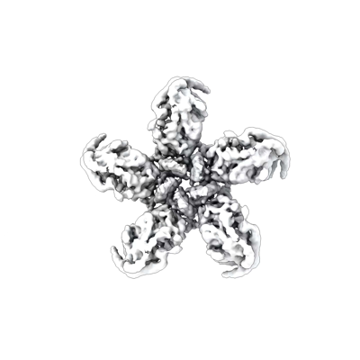 emd_36367.png
 http://ftp.pdbj.org/pub/emdb/structures/EMD-36367


 Z
Z Y
Y X
X


 試料の構成要素
 解析
解析 電子顕微鏡法
 FIELD EMISSION GUN