[English] 日本語
 Yorodumi
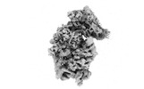
- EMDB-36332: Atomic structure of wheat ribosome reveals unique features of the... -
+ Open data
- Basic information
| Entry |  | |||||||||
|---|---|---|---|---|---|---|---|---|---|---|
| Title | Atomic structure of wheat ribosome reveals unique features of the plant ribosomes | |||||||||
 Map data Map data | ||||||||||
 Sample Sample |
| |||||||||
 Keywords Keywords | Protein Synthesis Machinery / Eukaryotic Ribosome / Plant / TRANSLATION | |||||||||
| Function / homology |  Function and homology information Function and homology informationnegative regulation of translational frameshifting / endonucleolytic cleavage to generate mature 3'-end of SSU-rRNA from (SSU-rRNA, 5.8S rRNA, LSU-rRNA) / endonucleolytic cleavage in ITS1 to separate SSU-rRNA from 5.8S rRNA and LSU-rRNA from tricistronic rRNA transcript (SSU-rRNA, 5.8S rRNA, LSU-rRNA) / translation regulator activity / rescue of stalled cytosolic ribosome / protein kinase C binding / maturation of SSU-rRNA from tricistronic rRNA transcript (SSU-rRNA, 5.8S rRNA, LSU-rRNA) / maturation of SSU-rRNA / small-subunit processome / rRNA processing ...negative regulation of translational frameshifting / endonucleolytic cleavage to generate mature 3'-end of SSU-rRNA from (SSU-rRNA, 5.8S rRNA, LSU-rRNA) / endonucleolytic cleavage in ITS1 to separate SSU-rRNA from 5.8S rRNA and LSU-rRNA from tricistronic rRNA transcript (SSU-rRNA, 5.8S rRNA, LSU-rRNA) / translation regulator activity / rescue of stalled cytosolic ribosome / protein kinase C binding / maturation of SSU-rRNA from tricistronic rRNA transcript (SSU-rRNA, 5.8S rRNA, LSU-rRNA) / maturation of SSU-rRNA / small-subunit processome / rRNA processing / ribosome binding / ribosomal small subunit biogenesis / ribosomal small subunit assembly / small ribosomal subunit / small ribosomal subunit rRNA binding / cytosolic small ribosomal subunit / cytoplasmic translation / rRNA binding / structural constituent of ribosome / ribosome / translation / ribonucleoprotein complex / mRNA binding / nucleolus / RNA binding / zinc ion binding / nucleus / cytosol Similarity search - Function | |||||||||
| Biological species |  | |||||||||
| Method | single particle reconstruction / cryo EM / Resolution: 2.88 Å | |||||||||
 Authors Authors | Mishra RK / Sharma P / Hussain T | |||||||||
| Funding support |  United Kingdom, 1 items United Kingdom, 1 items
| |||||||||
 Citation Citation |  Journal: Structure / Year: 2024 Journal: Structure / Year: 2024Title: Cryo-EM structure of wheat ribosome reveals unique features of the plant ribosomes. Authors: Rishi Kumar Mishra / Prafful Sharma / Faisal Tarique Khaja / Adwaith B Uday / Tanweer Hussain /  Abstract: Plants being sessile organisms exhibit unique features in ribosomes, which might aid in rapid gene expression and regulation in response to varying environmental conditions. Here, we present high- ...Plants being sessile organisms exhibit unique features in ribosomes, which might aid in rapid gene expression and regulation in response to varying environmental conditions. Here, we present high-resolution structures of the 60S and 80S ribosomes from wheat, a monocot staple crop plant (Triticum aestivum). While plant ribosomes have unique plant-specific rRNA modification (Cm1847) in the peptide exit tunnel (PET), the zinc-finger motif in eL34 is absent, and uL4 is extended, making an exclusive interaction network. We note differences in the eL15-helix 11 (25S) interaction, eL6-ES7 assembly, and certain rRNA chemical modifications between monocot and dicot ribosomes. In eukaryotes, we observe highly conserved rRNA modification (Gm75) in 5.8S rRNA and a flipped base (G1506) in PET. These features are likely involved in sensing or stabilizing nascent chain. Finally, we discuss the importance of the universal conservation of three consecutive rRNA modifications in all ribosomes for their interaction with A-site aminoacyl-tRNA. #1:  Journal: Acta Crystallogr., Sect. D: Biol. Crystallogr. / Year: 2018 Journal: Acta Crystallogr., Sect. D: Biol. Crystallogr. / Year: 2018Title: Real-space refinement in PHENIX for cryo-EM and crystallography Authors: Uday AB / Khaja FT | |||||||||
| History |
|
- Structure visualization
| Supplemental images |
|---|
- Downloads & links
-EMDB archive
| Map data |  emd_36332.map.gz emd_36332.map.gz | 267 MB |  EMDB map data format EMDB map data format | |
|---|---|---|---|---|
| Header (meta data) |  emd-36332-v30.xml emd-36332-v30.xml emd-36332.xml emd-36332.xml | 50.2 KB 50.2 KB | Display Display |  EMDB header EMDB header |
| FSC (resolution estimation) |  emd_36332_fsc.xml emd_36332_fsc.xml | 13.8 KB | Display |  FSC data file FSC data file |
| Images |  emd_36332.png emd_36332.png | 50.5 KB | ||
| Filedesc metadata |  emd-36332.cif.gz emd-36332.cif.gz | 11.5 KB | ||
| Others |  emd_36332_half_map_1.map.gz emd_36332_half_map_1.map.gz emd_36332_half_map_2.map.gz emd_36332_half_map_2.map.gz | 262.6 MB 262.6 MB | ||
| Archive directory |  http://ftp.pdbj.org/pub/emdb/structures/EMD-36332 http://ftp.pdbj.org/pub/emdb/structures/EMD-36332 ftp://ftp.pdbj.org/pub/emdb/structures/EMD-36332 ftp://ftp.pdbj.org/pub/emdb/structures/EMD-36332 | HTTPS FTP |
-Related structure data
| Related structure data |  8jiwMC  8jivC C: citing same article ( M: atomic model generated by this map |
|---|---|
| Similar structure data | Similarity search - Function & homology  F&H Search F&H Search |
- Links
Links
| EMDB pages |  EMDB (EBI/PDBe) / EMDB (EBI/PDBe) /  EMDataResource EMDataResource |
|---|---|
| Related items in Molecule of the Month |
- Map
Map
| File |  Download / File: emd_36332.map.gz / Format: CCP4 / Size: 282.6 MB / Type: IMAGE STORED AS FLOATING POINT NUMBER (4 BYTES) Download / File: emd_36332.map.gz / Format: CCP4 / Size: 282.6 MB / Type: IMAGE STORED AS FLOATING POINT NUMBER (4 BYTES) | ||||||||||||||||||||||||||||||||||||
|---|---|---|---|---|---|---|---|---|---|---|---|---|---|---|---|---|---|---|---|---|---|---|---|---|---|---|---|---|---|---|---|---|---|---|---|---|---|
| Projections & slices | Image control
Images are generated by Spider. | ||||||||||||||||||||||||||||||||||||
| Voxel size | X=Y=Z: 1.07 Å | ||||||||||||||||||||||||||||||||||||
| Density |
| ||||||||||||||||||||||||||||||||||||
| Symmetry | Space group: 1 | ||||||||||||||||||||||||||||||||||||
| Details | EMDB XML:
|
-Supplemental data
-Half map: #2
| File | emd_36332_half_map_1.map | ||||||||||||
|---|---|---|---|---|---|---|---|---|---|---|---|---|---|
| Projections & Slices |
| ||||||||||||
| Density Histograms |
-Half map: #1
| File | emd_36332_half_map_2.map | ||||||||||||
|---|---|---|---|---|---|---|---|---|---|---|---|---|---|
| Projections & Slices |
| ||||||||||||
| Density Histograms |
- Sample components
+Entire : The large subunit of wheat Ribosome
+Supramolecule #1: The large subunit of wheat Ribosome
+Macromolecule #1: 18S rRNA
+Macromolecule #2: 40S ribosomal protein SA
+Macromolecule #3: 40S ribosomal protein S3a
+Macromolecule #4: S5 DRBM domain-containing protein
+Macromolecule #5: 40S ribosomal protein S4
+Macromolecule #6: 40S ribosomal protein S6
+Macromolecule #7: 40S ribosomal protein S7
+Macromolecule #8: 40S ribosomal protein S8
+Macromolecule #9: 30S ribosomal protein S4, chloroplastic
+Macromolecule #10: 40S ribosomal protein S11 N-terminal domain-containing protein
+Macromolecule #11: Ribosomal protein S13/S15 N-terminal domain-containing protein
+Macromolecule #12: 40S ribosomal protein S14
+Macromolecule #13: 40S ribosomal protein S17
+Macromolecule #14: 40S ribosomal protein S21
+Macromolecule #15: 30S ribosomal protein S8, chloroplastic
+Macromolecule #16: 40S ribosomal protein S23
+Macromolecule #17: 40S ribosomal protein S24
+Macromolecule #18: 40S ribosomal protein S26
+Macromolecule #19: 40S ribosomal protein S27
+Macromolecule #20: 40S ribosomal protein S30
+Macromolecule #21: 60S ribosomal protein L41
+Macromolecule #22: KH type-2 domain-containing protein
+Macromolecule #23: Ribosomal protein S7 domain-containing protein
+Macromolecule #24: Plectin/S10 N-terminal domain-containing protein
+Macromolecule #25: 40S ribosomal protein S15
+Macromolecule #26: 40S ribosomal protein S16
+Macromolecule #27: 40S ribosomal protein S18
+Macromolecule #28: 40S ribosomal protein S19
+Macromolecule #29: Ribosomal protein S10 domain-containing protein
+Macromolecule #30: 40S ribosomal protein S28
+Macromolecule #31: 40S ribosomal protein S29
+Macromolecule #32: Mitogen-activated protein kinase
+Macromolecule #33: 40S ribosomal protein S25
+Macromolecule #34: POTASSIUM ION
+Macromolecule #35: MAGNESIUM ION
+Macromolecule #36: ZINC ION
-Experimental details
-Structure determination
| Method | cryo EM |
|---|---|
 Processing Processing | single particle reconstruction |
| Aggregation state | particle |
- Sample preparation
| Buffer | pH: 7.6 |
|---|---|
| Grid | Model: Quantifoil R2/2 / Material: COPPER / Mesh: 400 / Support film - Material: CARBON / Support film - topology: CONTINUOUS |
| Vitrification | Cryogen name: ETHANE / Chamber humidity: 100 % / Chamber temperature: 289 K / Instrument: FEI VITROBOT MARK IV |
| Details | The sample is the small subunit of wheat ribosome |
- Electron microscopy
| Microscope | FEI TITAN KRIOS |
|---|---|
| Image recording | Film or detector model: FEI FALCON III (4k x 4k) / Average electron dose: 44.6 e/Å2 |
| Electron beam | Acceleration voltage: 300 kV / Electron source:  FIELD EMISSION GUN FIELD EMISSION GUN |
| Electron optics | Calibrated magnification: 75000 / Illumination mode: OTHER / Imaging mode: OTHER / Cs: 2.7 mm / Nominal defocus max: 3.0 µm / Nominal defocus min: 1.5 µm |
| Sample stage | Specimen holder model: FEI TITAN KRIOS AUTOGRID HOLDER / Cooling holder cryogen: NITROGEN |
| Experimental equipment |  Model: Titan Krios / Image courtesy: FEI Company |
 Movie
Movie Controller


 Z (Sec.)
 Y (Row.)
 X (Col.)


