[English] 日本語
 Yorodumi
- EMDB-36323: Cryo-EM structure of the GLP-1R/GCGR dual agonist MEDI0382-bound ... -
+ Open data
- Basic information
| Entry |  | |||||||||||||||||||||||||||
|---|---|---|---|---|---|---|---|---|---|---|---|---|---|---|---|---|---|---|---|---|---|---|---|---|---|---|---|---|
| Title | Cryo-EM structure of the GLP-1R/GCGR dual agonist MEDI0382-bound human GLP-1R-Gs complex | |||||||||||||||||||||||||||
 Map data Map data | ||||||||||||||||||||||||||||
 Sample Sample |
| |||||||||||||||||||||||||||
 Keywords Keywords | G protein-coupled receptor / ligand recognition / receptor activation / unimolecular dual agonist / STRUCTURAL PROTEIN | |||||||||||||||||||||||||||
| Function / homology |  Function and homology information Function and homology informationglucagon-like peptide 1 receptor activity / glucagon receptor activity / positive regulation of blood pressure / hormone secretion / post-translational protein targeting to membrane, translocation / G-protein activation / Activation of the phototransduction cascade / Glucagon-type ligand receptors / Thromboxane signalling through TP receptor / Sensory perception of sweet, bitter, and umami (glutamate) taste ...glucagon-like peptide 1 receptor activity / glucagon receptor activity / positive regulation of blood pressure / hormone secretion / post-translational protein targeting to membrane, translocation / G-protein activation / Activation of the phototransduction cascade / Glucagon-type ligand receptors / Thromboxane signalling through TP receptor / Sensory perception of sweet, bitter, and umami (glutamate) taste / G beta:gamma signalling through PI3Kgamma / G beta:gamma signalling through CDC42 / Cooperation of PDCL (PhLP1) and TRiC/CCT in G-protein beta folding / Activation of G protein gated Potassium channels / Inhibition of voltage gated Ca2+ channels via Gbeta/gamma subunits / Ca2+ pathway / G alpha (z) signalling events / High laminar flow shear stress activates signaling by PIEZO1 and PECAM1:CDH5:KDR in endothelial cells / Glucagon-like Peptide-1 (GLP1) regulates insulin secretion / Vasopressin regulates renal water homeostasis via Aquaporins / Adrenaline,noradrenaline inhibits insulin secretion / ADP signalling through P2Y purinoceptor 12 / G alpha (q) signalling events / G alpha (i) signalling events / Thrombin signalling through proteinase activated receptors (PARs) / Activation of G protein gated Potassium channels / G-protein activation / G beta:gamma signalling through PI3Kgamma / Prostacyclin signalling through prostacyclin receptor / G beta:gamma signalling through PLC beta / ADP signalling through P2Y purinoceptor 1 / Thromboxane signalling through TP receptor / Presynaptic function of Kainate receptors / G beta:gamma signalling through CDC42 / Inhibition of voltage gated Ca2+ channels via Gbeta/gamma subunits / G alpha (12/13) signalling events / Glucagon-type ligand receptors / G beta:gamma signalling through BTK / ADP signalling through P2Y purinoceptor 12 / Adrenaline,noradrenaline inhibits insulin secretion / Cooperation of PDCL (PhLP1) and TRiC/CCT in G-protein beta folding / Ca2+ pathway / Thrombin signalling through proteinase activated receptors (PARs) / G alpha (z) signalling events / Extra-nuclear estrogen signaling / G alpha (s) signalling events / photoreceptor outer segment membrane / G alpha (q) signalling events / response to psychosocial stress / spectrin binding / G alpha (i) signalling events / Glucagon-like Peptide-1 (GLP1) regulates insulin secretion / High laminar flow shear stress activates signaling by PIEZO1 and PECAM1:CDH5:KDR in endothelial cells / Vasopressin regulates renal water homeostasis via Aquaporins / regulation of heart contraction / alkylglycerophosphoethanolamine phosphodiesterase activity / peptide hormone binding / photoreceptor outer segment / activation of adenylate cyclase activity / negative regulation of blood pressure / cardiac muscle cell apoptotic process / photoreceptor inner segment / adenylate cyclase-activating G protein-coupled receptor signaling pathway / Glucagon-type ligand receptors / transmembrane signaling receptor activity / Glucagon-like Peptide-1 (GLP1) regulates insulin secretion / cellular response to catecholamine stimulus / adenylate cyclase-activating dopamine receptor signaling pathway / G-protein beta-subunit binding / cellular response to prostaglandin E stimulus / heterotrimeric G-protein complex / sensory perception of taste / signaling receptor complex adaptor activity / positive regulation of cytosolic calcium ion concentration / retina development in camera-type eye / cell body / GTPase binding / cellular response to hypoxia / G alpha (s) signalling events / phospholipase C-activating G protein-coupled receptor signaling pathway / learning or memory / cell surface receptor signaling pathway / cell population proliferation / G protein-coupled receptor signaling pathway / GTPase activity / dendrite / synapse / protein-containing complex binding / membrane / plasma membrane / cytoplasm Similarity search - Function | |||||||||||||||||||||||||||
| Biological species |  Homo sapiens (human) / synthetic construct (others) / Homo sapiens (human) / synthetic construct (others) /    | |||||||||||||||||||||||||||
| Method | single particle reconstruction / cryo EM / Resolution: 2.85 Å | |||||||||||||||||||||||||||
 Authors Authors | Yang L / Zhou QT / Dai AT / Zhao FH / Chang RL / Ying TL / Wu BL / Yang DH / Wang MW / Cong ZT | |||||||||||||||||||||||||||
| Funding support |  China, 8 items China, 8 items
| |||||||||||||||||||||||||||
 Citation Citation |  Journal: Proc Natl Acad Sci U S A / Year: 2023 Journal: Proc Natl Acad Sci U S A / Year: 2023Title: Structural analysis of the dual agonism at GLP-1R and GCGR. Authors: Yang Li / Qingtong Zhou / Antao Dai / Fenghui Zhao / Rulue Chang / Tianlei Ying / Beili Wu / Dehua Yang / Ming-Wei Wang / Zhaotong Cong /   Abstract: Glucagon-like peptide-1 receptor (GLP-1R) and glucagon receptor (GCGR), two members of class B1 G protein-coupled receptors, play important roles in glucose homeostasis and energy metabolism. They ...Glucagon-like peptide-1 receptor (GLP-1R) and glucagon receptor (GCGR), two members of class B1 G protein-coupled receptors, play important roles in glucose homeostasis and energy metabolism. They share a high degree of sequence homology but have different functionalities. Unimolecular dual agonists of both receptors developed recently displayed better clinical efficacies than that of monotherapy. To study the underlying molecular mechanisms, we determined high-resolution cryo-electron microscopy structures of GLP-1R or GCGR in complex with heterotrimeric G protein and three GLP-1R/GCGR dual agonists including peptide 15, MEDI0382 (cotadutide) and SAR425899 with variable activating profiles at GLP-1R versus GCGR. Compared with related structures reported previously and supported by our published pharmacological data, key residues responsible for ligand recognition and dual agonism were identified. Analyses of peptide conformational features revealed a difference in side chain orientations within the first three residues, indicating that distinct engagements in the deep binding pocket are required to achieve receptor selectivity. The middle region recognizes extracellular loop 1 (ECL1), ECL2, and the top of transmembrane helix 1 (TM1) resulting in specific conformational changes of both ligand and receptor, especially the dual agonists reshaped ECL1 conformation of GLP-1R relative to that of GCGR, suggesting an important role of ECL1 interaction in executing dual agonism. Structural investigation of lipid modification showed a better interaction between lipid moiety of MEDI0382 and TM1-TM2 cleft, in line with its increased potency at GCGR than SAR425899. Together, the results provide insightful information for the design and development of improved therapeutics targeting these two receptors simultaneously. | |||||||||||||||||||||||||||
| History |
|
- Structure visualization
| Supplemental images |
|---|
- Downloads & links
-EMDB archive
| Map data |  emd_36323.map.gz emd_36323.map.gz | 46.5 MB |  EMDB map data format EMDB map data format | |
|---|---|---|---|---|
| Header (meta data) |  emd-36323-v30.xml emd-36323-v30.xml emd-36323.xml emd-36323.xml | 21.9 KB 21.9 KB | Display Display |  EMDB header EMDB header |
| Images |  emd_36323.png emd_36323.png | 67.3 KB | ||
| Filedesc metadata |  emd-36323.cif.gz emd-36323.cif.gz | 6.8 KB | ||
| Others |  emd_36323_half_map_1.map.gz emd_36323_half_map_1.map.gz emd_36323_half_map_2.map.gz emd_36323_half_map_2.map.gz | 84.5 MB 84.5 MB | ||
| Archive directory |  http://ftp.pdbj.org/pub/emdb/structures/EMD-36323 http://ftp.pdbj.org/pub/emdb/structures/EMD-36323 ftp://ftp.pdbj.org/pub/emdb/structures/EMD-36323 ftp://ftp.pdbj.org/pub/emdb/structures/EMD-36323 | HTTPS FTP |
-Related structure data
| Related structure data | 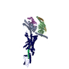 8jipMC 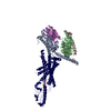 8jiqC 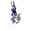 8jirC 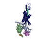 8jisC 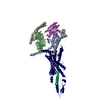 8jitC 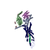 8jiuC M: atomic model generated by this map C: citing same article ( |
|---|---|
| Similar structure data | Similarity search - Function & homology  F&H Search F&H Search |
- Links
Links
| EMDB pages |  EMDB (EBI/PDBe) / EMDB (EBI/PDBe) /  EMDataResource EMDataResource |
|---|---|
| Related items in Molecule of the Month |
- Map
Map
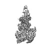
| File |  Download / File: emd_36323.map.gz / Format: CCP4 / Size: 91.1 MB / Type: IMAGE STORED AS FLOATING POINT NUMBER (4 BYTES) Download / File: emd_36323.map.gz / Format: CCP4 / Size: 91.1 MB / Type: IMAGE STORED AS FLOATING POINT NUMBER (4 BYTES) | ||||||||||||||||||||||||||||||||||||
|---|---|---|---|---|---|---|---|---|---|---|---|---|---|---|---|---|---|---|---|---|---|---|---|---|---|---|---|---|---|---|---|---|---|---|---|---|---|
| Projections & slices | Image control
Images are generated by Spider. | ||||||||||||||||||||||||||||||||||||
| Voxel size | X=Y=Z: 0.86 Å | ||||||||||||||||||||||||||||||||||||
| Density |
| ||||||||||||||||||||||||||||||||||||
| Symmetry | Space group: 1 | ||||||||||||||||||||||||||||||||||||
| Details | EMDB XML:
|
-Supplemental data
-Half map: #2
| File | emd_36323_half_map_1.map | ||||||||||||
|---|---|---|---|---|---|---|---|---|---|---|---|---|---|
| Projections & Slices |
| ||||||||||||
| Density Histograms |
-Half map: #1
| File | emd_36323_half_map_2.map | ||||||||||||
|---|---|---|---|---|---|---|---|---|---|---|---|---|---|
| Projections & Slices |
| ||||||||||||
| Density Histograms |
- Sample components
-Entire : Cryo-EM structure of the GLP-1R/GCGR dual agonist MEDI0382-bound ...
| Entire | Name: Cryo-EM structure of the GLP-1R/GCGR dual agonist MEDI0382-bound human GLP-1R-Gs complex |
|---|---|
| Components |
|
-Supramolecule #1: Cryo-EM structure of the GLP-1R/GCGR dual agonist MEDI0382-bound ...
| Supramolecule | Name: Cryo-EM structure of the GLP-1R/GCGR dual agonist MEDI0382-bound human GLP-1R-Gs complex type: complex / ID: 1 / Parent: 0 / Macromolecule list: #1-#6 |
|---|---|
| Source (natural) | Organism:  Homo sapiens (human) Homo sapiens (human) |
-Macromolecule #1: Glucagon-like peptide 1 receptor
| Macromolecule | Name: Glucagon-like peptide 1 receptor / type: protein_or_peptide / ID: 1 / Number of copies: 1 / Enantiomer: LEVO |
|---|---|
| Source (natural) | Organism:  Homo sapiens (human) Homo sapiens (human) |
| Molecular weight | Theoretical: 50.860801 KDa |
| Recombinant expression | Organism:  |
| Sequence | String: RPQGATVSLW ETVQKWREYR RQCQRSLTED PPPATDLFCN RTFDEYACWP DGEPGSFVNV SCPWYLPWAS SVPQGHVYRF CTAEGLWLQ KDNSSLPWRD LSECEESKRG ERSSPEEQLL FLYIIYTVGY ALSFSALVIA SAILLGFRHL HCTRNYIHLN L FASFILRA ...String: RPQGATVSLW ETVQKWREYR RQCQRSLTED PPPATDLFCN RTFDEYACWP DGEPGSFVNV SCPWYLPWAS SVPQGHVYRF CTAEGLWLQ KDNSSLPWRD LSECEESKRG ERSSPEEQLL FLYIIYTVGY ALSFSALVIA SAILLGFRHL HCTRNYIHLN L FASFILRA LSVFIKDAAL KWMYSTAAQQ HQWDGLLSYQ DSLSCRLVFL LMQYCVAANY YWLLVEGVYL YTLLAFSVLS EQ WIFRLYV SIGWGVPLLF VVPWGIVKYL YEDEGCWTRN SNMNYWLIIR LPILFAIGVN FLIFVRVICI VVSKLKANLM CKT DIKCRL AKSTLTLIPL LGTHEVIFAF VMDEHARGTL RFIKLFTELS FTSFQGLMVA ILYCFVNNEV QLEFRKSWER WRLE HLHIQ RDSSMKPLKC PTSSLSSGAT AGSSMYTATC QASCS UniProtKB: Glucagon-like peptide 1 receptor |
-Macromolecule #2: Guanine nucleotide-binding protein G(s) subunit alpha isoforms short
| Macromolecule | Name: Guanine nucleotide-binding protein G(s) subunit alpha isoforms short type: protein_or_peptide / ID: 2 Details: There is no an appropriate UniProt/GenBank entry for entity 2 because the protein sequence (P63092) was modified. Number of copies: 1 / Enantiomer: LEVO |
|---|---|
| Source (natural) | Organism:  Homo sapiens (human) Homo sapiens (human) |
| Molecular weight | Theoretical: 41.907516 KDa |
| Recombinant expression | Organism:  |
| Sequence | String: MGCTLSAEDK AAVERSKMIE KQLQKDKQVY RATHRLLLLG ADNSGKSTIV KQMRIYHVNG YSEEECKQYK AVVYSNTIQS IIAIIRAMG RLKIDFGDSA RADDARQLFV LAGAAEEGFM TAELAGVIKR LWKDSGVQAC FNRSREYQLN DSAAYYLNDL D RIAQPNYI ...String: MGCTLSAEDK AAVERSKMIE KQLQKDKQVY RATHRLLLLG ADNSGKSTIV KQMRIYHVNG YSEEECKQYK AVVYSNTIQS IIAIIRAMG RLKIDFGDSA RADDARQLFV LAGAAEEGFM TAELAGVIKR LWKDSGVQAC FNRSREYQLN DSAAYYLNDL D RIAQPNYI PTQQDVLRTR VKTSGIFETK FQVDKVNFHM FDVGAQRDER RKWIQCFNDV TAIIFVVDSS DYNRLQEALN DF KSIWNNR WLRTISVILF LNKQDLLAEK VLAGKSKIED YFPEFARYTT PEDATPEPGE DPRVTRAKYF IRDEFLRIST ASG DGRHYC YPHFTCSVDT ENIRRVFNDC RDIIQRMHLR QYELL |
-Macromolecule #3: MEDI0382
| Macromolecule | Name: MEDI0382 / type: protein_or_peptide / ID: 3 / Number of copies: 1 / Enantiomer: LEVO |
|---|---|
| Source (natural) | Organism: synthetic construct (others) |
| Molecular weight | Theoretical: 3.278431 KDa |
| Sequence | String: HSQGTFTSDK SEYLDSERAQ DFVAWLEAG |
-Macromolecule #4: Guanine nucleotide-binding protein G(I)/G(S)/G(T) subunit beta-1
| Macromolecule | Name: Guanine nucleotide-binding protein G(I)/G(S)/G(T) subunit beta-1 type: protein_or_peptide / ID: 4 / Number of copies: 1 / Enantiomer: LEVO |
|---|---|
| Source (natural) | Organism:  |
| Molecular weight | Theoretical: 37.915496 KDa |
| Recombinant expression | Organism:  |
| Sequence | String: MGSLLQSELD QLRQEAEQLK NQIRDARKAC ADATLSQITN NIDPVGRIQM RTRRTLRGHL AKIYAMHWGT DSRLLVSASQ DGKLIIWDS YTTNKVHAIP LRSSWVMTCA YAPSGNYVAC GGLDNICSIY NLKTREGNVR VSRELAGHTG YLSCCRFLDD N QIVTSSGD ...String: MGSLLQSELD QLRQEAEQLK NQIRDARKAC ADATLSQITN NIDPVGRIQM RTRRTLRGHL AKIYAMHWGT DSRLLVSASQ DGKLIIWDS YTTNKVHAIP LRSSWVMTCA YAPSGNYVAC GGLDNICSIY NLKTREGNVR VSRELAGHTG YLSCCRFLDD N QIVTSSGD TTCALWDIET GQQTTTFTGH TGDVMSLSLA PDTRLFVSGA CDASAKLWDV REGMCRQTFT GHESDINAIC FF PNGNAFA TGSDDATCRL FDLRADQELM TYSHDNIICG ITSVSFSKSG RLLLAGYDDF NCNVWDALKA DRAGVLAGHD NRV SCLGVT DDGMAVATGS WDSFLKIWN UniProtKB: Guanine nucleotide-binding protein G(I)/G(S)/G(T) subunit beta-1 |
-Macromolecule #5: Guanine nucleotide-binding protein G(I)/G(S)/G(O) subunit gamma-2
| Macromolecule | Name: Guanine nucleotide-binding protein G(I)/G(S)/G(O) subunit gamma-2 type: protein_or_peptide / ID: 5 / Number of copies: 1 / Enantiomer: LEVO |
|---|---|
| Source (natural) | Organism:  |
| Molecular weight | Theoretical: 7.729947 KDa |
| Recombinant expression | Organism:  |
| Sequence | String: ASNNTASIAQ ARKLVEQLKM EANIDRIKVS KAAADLMAYC EAHAKEDPLL TPVPASENPF REKKFFCAIL UniProtKB: Guanine nucleotide-binding protein G(I)/G(S)/G(O) subunit gamma-2 |
-Macromolecule #6: Nanobody 35
| Macromolecule | Name: Nanobody 35 / type: protein_or_peptide / ID: 6 / Number of copies: 1 / Enantiomer: LEVO |
|---|---|
| Source (natural) | Organism:  |
| Molecular weight | Theoretical: 15.343019 KDa |
| Recombinant expression | Organism:  |
| Sequence | String: MAQVQLQESG GGLVQPGGSL RLSCAASGFT FSNYKMNWVR QAPGKGLEWV SDISQSGASI SYTGSVKGRF TISRDNAKNT LYLQMNSLK PEDTAVYYCA RCPAPFTRDC FDVTSTTYAY RGQGTQVTVS SHHHHHHEPE A |
-Macromolecule #7: N-hexadecanoyl-L-glutamic acid
| Macromolecule | Name: N-hexadecanoyl-L-glutamic acid / type: ligand / ID: 7 / Number of copies: 1 / Formula: D6M |
|---|---|
| Molecular weight | Theoretical: 385.538 Da |
| Chemical component information |  ChemComp-D6M: |
-Experimental details
-Structure determination
| Method | cryo EM |
|---|---|
 Processing Processing | single particle reconstruction |
| Aggregation state | particle |
- Sample preparation
| Buffer | pH: 7.4 |
|---|---|
| Vitrification | Cryogen name: ETHANE |
- Electron microscopy
| Microscope | FEI TITAN KRIOS |
|---|---|
| Image recording | Film or detector model: GATAN K3 (6k x 4k) / Average electron dose: 80.0 e/Å2 |
| Electron beam | Acceleration voltage: 300 kV / Electron source: OTHER |
| Electron optics | Illumination mode: OTHER / Imaging mode: BRIGHT FIELD / Nominal defocus max: 2.2 µm / Nominal defocus min: 1.2 µm |
| Experimental equipment |  Model: Titan Krios / Image courtesy: FEI Company |
- Image processing
| Startup model | Type of model: PDB ENTRY PDB model - PDB ID: |
|---|---|
| Final reconstruction | Resolution.type: BY AUTHOR / Resolution: 2.85 Å / Resolution method: FSC 0.143 CUT-OFF / Number images used: 796065 |
| Initial angle assignment | Type: OTHER |
| Final angle assignment | Type: OTHER |
 Movie
Movie Controller




 Z (Sec.)
 Y (Row.)
 X (Col.)


