National Natural Science Foundation of China (NSFC)
31970040
China
Citation
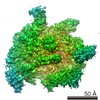
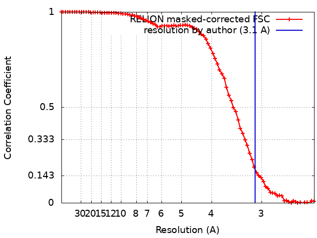
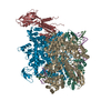
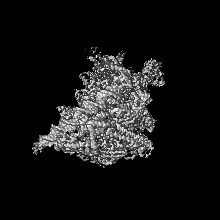
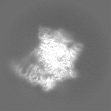
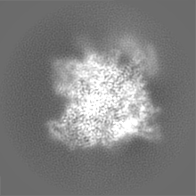
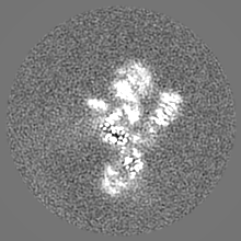
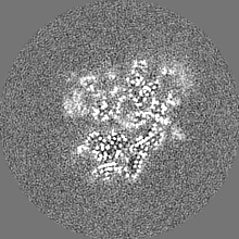
Journal: Structure / Year: 2023 Title: Structural basis of λCII-dependent transcription activation. Authors: Minxing Zhao / Bo Gao / Aijia Wen / Yu Feng / Yuan-Qiang Lu / Abstract: The CII protein of bacteriophage λ activates transcription from the phage promoters P, P, and P by binding to two direct repeats that straddle the promoter -35 element. Although genetic, ...The CII protein of bacteriophage λ activates transcription from the phage promoters P, P, and P by binding to two direct repeats that straddle the promoter -35 element. Although genetic, biochemical, and structural studies have elucidated many aspects of λCII-mediated transcription activation, no precise structure of the transcription machinery in the process is available. Here, we report a 3.1-Å cryo-electron microscopy (cryo-EM) structure of an intact λCII-dependent transcription activation complex (TAC-λCII), which comprises λCII, E. coli RNAP-σ holoenzyme, and the phage promoter P. The structure reveals the interactions between λCII and the direct repeats responsible for promoter specificity and the interactions between λCII and RNAP α subunit C-terminal domain responsible for transcription activation. We also determined a 3.4-Å cryo-EM structure of an RNAP-promoter open complex (RPo-P) from the same dataset. Structural comparison between TAC-λCII and RPo-P provides new insights into λCII-dependent transcription activation.
In the structure databanks used in Yorodumi, some data are registered as the other names, "COVID-19 virus" and "2019-nCoV". Here are the details of the virus and the list of structure data.
Jan 31, 2019. EMDB accession codes are about to change! (news from PDBe EMDB page)
EMDB accession codes are about to change! (news from PDBe EMDB page)
The allocation of 4 digits for EMDB accession codes will soon come to an end. Whilst these codes will remain in use, new EMDB accession codes will include an additional digit and will expand incrementally as the available range of codes is exhausted. The current 4-digit format prefixed with “EMD-” (i.e. EMD-XXXX) will advance to a 5-digit format (i.e. EMD-XXXXX), and so on. It is currently estimated that the 4-digit codes will be depleted around Spring 2019, at which point the 5-digit format will come into force.
The EM Navigator/Yorodumi systems omit the EMD- prefix.
Related info.:Q: What is EMD? / ID/Accession-code notation in Yorodumi/EM Navigator
Yorodumi is a browser for structure data from EMDB, PDB, SASBDB, etc.
This page is also the successor to EM Navigator detail page, and also detail information page/front-end page for Omokage search.
The word "yorodu" (or yorozu) is an old Japanese word meaning "ten thousand". "mi" (miru) is to see.
Related info.:EMDB / PDB / SASBDB / Comparison of 3 databanks / Yorodumi Search / Aug 31, 2016. New EM Navigator & Yorodumi / Yorodumi Papers / Jmol/JSmol / Function and homology information / Changes in new EM Navigator and Yorodumi
 Movie
Movie Controller

 Yorodumi
 Open data
 Basic information
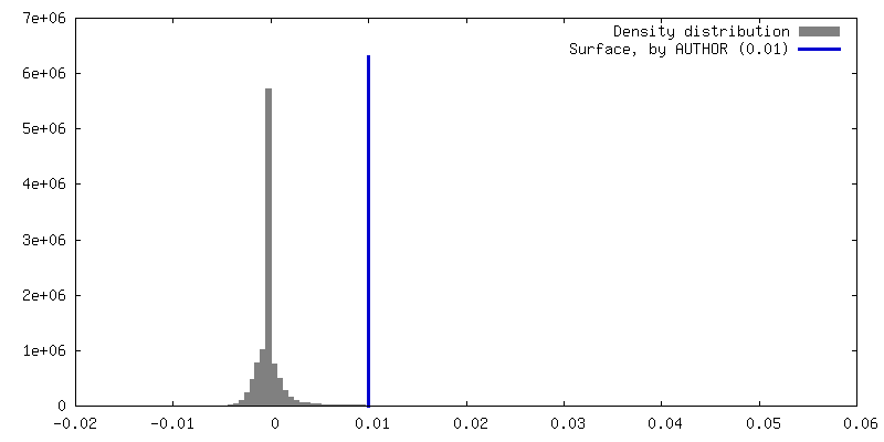
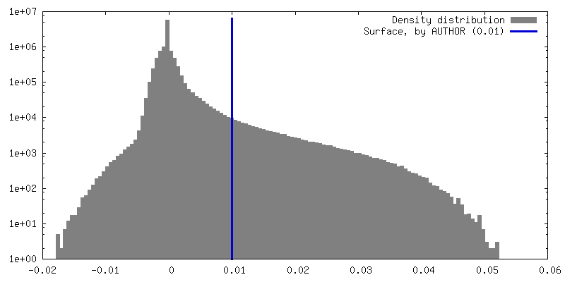
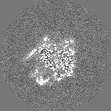
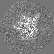
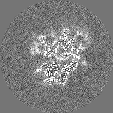
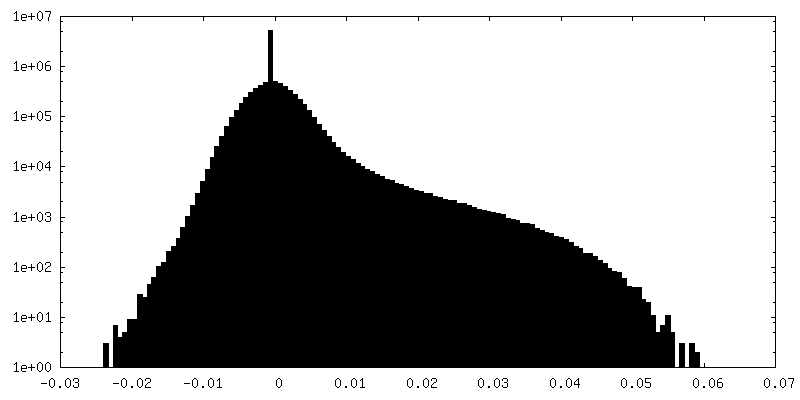
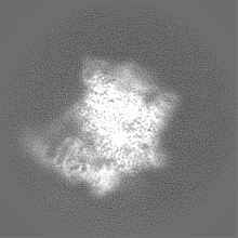
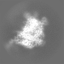
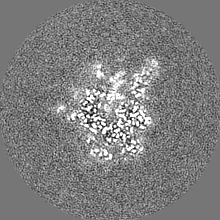
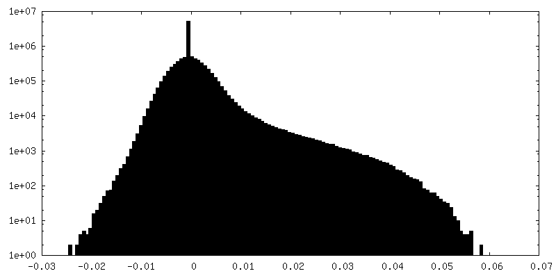
 Map data
 Sample
 Keywords
 Function and homology information
 Escherichia phage Lambda (virus)
 Authors
 China, 1 items
  Citation
 Structure visualization
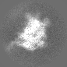
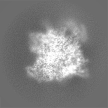
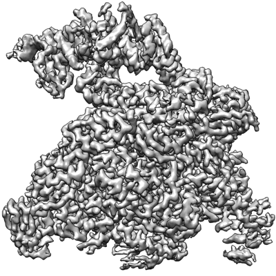
 Downloads & links
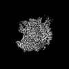
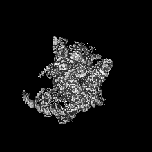
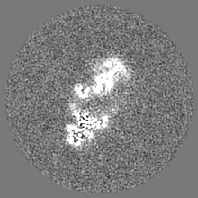
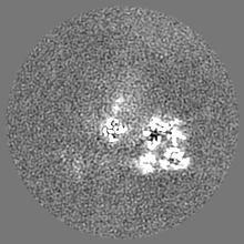
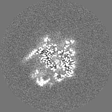
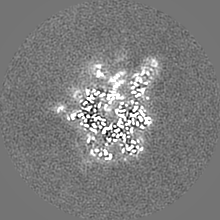
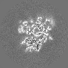
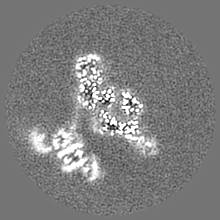
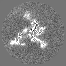
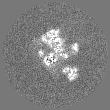
 emd_35438.png
 http://ftp.pdbj.org/pub/emdb/structures/EMD-35438
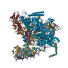


 Z (Sec.)
 Y (Row.)
 X (Col.)


 Sample components
 Processing
 Electron microscopy
 FIELD EMISSION GUN