 Movie
Movie Controller

[English] 日本語
 Yorodumi
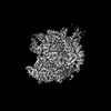
- PDB-8igr: Cryo-EM structure of CII-dependent transcription activation complex -
+ Open data

- Basic information
| Entry | Database: PDB / ID: 8igr | ||||||
|---|---|---|---|---|---|---|---|
| Title | Cryo-EM structure of CII-dependent transcription activation complex | ||||||
 Components Components |
| ||||||
 Keywords Keywords | TRANSCRIPTION / RNA polymerase / transcription activation / bacteriophage / CII | ||||||
| Function / homology |  Function and homology information Function and homology informationviral latency / sigma factor antagonist complex / RNA polymerase complex / submerged biofilm formation / cellular response to cell envelope stress / regulation of DNA-templated transcription initiation / sigma factor activity / bacterial-type flagellum assembly / bacterial-type RNA polymerase core enzyme binding / cytosolic DNA-directed RNA polymerase complex ...viral latency / sigma factor antagonist complex / RNA polymerase complex / submerged biofilm formation / cellular response to cell envelope stress / regulation of DNA-templated transcription initiation / sigma factor activity / bacterial-type flagellum assembly / bacterial-type RNA polymerase core enzyme binding / cytosolic DNA-directed RNA polymerase complex / bacterial-type flagellum-dependent cell motility / nitrate assimilation / regulation of DNA-templated transcription elongation / transcription elongation factor complex / transcription antitermination / cell motility / DNA-templated transcription initiation / ribonucleoside binding / DNA-directed RNA polymerase / DNA-directed RNA polymerase activity / response to heat / protein-containing complex assembly / intracellular iron ion homeostasis / protein dimerization activity / response to antibiotic / negative regulation of DNA-templated transcription / regulation of DNA-templated transcription / magnesium ion binding / DNA binding / zinc ion binding / membrane / cytosol / cytoplasm Similarity search - Function | ||||||
| Biological species |   Escherichia phage Lambda (virus) Escherichia phage Lambda (virus) | ||||||
| Method | ELECTRON MICROSCOPY / single particle reconstruction / cryo EM / Resolution: 3.1 Å | ||||||
 Authors Authors | Zhao, M. / Gao, B. / Wen, A. / Feng, Y. / Lu, Y. | ||||||
| Funding support |  China, 1items China, 1items
| ||||||
 Citation Citation | Journal: Structure / Year: 2023 Title: Structural basis of λCII-dependent transcription activation. Authors: Minxing Zhao / Bo Gao / Aijia Wen / Yu Feng / Yuan-Qiang Lu / Abstract: The CII protein of bacteriophage λ activates transcription from the phage promoters P, P, and P by binding to two direct repeats that straddle the promoter -35 element. Although genetic, ...The CII protein of bacteriophage λ activates transcription from the phage promoters P, P, and P by binding to two direct repeats that straddle the promoter -35 element. Although genetic, biochemical, and structural studies have elucidated many aspects of λCII-mediated transcription activation, no precise structure of the transcription machinery in the process is available. Here, we report a 3.1-Å cryo-electron microscopy (cryo-EM) structure of an intact λCII-dependent transcription activation complex (TAC-λCII), which comprises λCII, E. coli RNAP-σ holoenzyme, and the phage promoter P. The structure reveals the interactions between λCII and the direct repeats responsible for promoter specificity and the interactions between λCII and RNAP α subunit C-terminal domain responsible for transcription activation. We also determined a 3.4-Å cryo-EM structure of an RNAP-promoter open complex (RPo-P) from the same dataset. Structural comparison between TAC-λCII and RPo-P provides new insights into λCII-dependent transcription activation. | ||||||
| History |
|
- Structure visualization
| Structure viewer | Molecule: MolmilJmol/JSmol |
|---|
- Downloads & links
-Download
| PDBx/mmCIF format | 8igr.cif.gz | 768 KB | Display | PDBx/mmCIF format |
|---|---|---|---|---|
| PDB format | pdb8igr.ent.gz | 600.4 KB | Display | PDB format |
| PDBx/mmJSON format | 8igr.json.gz | Tree view | PDBx/mmJSON format | |
| Others |  Other downloads Other downloads |
-Validation report
| Summary document | 8igr_validation.pdf.gz | 436.1 KB | Display | wwPDB validaton report |
|---|---|---|---|---|
| Full document | 8igr_full_validation.pdf.gz | 467 KB | Display | |
| Data in XML | 8igr_validation.xml.gz | 64.8 KB | Display | |
| Data in CIF | 8igr_validation.cif.gz | 100.7 KB | Display | |
| Arichive directory | https://data.pdbj.org/pub/pdb/validation_reports/ig/8igrftp://data.pdbj.org/pub/pdb/validation_reports/ig/8igr | HTTPS FTP |
-Related structure data
| Related structure data | 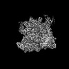 35438MC 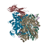 8igsC M: map data used to model this data C: citing same article ( |
|---|---|
| Similar structure data |
-Links
 PDBj
PDBj


- Assembly
| Deposited unit | 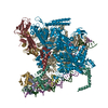
|
|---|---|
| 1 |
|
-Components
-DNA-directed RNA polymerase subunit ... , 4 types, 5 molecules GHIJK
| #1: Protein | Mass: 36558.680 Da / Num. of mol.: 2 Source method: isolated from a genetically manipulated source Source: (gene. exp.) Gene: rpoA, pez, phs, sez, b3295, JW3257 / Production host: #2: Protein | | Mass: 150820.875 Da / Num. of mol.: 1 Source method: isolated from a genetically manipulated source Source: (gene. exp.) Gene: rpoB, groN, nitB, rif, ron, stl, stv, tabD, b3987, JW3950 Production host: #3: Protein | | Mass: 155366.781 Da / Num. of mol.: 1 Source method: isolated from a genetically manipulated source Source: (gene. exp.) Gene: rpoC, tabB, b3988, JW3951 / Production host: #4: Protein | | Mass: 10249.547 Da / Num. of mol.: 1 Source method: isolated from a genetically manipulated source Source: (gene. exp.) Gene: rpoZ, b3649, JW3624 / Production host: |
|---|
-Protein , 2 types, 5 molecules LABCD
| #5: Protein | Mass: 70352.242 Da / Num. of mol.: 1 Source method: isolated from a genetically manipulated source Source: (gene. exp.) Gene: rpoD, alt, b3067, JW3039 / Production host: |
|---|---|
| #6: Protein | Mass: 11072.907 Da / Num. of mol.: 4 Source method: isolated from a genetically manipulated source Source: (gene. exp.) Escherichia phage Lambda (virus) / Gene: cII, lambdap59 / Production host: |
-DNA chain , 2 types, 2 molecules NT
| #7: DNA chain | Mass: 26185.760 Da / Num. of mol.: 1 / Source method: obtained synthetically / Source: (synth.) |
|---|---|
| #8: DNA chain | Mass: 26235.889 Da / Num. of mol.: 1 / Source method: obtained synthetically / Source: (synth.) |
-Non-polymers , 2 types, 3 molecules 

| #9: Chemical | ChemComp-MG /  Mass: 24.305 Da / Num. of mol.: 1 / Source method: obtained synthetically / Formula: Mg Mass: 24.305 Da / Num. of mol.: 1 / Source method: obtained synthetically / Formula: Mg |
|---|---|
| #10: Chemical |  Mass: 65.409 Da / Num. of mol.: 2 / Source method: obtained synthetically / Formula: Zn Mass: 65.409 Da / Num. of mol.: 2 / Source method: obtained synthetically / Formula: Zn |
-Details
| Has ligand of interest | N |
|---|
-Experimental details
-Experiment
| Experiment | Method: ELECTRON MICROSCOPY |
|---|---|
| EM experiment | Aggregation state: PARTICLE / 3D reconstruction method: single particle reconstruction |
- Sample preparation
| Component | Name: CII-dependent transcription activation complex / Type: COMPLEX / Entity ID: #1-#8 / Source: MULTIPLE SOURCES |
|---|---|
| Source (natural) | Organism: |
| Source (recombinant) | Organism: |
| Buffer solution | pH: 7.5 |
| Specimen | Embedding applied: NO / Shadowing applied: NO / Staining applied: NO / Vitrification applied: YES |
| Vitrification | Cryogen name: ETHANE |
- Electron microscopy imaging
| Experimental equipment |  Model: Titan Krios / Image courtesy: FEI Company |
|---|---|
| Microscopy | Model: FEI TITAN KRIOS |
| Electron gun | Electron source:  FIELD EMISSION GUN / Accelerating voltage: 300 kV / Illumination mode: FLOOD BEAM FIELD EMISSION GUN / Accelerating voltage: 300 kV / Illumination mode: FLOOD BEAM |
| Electron lens | Mode: BRIGHT FIELD / Nominal defocus max: 1700 nm / Nominal defocus min: 800 nm |
| Image recording | Electron dose: 51.7 e/Å2 / Film or detector model: FEI FALCON IV (4k x 4k) |
- Processing
| CTF correction | Type: PHASE FLIPPING AND AMPLITUDE CORRECTION | ||||||||||||||||||||||||
|---|---|---|---|---|---|---|---|---|---|---|---|---|---|---|---|---|---|---|---|---|---|---|---|---|---|
| 3D reconstruction | Resolution: 3.1 Å / Resolution method: FSC 0.143 CUT-OFF / Num. of particles: 103401 / Symmetry type: POINT | ||||||||||||||||||||||||
| Refine LS restraints |
|
