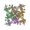
- EMDB-33937: Structure of recombinant RyR2 (EGTA dataset, class 2, closed state) -
+
Open data
ID or keywords:
Loading...
-
Basic information
Entry
Database: EMDB / ID: EMD-33937
Title
Structure of recombinant RyR2 (EGTA dataset, class 2, closed state)
Map data
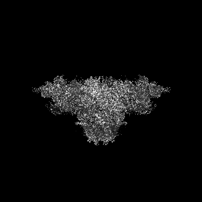
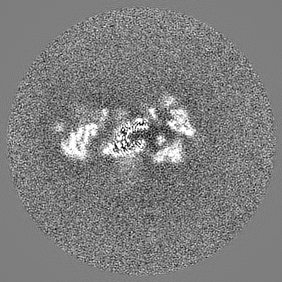
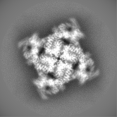
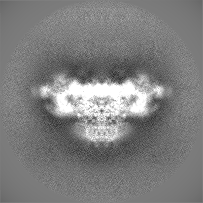
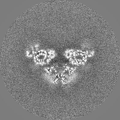
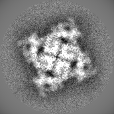
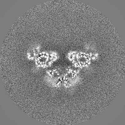
Structure of recombinant RyR2 (EGTA dataset, class 2, closed state)
Sample
Complex: Recombinant RyR2 in the presence of EGTA
Complex: Ryanodine receptor 2
Complex: FKBP1B
Function / homology
Function and homology information
establishment of protein localization to endoplasmic reticulum / manganese ion transmembrane transport / : / type B pancreatic cell apoptotic process / Purkinje myocyte to ventricular cardiac muscle cell signaling / regulation of atrial cardiac muscle cell action potential / left ventricular cardiac muscle tissue morphogenesis / suramin binding / regulation of AV node cell action potential / sarcoplasmic reticulum calcium ion transport ...establishment of protein localization to endoplasmic reticulum / manganese ion transmembrane transport / : / type B pancreatic cell apoptotic process / Purkinje myocyte to ventricular cardiac muscle cell signaling / regulation of atrial cardiac muscle cell action potential / left ventricular cardiac muscle tissue morphogenesis / suramin binding / regulation of AV node cell action potential / sarcoplasmic reticulum calcium ion transport / regulation of SA node cell action potential / calcium-induced calcium release activity / calcium ion transport into cytosol / A band / embryonic heart tube morphogenesis / Stimuli-sensing channels / ventricular cardiac muscle cell action potential / negative regulation of calcium-mediated signaling / cardiac muscle hypertrophy / Ion homeostasis / regulation of ventricular cardiac muscle cell action potential / ryanodine-sensitive calcium-release channel activity / negative regulation of release of sequestered calcium ion into cytosol / response to redox state / release of sequestered calcium ion into cytosol by sarcoplasmic reticulum / negative regulation of heart rate / response to caffeine / regulation of cardiac muscle contraction by calcium ion signaling / extrinsic component of cytoplasmic side of plasma membrane / response to muscle activity / cellular response to caffeine / 'de novo' protein folding / FK506 binding / calcium ion transmembrane import into cytosol / protein kinase A regulatory subunit binding / protein kinase A catalytic subunit binding / negative regulation of cytosolic calcium ion concentration / positive regulation of the force of heart contraction / smooth endoplasmic reticulum / response to muscle stretch / intracellularly gated calcium channel activity / regulation of ryanodine-sensitive calcium-release channel activity / response to magnesium ion / detection of calcium ion / regulation of cardiac muscle contraction / regulation of cytosolic calcium ion concentration / positive regulation of heart rate / cardiac muscle contraction / calcium channel inhibitor activity / release of sequestered calcium ion into cytosol / regulation of release of sequestered calcium ion into cytosol by sarcoplasmic reticulum / Ion homeostasis / regulation of heart rate / regulation of cardiac muscle contraction by regulation of the release of sequestered calcium ion / cellular response to epinephrine stimulus / calcium channel complex / sarcoplasmic reticulum membrane / calcium-mediated signaling / peptidylprolyl isomerase / sarcomere / sarcoplasmic reticulum / striated muscle contraction / peptidyl-prolyl cis-trans isomerase activity / calcium channel regulator activity / protein maturation / response to calcium ion / protein refolding / sarcolemma / intracellular calcium ion homeostasis / Stimuli-sensing channels / Z disc / calcium ion transmembrane transport / calcium channel activity / calcium ion transport / nuclear envelope / protein folding / scaffold protein binding / monoatomic ion transmembrane transport / response to hypoxia / transmembrane transporter binding / calmodulin binding / signaling receptor binding / calcium ion binding / protein kinase binding / enzyme binding / protein-containing complex / membrane / identical protein binding / cytoplasm Similarity search - Function
Japan Agency for Medical Research and Development (AMED)
JP21am0101080
Japan
Japan Agency for Medical Research and Development (AMED)
19ek0109202
Japan
Citation
Journal: Nat Commun / Year: 2022 Title: Molecular basis for gating of cardiac ryanodine receptor explains the mechanisms for gain- and loss-of function mutations. Authors: Takuya Kobayashi / Akihisa Tsutsumi / Nagomi Kurebayashi / Kei Saito / Masami Kodama / Takashi Sakurai / Masahide Kikkawa / Takashi Murayama / Haruo Ogawa / Abstract: Cardiac ryanodine receptor (RyR2) is a large Ca release channel in the sarcoplasmic reticulum and indispensable for excitation-contraction coupling in the heart. RyR2 is activated by Ca and RyR2 ...Cardiac ryanodine receptor (RyR2) is a large Ca release channel in the sarcoplasmic reticulum and indispensable for excitation-contraction coupling in the heart. RyR2 is activated by Ca and RyR2 mutations are implicated in severe arrhythmogenic diseases. Yet, the structural basis underlying channel opening and how mutations affect the channel remains unknown. Here, we address the gating mechanism of RyR2 by combining high-resolution structures determined by cryo-electron microscopy with quantitative functional analysis of channels carrying various mutations in specific residues. We demonstrated two fundamental mechanisms for channel gating: interactions close to the channel pore stabilize the channel to prevent hyperactivity and a series of interactions in the surrounding regions is necessary for channel opening upon Ca binding. Mutations at the residues involved in the former and the latter mechanisms cause gain-of-function and loss-of-function, respectively. Our results reveal gating mechanisms of the RyR2 channel and alterations by pathogenic mutations at the atomic level.
In the structure databanks used in Yorodumi, some data are registered as the other names, "COVID-19 virus" and "2019-nCoV". Here are the details of the virus and the list of structure data.
Jan 31, 2019. EMDB accession codes are about to change! (news from PDBe EMDB page)
EMDB accession codes are about to change! (news from PDBe EMDB page)
The allocation of 4 digits for EMDB accession codes will soon come to an end. Whilst these codes will remain in use, new EMDB accession codes will include an additional digit and will expand incrementally as the available range of codes is exhausted. The current 4-digit format prefixed with “EMD-” (i.e. EMD-XXXX) will advance to a 5-digit format (i.e. EMD-XXXXX), and so on. It is currently estimated that the 4-digit codes will be depleted around Spring 2019, at which point the 5-digit format will come into force.
The EM Navigator/Yorodumi systems omit the EMD- prefix.
Related info.:Q: What is EMD? / ID/Accession-code notation in Yorodumi/EM Navigator
Yorodumi is a browser for structure data from EMDB, PDB, SASBDB, etc.
This page is also the successor to EM Navigator detail page, and also detail information page/front-end page for Omokage search.
The word "yorodu" (or yorozu) is an old Japanese word meaning "ten thousand". "mi" (miru) is to see.
Related info.:EMDB / PDB / SASBDB / Comparison of 3 databanks / Yorodumi Search / Aug 31, 2016. New EM Navigator & Yorodumi / Yorodumi Papers / Jmol/JSmol / Function and homology information / Changes in new EM Navigator and Yorodumi
 Movie
Movie Controller

 Yorodumi
 Open data
 Basic information
 Map data
 Sample
 Function and homology information
 Mus musculus (house mouse) /
  Homo sapiens (human)
 Authors
 Japan, 6 items
  Citation
 Structure visualization
 Downloads & links
 emd_33937.png
 http://ftp.pdbj.org/pub/emdb/structures/EMD-33937







 Z (Sec.)
 Y (Row.)
 X (Col.)












 Sample components
 Escherichia coli (E. coli)
 Processing
 Electron microscopy
 FIELD EMISSION GUN