 Movie
Movie Controller

[English] 日本語
 Yorodumi
- EMDB-33633: Cryo-EM structure of C5a-bound C5aR1 in complex with Gi protein -
+ Open data

- Basic information
| Entry |  | |||||||||
|---|---|---|---|---|---|---|---|---|---|---|
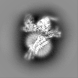
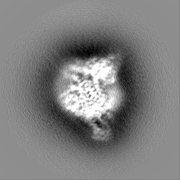
| Title | Cryo-EM structure of C5a-bound C5aR1 in complex with Gi protein | |||||||||
 Map data Map data | ||||||||||
 Sample Sample |
| |||||||||
 Keywords Keywords | Complex / Agonist / MEMBRANE PROTEIN | |||||||||
| Function / homology |  Function and homology information Function and homology informationpresynapse organization / complement component C5a signaling pathway / Terminal pathway of complement / complement component C5a receptor activity / membrane attack complex / response to peptidoglycan / Activation of C3 and C5 / complement activation, GZMK pathway / negative regulation of macrophage chemotaxis / sensory perception of chemical stimulus ...presynapse organization / complement component C5a signaling pathway / Terminal pathway of complement / complement component C5a receptor activity / membrane attack complex / response to peptidoglycan / Activation of C3 and C5 / complement activation, GZMK pathway / negative regulation of macrophage chemotaxis / sensory perception of chemical stimulus / other organism cell membrane / complement receptor mediated signaling pathway / complement activation, alternative pathway / positive regulation of neutrophil chemotaxis / chemokine activity / endopeptidase inhibitor activity / positive regulation of macrophage chemotaxis / amyloid-beta clearance / complement activation, classical pathway / positive regulation of vascular endothelial growth factor production / cellular defense response / positive regulation of chemokine production / neutrophil chemotaxis / adenylate cyclase inhibitor activity / positive regulation of protein localization to cell cortex / T cell migration / positive regulation of relaxation of smooth muscle / Adenylate cyclase inhibitory pathway / D2 dopamine receptor binding / adenylate cyclase-inhibiting serotonin receptor signaling pathway / G protein-coupled serotonin receptor binding / astrocyte activation / Regulation of Complement cascade / secretory granule membrane / cellular response to forskolin / positive regulation of epithelial cell proliferation / Peptide ligand-binding receptors / regulation of mitotic spindle organization / chemokine-mediated signaling pathway / Regulation of insulin secretion / neuropeptide signaling pathway / response to prostaglandin E / microglial cell activation / positive regulation of cholesterol biosynthetic process / negative regulation of insulin secretion / mRNA transcription by RNA polymerase II / G protein-coupled receptor binding / response to peptide hormone / cognition / G protein-coupled receptor activity / chemotaxis / centriolar satellite / G-protein beta/gamma-subunit complex binding / positive regulation of angiogenesis / adenylate cyclase-modulating G protein-coupled receptor signaling pathway / apical part of cell / adenylate cyclase-inhibiting G protein-coupled receptor signaling pathway / Olfactory Signaling Pathway / Activation of the phototransduction cascade / G protein-coupled acetylcholine receptor signaling pathway / G beta:gamma signalling through PLC beta / Presynaptic function of Kainate receptors / Thromboxane signalling through TP receptor / Activation of G protein gated Potassium channels / Inhibition of voltage gated Ca2+ channels via Gbeta/gamma subunits / G-protein activation / Glucagon signaling in metabolic regulation / Prostacyclin signalling through prostacyclin receptor / G beta:gamma signalling through CDC42 / Synthesis, secretion, and inactivation of Glucagon-like Peptide-1 (GLP-1) / G beta:gamma signalling through BTK / photoreceptor disc membrane / ADP signalling through P2Y purinoceptor 12 / Sensory perception of sweet, bitter, and umami (glutamate) taste / Glucagon-type ligand receptors / GDP binding / Adrenaline,noradrenaline inhibits insulin secretion / Vasopressin regulates renal water homeostasis via Aquaporins / Glucagon-like Peptide-1 (GLP1) regulates insulin secretion / G alpha (z) signalling events / cellular response to catecholamine stimulus / ADP signalling through P2Y purinoceptor 1 / ADORA2B mediated anti-inflammatory cytokines production / G beta:gamma signalling through PI3Kgamma / adenylate cyclase-activating dopamine receptor signaling pathway / Cooperation of PDCL (PhLP1) and TRiC/CCT in G-protein beta folding / GPER1 signaling / cellular response to prostaglandin E stimulus / heterotrimeric G-protein complex / G alpha (12/13) signalling events / Inactivation, recovery and regulation of the phototransduction cascade / G-protein beta-subunit binding / extracellular vesicle / sensory perception of taste / sperm principal piece / Thrombin signalling through proteinase activated receptors (PARs) / signaling receptor complex adaptor activity / adenylate cyclase-activating G protein-coupled receptor signaling pathway / positive regulation of cytosolic calcium ion concentration / retina development in camera-type eye Similarity search - Function | |||||||||
| Biological species |  Homo sapiens (human) / Homo sapiens (human) /  | |||||||||
| Method | single particle reconstruction / cryo EM / Resolution: 2.9 Å | |||||||||
 Authors Authors | Feng YY / Zhao C / Yan W / Shao ZH | |||||||||
| Funding support |  China, 1 items China, 1 items
| |||||||||
 Citation Citation | Journal: Cell Res / Year: 2023 Title: Mechanism of activation and biased signaling in complement receptor C5aR1. Authors: Yuying Feng / Chang Zhao / Yue Deng / Heli Wang / Liang Ma / Sicen Liu / Xiaowen Tian / Bo Wang / Yan Bin / Peipei Chen / Wei Yan / Ping Fu / Zhenhua Shao / Abstract: The complement system plays an important role in the innate immune response to invading pathogens. The complement fragment C5a is one of its important effector components and exerts diverse ...The complement system plays an important role in the innate immune response to invading pathogens. The complement fragment C5a is one of its important effector components and exerts diverse physiological functions through activation of the C5a receptor 1 (C5aR1) and associated downstream G protein and β-arrestin signaling pathways. Dysfunction of the C5a-C5aR1 axis is linked to numerous inflammatory and immune-mediated diseases, but the structural basis for activation and biased signaling of C5aR1 remains elusive. Here, we present cryo-electron microscopy structures of the activated wild-type C5aR1-G protein complex bound to each of the following: C5a, the hexapeptidic agonist C5a, and the G protein-biased agonist BM213. The structures reveal the landscape of the C5a-C5aR1 interaction as well as a common motif for the recognition of diverse orthosteric ligands. Moreover, combined with mutagenesis studies and cell-based pharmacological assays, we deciphered a framework for biased signaling using different peptide analogs and provided insight into the activation mechanism of C5aR1 by solving the structure of C5aR1 mutant-G signaling activation complex induced by C089, which exerts antagonism on wild-type C5aR1. In addition, unusual conformational changes in the intracellular end of transmembrane domain 7 and helix 8 upon agonist binding suggest a differential signal transduction process. Collectively, our study provides mechanistic understanding into the ligand recognition, biased signaling modulation, activation, and G protein coupling of C5aR1, which may facilitate the future design of therapeutic agents. | |||||||||
| History |
|
- Structure visualization
| Supplemental images |
|---|

- Downloads & links
-EMDB archive
| Map data | emd_33633.map.gz | 55.3 MB | EMDB map data format | |
|---|---|---|---|---|
| Header (meta data) | emd-33633-v30.xmlemd-33633.xml | 21.5 KB 21.5 KB | Display Display | EMDB header |
| Images |  emd_33633.png emd_33633.png | 34.6 KB | ||
| Filedesc metadata | emd-33633.cif.gz | 6.3 KB | ||
| Others | emd_33633_half_map_1.map.gzemd_33633_half_map_2.map.gz | 49.5 MB 49.5 MB | ||
| Archive directory |  http://ftp.pdbj.org/pub/emdb/structures/EMD-33633ftp://ftp.pdbj.org/pub/emdb/structures/EMD-33633 http://ftp.pdbj.org/pub/emdb/structures/EMD-33633ftp://ftp.pdbj.org/pub/emdb/structures/EMD-33633 | HTTPS FTP |
-Related structure data
| Related structure data |  7y64MC  7y65C  7y66C  7y67C M: atomic model generated by this map C: citing same article ( |
|---|---|
| Similar structure data |
-Links
| EMDB pages | EMDB (EBI/PDBe) / EMDataResource |
|---|---|
| Related items in Molecule of the Month |


-Map
| File | Download / File: emd_33633.map.gz / Format: CCP4 / Size: 64 MB / Type: IMAGE STORED AS FLOATING POINT NUMBER (4 BYTES) | ||||||||||||||||||||||||||||||||||||
|---|---|---|---|---|---|---|---|---|---|---|---|---|---|---|---|---|---|---|---|---|---|---|---|---|---|---|---|---|---|---|---|---|---|---|---|---|---|
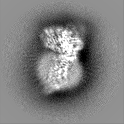
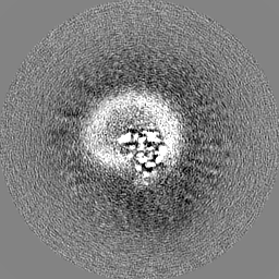
| Projections & slices | Image control
Images are generated by Spider. | ||||||||||||||||||||||||||||||||||||
| Voxel size | X=Y=Z: 0.85 Å | ||||||||||||||||||||||||||||||||||||
| Density |
| ||||||||||||||||||||||||||||||||||||
| Symmetry | Space group: 1 | ||||||||||||||||||||||||||||||||||||
| Details | EMDB XML:
|
 Z (Sec.)
 Y (Row.)
 X (Col.)


-Supplemental data
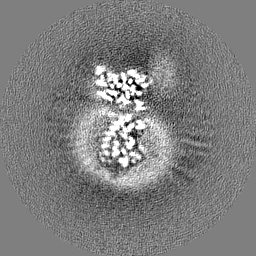
-Half map: #1
| File | emd_33633_half_map_1.map | ||||||||||||
|---|---|---|---|---|---|---|---|---|---|---|---|---|---|
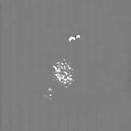
| Projections & Slices |
| ||||||||||||
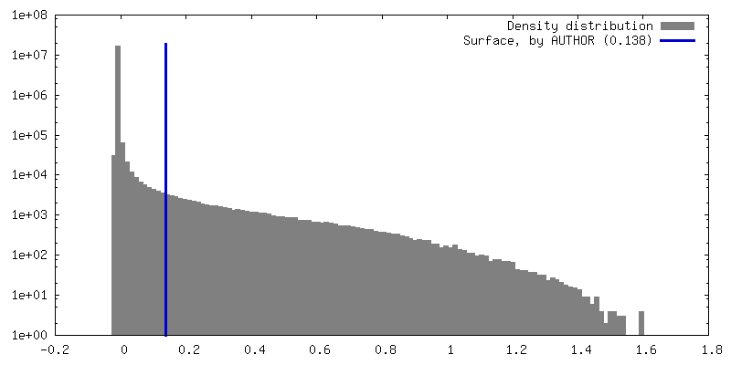
| Density Histograms |


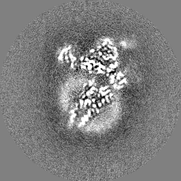
-Half map: #2
| File | emd_33633_half_map_2.map | ||||||||||||
|---|---|---|---|---|---|---|---|---|---|---|---|---|---|
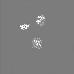
| Projections & Slices |
| ||||||||||||
| Density Histograms |


- Sample components
+Entire : C5a-bound C5aR1 in complex with Gi heterotrimer
+Supramolecule #1: C5a-bound C5aR1 in complex with Gi heterotrimer
+Supramolecule #2: Gi heterotrimer
+Supramolecule #3: scFV16
+Supramolecule #4: C5a-bound C5aR1
+Macromolecule #1: Guanine nucleotide-binding protein G(i) subunit alpha-1

 Spodoptera frugiperda (fall armyworm)
+Macromolecule #2: Guanine nucleotide-binding protein G(I)/G(S)/G(T) subunit beta-1
+Macromolecule #3: Guanine nucleotide-binding protein G(I)/G(S)/G(O) subunit gamma-2
+Macromolecule #4: scFV16
+Macromolecule #5: C5a anaphylatoxin chemotactic receptor 1
+Macromolecule #6: C5a anaphylatoxin

-Experimental details
-Structure determination
| Method | cryo EM |
|---|---|
 Processing Processing | single particle reconstruction |
| Aggregation state | particle |
-Sample preparation
| Buffer | pH: 7.5 |
|---|---|
| Vitrification | Cryogen name: ETHANE |
- Electron microscopy
| Microscope | FEI TITAN KRIOS |
|---|---|
| Image recording | Film or detector model: GATAN K2 SUMMIT (4k x 4k) / Average electron dose: 66.0 e/Å2 |
| Electron beam | Acceleration voltage: 300 kV / Electron source:  FIELD EMISSION GUN FIELD EMISSION GUN |
| Electron optics | Illumination mode: FLOOD BEAM / Imaging mode: BRIGHT FIELD / Cs: 2.7 mm / Nominal defocus max: 1.8 µm / Nominal defocus min: 1.0 µm |
| Experimental equipment |  Model: Titan Krios / Image courtesy: FEI Company |
-Image processing
| Startup model | Type of model: NONE |
|---|---|
| Final reconstruction | Resolution.type: BY AUTHOR / Resolution: 2.9 Å / Resolution method: FSC 0.143 CUT-OFF / Number images used: 623262 |
| Initial angle assignment | Type: MAXIMUM LIKELIHOOD |
| Final angle assignment | Type: MAXIMUM LIKELIHOOD |
