ムービー
ムービー コントローラー


+ データを開く

- 基本情報
基本情報
| 登録情報 | 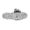 | |||||||||
|---|---|---|---|---|---|---|---|---|---|---|
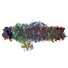
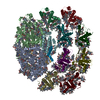
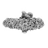
| タイトル | PSI-LHCI from Chlamydomonas reinhardtii with bound ferredoxin | |||||||||
 マップデータ マップデータ | PSI-LHCI from Chlamydomonas reinhardtii with bound ferredoxin | |||||||||
 試料 試料 |
| |||||||||
 キーワード キーワード | membrane supercomplex / light harvesting / electron transport / green algae / PHOTOSYNTHESIS | |||||||||
| 機能・相同性 |  機能・相同性情報 機能・相同性情報photosynthesis, light harvesting / chloroplast thylakoid lumen / photosynthesis, light harvesting in photosystem I / photosystem I reaction center / photosystem I / photosystem I / photosystem II / chlorophyll binding / iron-sulfur cluster binding / photosynthetic electron transport in photosystem I ...photosynthesis, light harvesting / chloroplast thylakoid lumen / photosynthesis, light harvesting in photosystem I / photosystem I reaction center / photosystem I / photosystem I / photosystem II / chlorophyll binding / iron-sulfur cluster binding / photosynthetic electron transport in photosystem I / chloroplast thylakoid membrane / response to light stimulus / photosynthesis / chloroplast / electron transport chain / 2 iron, 2 sulfur cluster binding / 4 iron, 4 sulfur cluster binding / oxidoreductase activity / electron transfer activity / magnesium ion binding / metal ion binding 類似検索 - 分子機能 | |||||||||
| 生物種 |   Chlamydomonas reinhardtii (クラミドモナス) Chlamydomonas reinhardtii (クラミドモナス) | |||||||||
| 手法 | 単粒子再構成法 / クライオ電子顕微鏡法 / 解像度: 4.9 Å | |||||||||
 データ登録者 データ登録者 | Kurisu G / Gerle C | |||||||||
| 資金援助 |  日本, 2件 日本, 2件
| |||||||||
 引用 引用 | ジャーナル: Biochim Biophys Acta Bioenerg / 年: 2023 タイトル: Three structures of PSI-LHCI from Chlamydomonas reinhardtii suggest a resting state re-activated by ferredoxin. 著者: Christoph Gerle / Yuko Misumi / Akihiro Kawamoto / Hideaki Tanaka / Hisako Kubota-Kawai / Ryutaro Tokutsu / Eunchul Kim / Dror Chorev / Kazuhiro Abe / Carol V Robinson / Kaoru Mitsuoka / Jun ...著者: Christoph Gerle / Yuko Misumi / Akihiro Kawamoto / Hideaki Tanaka / Hisako Kubota-Kawai / Ryutaro Tokutsu / Eunchul Kim / Dror Chorev / Kazuhiro Abe / Carol V Robinson / Kaoru Mitsuoka / Jun Minagawa / Genji Kurisu /  要旨: Photosystem I (PSI) from the green alga Chlamydomonas reinhardtii, with various numbers of membrane bound antenna complexes (LHCI), has been described in great detail. In contrast, structural ...Photosystem I (PSI) from the green alga Chlamydomonas reinhardtii, with various numbers of membrane bound antenna complexes (LHCI), has been described in great detail. In contrast, structural characterization of soluble binding partners is less advanced. Here, we used X-ray crystallography and single particle cryo-EM to investigate three structures of the PSI-LHCI supercomplex from Chlamydomonas reinhardtii. An X-ray structure demonstrates the absence of six chlorophylls from the luminal side of the LHCI belts, suggesting these pigments were either physically absent or less stably associated with the complex, potentially influencing excitation transfer significantly. CryoEM revealed extra densities on luminal and stromal sides of the supercomplex, situated in the vicinity of the electron transfer sites. These densities disappeared after the binding of oxidized ferredoxin to PSI-LHCI. Based on these structures, we propose the existence of a PSI-LHCI resting state with a reduced active chlorophyll content, electron donors docked in waiting positions and regulatory binding partners positioned at the electron acceptor site. The resting state PSI-LHCI supercomplex would be recruited to its active form by the availability of oxidized ferredoxin. | |||||||||
| 履歴 |
|
- 構造の表示
構造の表示
| 添付画像 |
|---|

- ダウンロードとリンク
-EMDBアーカイブ
| マップデータ | emd_32907.map.gz | 217 MB | EMDBマップデータ形式 | |
|---|---|---|---|---|
| ヘッダ (付随情報) | emd-32907-v30.xmlemd-32907.xml | 36.7 KB 36.7 KB | 表示 表示 | EMDBヘッダ |
| FSC (解像度算出) | emd_32907_fsc.xml | 13.6 KB | 表示 | FSCデータファイル |
| 画像 |  emd_32907.png emd_32907.png | 42.7 KB | ||
| Filedesc metadata | emd-32907.cif.gz | 9.9 KB | ||
| アーカイブディレクトリ |  http://ftp.pdbj.org/pub/emdb/structures/EMD-32907ftp://ftp.pdbj.org/pub/emdb/structures/EMD-32907 http://ftp.pdbj.org/pub/emdb/structures/EMD-32907ftp://ftp.pdbj.org/pub/emdb/structures/EMD-32907 | HTTPS FTP |
-検証レポート
| 文書・要旨 | emd_32907_validation.pdf.gz | 520.6 KB | 表示 | EMDB検証レポート |
|---|---|---|---|---|
| 文書・詳細版 | emd_32907_full_validation.pdf.gz | 520.2 KB | 表示 | |
| XML形式データ | emd_32907_validation.xml.gz | 13.8 KB | 表示 | |
| CIF形式データ | emd_32907_validation.cif.gz | 18.6 KB | 表示 | |
| アーカイブディレクトリ | https://ftp.pdbj.org/pub/emdb/validation_reports/EMD-32907ftp://ftp.pdbj.org/pub/emdb/validation_reports/EMD-32907 | HTTPS FTP |
-関連構造データ

-リンク
| EMDBのページ | EMDB (EBI/PDBe) / EMDataResource |
|---|---|
| 「今月の分子」の関連する項目 |


-マップ
| ファイル | ダウンロード / ファイル: emd_32907.map.gz / 形式: CCP4 / 大きさ: 229.8 MB / タイプ: IMAGE STORED AS FLOATING POINT NUMBER (4 BYTES) | ||||||||||||||||||||
|---|---|---|---|---|---|---|---|---|---|---|---|---|---|---|---|---|---|---|---|---|---|
| 注釈 | PSI-LHCI from Chlamydomonas reinhardtii with bound ferredoxin | ||||||||||||||||||||
| ボクセルのサイズ | X=Y=Z: 1.1 Å | ||||||||||||||||||||
| 密度 |
| ||||||||||||||||||||
| 対称性 | 空間群: 1 | ||||||||||||||||||||
| 詳細 | EMDB XML:
|
-添付データ
- 試料の構成要素
+全体 : Photosystem I from Chlamydomonas reinhardtii with bound ferredoxin.
+超分子 #1: Photosystem I from Chlamydomonas reinhardtii with bound ferredoxin.
+超分子 #2: Membrane supercomplex
+超分子 #3: ferredoxin
+分子 #1: Photosystem I P700 chlorophyll a apoprotein A1
+分子 #2: Photosystem I P700 chlorophyll a apoprotein A2
+分子 #3: Photosystem I iron-sulfur center
+分子 #4: Photosystem I reaction center subunit II, chloroplastic
+分子 #5: Photosystem I reaction center subunit IV, chloroplastic
+分子 #6: Photosystem I reaction center subunit III, chloroplastic
+分子 #7: Photosystem I reaction center subunit IX
+分子 #8: Chlorophyll a-b binding protein, chloroplastic
+分子 #9: Chlorophyll a-b binding protein, chloroplastic
+分子 #10: Chlorophyll a-b binding protein, chloroplastic
+分子 #11: Chlorophyll a-b binding protein, chloroplastic
+分子 #12: Chlorophyll a-b binding protein, chloroplastic
+分子 #13: Chlorophyll a-b binding protein, chloroplastic
+分子 #14: Chlorophyll a-b binding protein, chloroplastic
+分子 #15: Chlorophyll a-b binding protein, chloroplastic
+分子 #16: Ferredoxin, chloroplastic

+分子 #17: CHLOROPHYLL A ISOMER

+分子 #18: CHLOROPHYLL A

+分子 #19: PHYLLOQUINONE

+分子 #20: IRON/SULFUR CLUSTER

+分子 #21: CHLOROPHYLL B

+分子 #22: FE2/S2 (INORGANIC) CLUSTER

-実験情報
-構造解析
| 手法 | クライオ電子顕微鏡法 |
|---|---|
 解析 解析 | 単粒子再構成法 |
| 試料の集合状態 | particle |
-試料調製
| 濃度 | 7.0 mg/mL | |||||||||
|---|---|---|---|---|---|---|---|---|---|---|
| 緩衝液 | pH: 6.5 構成要素:
| |||||||||
| グリッド | モデル: Quantifoil R1.2/1.3 / 材質: COPPER / メッシュ: 200 詳細: Glow discharge on both sides at 5 mA for 90 seconds each. | |||||||||
| 凍結 | 凍結剤: ETHANE / チャンバー内湿度: 95 % / チャンバー内温度: 277 K / 装置: FEI VITROBOT MARK IV 詳細: 2.6 microliter of sample were applied to cryo-grids and blotted for 3.5 seconds using Whatman #1 before plunge freezing.. | |||||||||
| 詳細 | The sample was monodisperse with a low level of contaminants and excess free detergent removed using the GraDeR approach. |
- 電子顕微鏡法
| 顕微鏡 | FEI TITAN KRIOS |
|---|---|
| 詳細 | Preliminary grid screening was performed manually using a JEOL JEM-3000SFF microscope. |
| 撮影 | フィルム・検出器のモデル: FEI FALCON II (4k x 4k) 検出モード: INTEGRATING / デジタル化 - サイズ - 横: 4096 pixel / デジタル化 - サイズ - 縦: 4096 pixel / デジタル化 - 画像ごとのフレーム数: 1-16 / 撮影したグリッド数: 1 / 実像数: 2646 / 平均露光時間: 2.0 sec. / 平均電子線量: 91.0 e/Å2 |
| 電子線 | 加速電圧: 300 kV / 電子線源:  FIELD EMISSION GUN FIELD EMISSION GUN |
| 電子光学系 | C2レンズ絞り径: 100.0 µm / 照射モード: FLOOD BEAM / 撮影モード: BRIGHT FIELD / Cs: 2.7 mm / 最大 デフォーカス(公称値): 2.5 µm / 最小 デフォーカス(公称値): 1.25 µm / 倍率(公称値): 75000 |
| 試料ステージ | 試料ホルダーモデル: FEI TITAN KRIOS AUTOGRID HOLDER ホルダー冷却材: NITROGEN |
| 実験機器 |  モデル: Titan Krios / 画像提供: FEI Company |