 Movie
Movie Controller

[English] 日本語
 Yorodumi
- PDB-8h2u: X-ray Structure of photosystem I-LHCI super complex from Chlamydo... -
+ Open data

- Basic information
| Entry | Database: PDB / ID: 8h2u | |||||||||
|---|---|---|---|---|---|---|---|---|---|---|
| Title | X-ray Structure of photosystem I-LHCI super complex from Chlamydomonas reinhardtii. | |||||||||
 Components Components |
| |||||||||
 Keywords Keywords | PHOTOSYNTHESIS / Photosystem I / light-harvesting chlorophyll protein complex I | |||||||||
| Function / homology |  Function and homology information Function and homology informationphotosynthesis, light harvesting in photosystem I / photosynthesis, light harvesting / chloroplast thylakoid lumen / photosystem I reaction center / photosystem I / photosynthetic electron transport in photosystem I / photosystem I / photosystem II / chlorophyll binding / chloroplast thylakoid membrane ...photosynthesis, light harvesting in photosystem I / photosynthesis, light harvesting / chloroplast thylakoid lumen / photosystem I reaction center / photosystem I / photosynthetic electron transport in photosystem I / photosystem I / photosystem II / chlorophyll binding / chloroplast thylakoid membrane / response to light stimulus / photosynthesis / 4 iron, 4 sulfur cluster binding / electron transfer activity / oxidoreductase activity / magnesium ion binding / metal ion binding Similarity search - Function | |||||||||
| Biological species |   Chlamydomonas reinhardtii (plant) Chlamydomonas reinhardtii (plant) | |||||||||
| Method |  X-RAY DIFFRACTION / SYNCHROTRON / MOLECULAR REPLACEMENT / Resolution: 3.4 Å X-RAY DIFFRACTION / SYNCHROTRON / MOLECULAR REPLACEMENT / Resolution: 3.4 Å | |||||||||
 Authors Authors | Tanaka, H. / Kubota-Kawai, H. / Misumi, Y. / Kurisu, G. | |||||||||
| Funding support |  Japan, 2items Japan, 2items
| |||||||||
 Citation Citation | Journal: Biochim Biophys Acta Bioenerg / Year: 2023 Title: Three structures of PSI-LHCI from Chlamydomonas reinhardtii suggest a resting state re-activated by ferredoxin. Authors: Christoph Gerle / Yuko Misumi / Akihiro Kawamoto / Hideaki Tanaka / Hisako Kubota-Kawai / Ryutaro Tokutsu / Eunchul Kim / Dror Chorev / Kazuhiro Abe / Carol V Robinson / Kaoru Mitsuoka / Jun ...Authors: Christoph Gerle / Yuko Misumi / Akihiro Kawamoto / Hideaki Tanaka / Hisako Kubota-Kawai / Ryutaro Tokutsu / Eunchul Kim / Dror Chorev / Kazuhiro Abe / Carol V Robinson / Kaoru Mitsuoka / Jun Minagawa / Genji Kurisu /  Abstract: Photosystem I (PSI) from the green alga Chlamydomonas reinhardtii, with various numbers of membrane bound antenna complexes (LHCI), has been described in great detail. In contrast, structural ...Photosystem I (PSI) from the green alga Chlamydomonas reinhardtii, with various numbers of membrane bound antenna complexes (LHCI), has been described in great detail. In contrast, structural characterization of soluble binding partners is less advanced. Here, we used X-ray crystallography and single particle cryo-EM to investigate three structures of the PSI-LHCI supercomplex from Chlamydomonas reinhardtii. An X-ray structure demonstrates the absence of six chlorophylls from the luminal side of the LHCI belts, suggesting these pigments were either physically absent or less stably associated with the complex, potentially influencing excitation transfer significantly. CryoEM revealed extra densities on luminal and stromal sides of the supercomplex, situated in the vicinity of the electron transfer sites. These densities disappeared after the binding of oxidized ferredoxin to PSI-LHCI. Based on these structures, we propose the existence of a PSI-LHCI resting state with a reduced active chlorophyll content, electron donors docked in waiting positions and regulatory binding partners positioned at the electron acceptor site. The resting state PSI-LHCI supercomplex would be recruited to its active form by the availability of oxidized ferredoxin. | |||||||||
| History |
|
- Structure visualization
| Structure viewer | Molecule: MolmilJmol/JSmol |
|---|
- Downloads & links
-Download
| PDBx/mmCIF format | 8h2u.cif.gz | 1.1 MB | Display | PDBx/mmCIF format |
|---|---|---|---|---|
| PDB format | pdb8h2u.ent.gz | 940 KB | Display | PDB format |
| PDBx/mmJSON format | 8h2u.json.gz | Tree view | PDBx/mmJSON format | |
| Others |  Other downloads Other downloads |
-Validation report
| Arichive directory | https://data.pdbj.org/pub/pdb/validation_reports/h2/8h2uftp://data.pdbj.org/pub/pdb/validation_reports/h2/8h2u | HTTPS FTP |
|---|
-Related structure data
| Related structure data |  7wyiC 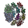 7wznC  4xk8S S: Starting model for refinement C: citing same article ( |
|---|---|
| Similar structure data |
-Links
 PDBj
PDBj


- Assembly
| Deposited unit | 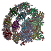
| ||||||||
|---|---|---|---|---|---|---|---|---|---|
| 1 |
| ||||||||
| Unit cell |
|
-Components
-Photosystem I P700 chlorophyll a apoprotein ... , 2 types, 2 molecules AB
| #1: Protein | Mass: 83239.203 Da / Num. of mol.: 1 / Source method: isolated from a natural source / Source: (natural) Chlamydomonas reinhardtii (plant) / References: UniProt: P12154, photosystem I |
|---|---|
| #2: Protein | Mass: 82184.266 Da / Num. of mol.: 1 / Source method: isolated from a natural source / Source: (natural) Chlamydomonas reinhardtii (plant) / References: UniProt: P09144, photosystem I |
-Protein , 3 types, 3 molecules CL3
| #3: Protein | Mass: 8869.325 Da / Num. of mol.: 1 / Source method: isolated from a natural source / Source: (natural) Chlamydomonas reinhardtii (plant) / References: UniProt: Q00914, photosystem I |
|---|---|
| #12: Protein | Mass: 20300.539 Da / Num. of mol.: 1 / Source method: isolated from a natural source / Source: (natural) Chlamydomonas reinhardtii (plant) / References: UniProt: A8IL32 |
| #16: Protein | Mass: 32629.486 Da / Num. of mol.: 1 / Source method: isolated from a natural source Details: THE GENEBANK ACCESSION NUMBER IS 6IJJ_3 for Lhca3. (www.ncbi.nlm.nih.gov/protein/6IJJ_3) Source: (natural) Chlamydomonas reinhardtii (plant) |
-Photosystem I reaction center subunit ... , 8 types, 8 molecules DEFGHIJK
| #4: Protein | Mass: 21372.887 Da / Num. of mol.: 1 / Source method: isolated from a natural source / Source: (natural) Chlamydomonas reinhardtii (plant) / References: UniProt: Q39615 |
|---|---|
| #5: Protein | Mass: 10786.395 Da / Num. of mol.: 1 / Source method: isolated from a natural source / Source: (natural) Chlamydomonas reinhardtii (plant) / References: UniProt: P12352 |
| #6: Protein | Mass: 24088.936 Da / Num. of mol.: 1 / Source method: isolated from a natural source / Source: (natural) Chlamydomonas reinhardtii (plant) / References: UniProt: P12356 |
| #7: Protein | Mass: 13236.007 Da / Num. of mol.: 1 / Source method: isolated from a natural source / Source: (natural) Chlamydomonas reinhardtii (plant) / References: UniProt: P14224 |
| #8: Protein | Mass: 14173.131 Da / Num. of mol.: 1 / Source method: isolated from a natural source / Source: (natural) Chlamydomonas reinhardtii (plant) / References: UniProt: P13352 |
| #9: Protein | Mass: 10586.388 Da / Num. of mol.: 1 / Source method: isolated from a natural source / Source: (natural) Chlamydomonas reinhardtii (plant) / References: UniProt: A8IFG7 |
| #10: Protein/peptide | Mass: 4750.509 Da / Num. of mol.: 1 / Source method: isolated from a natural source / Source: (natural) Chlamydomonas reinhardtii (plant) / References: UniProt: P59777 |
| #11: Protein | Mass: 11214.084 Da / Num. of mol.: 1 / Source method: isolated from a natural source / Source: (natural) Chlamydomonas reinhardtii (plant) / References: UniProt: P14225 |
-Chlorophyll a-b binding protein, ... , 6 types, 7 molecules 0187465
| #13: Protein | Mass: 23923.205 Da / Num. of mol.: 2 / Source method: isolated from a natural source / Source: (natural) Chlamydomonas reinhardtii (plant) / References: UniProt: Q05093#14: Protein | | Mass: 25948.812 Da / Num. of mol.: 1 / Source method: isolated from a natural source / Source: (natural) Chlamydomonas reinhardtii (plant) / References: UniProt: Q75VY7#15: Protein | | Mass: 26249.812 Da / Num. of mol.: 1 / Source method: isolated from a natural source / Source: (natural) Chlamydomonas reinhardtii (plant) / References: UniProt: Q75VY4#17: Protein | | Mass: 28729.822 Da / Num. of mol.: 1 / Source method: isolated from a natural source / Source: (natural) Chlamydomonas reinhardtii (plant) / References: UniProt: Q75VZ0#18: Protein | | Mass: 27812.373 Da / Num. of mol.: 1 / Source method: isolated from a natural source / Source: (natural) Chlamydomonas reinhardtii (plant) / References: UniProt: Q75VY6#19: Protein | | Mass: 28257.555 Da / Num. of mol.: 1 / Source method: isolated from a natural source / Source: (natural) Chlamydomonas reinhardtii (plant) / References: UniProt: Q75VY8 |
|---|
-Sugars , 2 types, 4 molecules 

| #25: Sugar |  Type: saccharide / Mass: 949.299 Da / Num. of mol.: 2 / Source method: obtained synthetically / Formula: C51H96O15 / Feature type: SUBJECT OF INVESTIGATION Type: saccharide / Mass: 949.299 Da / Num. of mol.: 2 / Source method: obtained synthetically / Formula: C51H96O15 / Feature type: SUBJECT OF INVESTIGATION#26: Sugar |  Type: D-saccharide / Mass: 510.615 Da / Num. of mol.: 2 / Source method: obtained synthetically / Formula: C24H46O11 / Feature type: SUBJECT OF INVESTIGATION / Comment: detergent*YM Type: D-saccharide / Mass: 510.615 Da / Num. of mol.: 2 / Source method: obtained synthetically / Formula: C24H46O11 / Feature type: SUBJECT OF INVESTIGATION / Comment: detergent*YM |
|---|
-Non-polymers , 9 types, 274 molecules 


| #20: Chemical | ChemComp-CLA /  Mass: 893.489 Da / Num. of mol.: 185 / Source method: obtained synthetically / Formula: C55H72MgN4O5 / Feature type: SUBJECT OF INVESTIGATION Mass: 893.489 Da / Num. of mol.: 185 / Source method: obtained synthetically / Formula: C55H72MgN4O5 / Feature type: SUBJECT OF INVESTIGATION#21: Chemical |  Mass: 351.640 Da / Num. of mol.: 3 / Source method: obtained synthetically / Formula: Fe4S4 / Feature type: SUBJECT OF INVESTIGATION Mass: 351.640 Da / Num. of mol.: 3 / Source method: obtained synthetically / Formula: Fe4S4 / Feature type: SUBJECT OF INVESTIGATION#22: Chemical |  Mass: 450.696 Da / Num. of mol.: 2 / Source method: obtained synthetically / Formula: C31H46O2 / Feature type: SUBJECT OF INVESTIGATION Mass: 450.696 Da / Num. of mol.: 2 / Source method: obtained synthetically / Formula: C31H46O2 / Feature type: SUBJECT OF INVESTIGATION#23: Chemical | ChemComp-LHG /  Mass: 722.970 Da / Num. of mol.: 7 / Source method: obtained synthetically / Formula: C38H75O10P / Feature type: SUBJECT OF INVESTIGATION / Comment: phospholipid*YM Mass: 722.970 Da / Num. of mol.: 7 / Source method: obtained synthetically / Formula: C38H75O10P / Feature type: SUBJECT OF INVESTIGATION / Comment: phospholipid*YM#24: Chemical | ChemComp-BCR /  Mass: 536.873 Da / Num. of mol.: 27 / Source method: obtained synthetically / Formula: C40H56 / Feature type: SUBJECT OF INVESTIGATION Mass: 536.873 Da / Num. of mol.: 27 / Source method: obtained synthetically / Formula: C40H56 / Feature type: SUBJECT OF INVESTIGATION#27: Chemical | ChemComp-LMG /  Mass: 787.158 Da / Num. of mol.: 7 / Source method: obtained synthetically / Formula: C45H86O10 / Feature type: SUBJECT OF INVESTIGATION Mass: 787.158 Da / Num. of mol.: 7 / Source method: obtained synthetically / Formula: C45H86O10 / Feature type: SUBJECT OF INVESTIGATION#28: Chemical | ChemComp-LUT / (  Mass: 568.871 Da / Num. of mol.: 12 / Source method: obtained synthetically / Formula: C40H56O2 / Feature type: SUBJECT OF INVESTIGATION Mass: 568.871 Da / Num. of mol.: 12 / Source method: obtained synthetically / Formula: C40H56O2 / Feature type: SUBJECT OF INVESTIGATION#29: Chemical | ChemComp-CHL /  Mass: 907.472 Da / Num. of mol.: 26 / Source method: obtained synthetically / Formula: C55H70MgN4O6 / Feature type: SUBJECT OF INVESTIGATION Mass: 907.472 Da / Num. of mol.: 26 / Source method: obtained synthetically / Formula: C55H70MgN4O6 / Feature type: SUBJECT OF INVESTIGATION#30: Chemical | ChemComp-XAT / (  Mass: 600.870 Da / Num. of mol.: 5 / Source method: obtained synthetically / Formula: C40H56O4 / Feature type: SUBJECT OF INVESTIGATION Mass: 600.870 Da / Num. of mol.: 5 / Source method: obtained synthetically / Formula: C40H56O4 / Feature type: SUBJECT OF INVESTIGATION |
|---|
-Details
| Has ligand of interest | Y |
|---|---|
| Has protein modification | Y |
-Experimental details
-Experiment
| Experiment | Method: X-RAY DIFFRACTION / Number of used crystals: 1 |
|---|
- Sample preparation
| Crystal | Density Matthews: 3.83 Å3/Da / Density % sol: 67.87 % |
|---|---|
| Crystal grow | Temperature: 277 K / Method: vapor diffusion, hanging drop Details: 50 mM Tris-HCl (pH 7.0), 50 mM Li2SO4 and 4.5-7.0 % (w/v) polyethylene glycol (PEG) 6,000 |
-Data collection
| Diffraction | Mean temperature: 100 K / Serial crystal experiment: N |
|---|---|
| Diffraction source | Source: SYNCHROTRON / Site: SPring-8 / Beamline: BL44XU / Wavelength: 0.9 Å |
| Detector | Type: RAYONIX MX300HE / Detector: CCD / Date: Nov 7, 2014 |
| Radiation | Protocol: SINGLE WAVELENGTH / Monochromatic (M) / Laue (L): M / Scattering type: x-ray |
| Radiation wavelength | Wavelength: 0.9 Å / Relative weight: 1 |
| Reflection | Resolution: 3.25→47.74 Å / Num. obs: 123685 / % possible obs: 98.9 % / Redundancy: 5.7 % / CC1/2: 0.97 / Rmerge(I) obs: 0.145 / Net I/σ(I): 11.98 |
| Reflection shell | Resolution: 3.25→3.32 Å / Rmerge(I) obs: 2.299 / Num. unique obs: 9090 / CC1/2: 0.552 |
- Processing
| Software |
| ||||||||||||||||
|---|---|---|---|---|---|---|---|---|---|---|---|---|---|---|---|---|---|
| Refinement | Method to determine structure: MOLECULAR REPLACEMENT Starting model: 4XK8 Resolution: 3.4→47.74 Å / Cross valid method: FREE R-VALUE
| ||||||||||||||||
| Refinement step | Cycle: LAST / Resolution: 3.4→47.74 Å
| ||||||||||||||||
| LS refinement shell | Resolution: 3.4→3.49 Å
|
