Japan Agency for Medical Research and Development (AMED)
JP21am0101072 (support number 1113), JP19am0101115 (support number 2365)
Japan
Japan Agency for Medical Research and Development (AMED)
JP21gm0910007
Japan
Japan Agency for Medical Research and Development (AMED)
JP21am0401020
Japan
Japan Society for the Promotion of Science (JSPS)
15K08268, 19H03428,20H03434
Japan
Ministry of Education, Culture, Sports, Science and Technology (Japan)
21H05112
Japan
Japan Agency for Medical Research and Development (AMED)
JP20ak010110
Japan
Citation
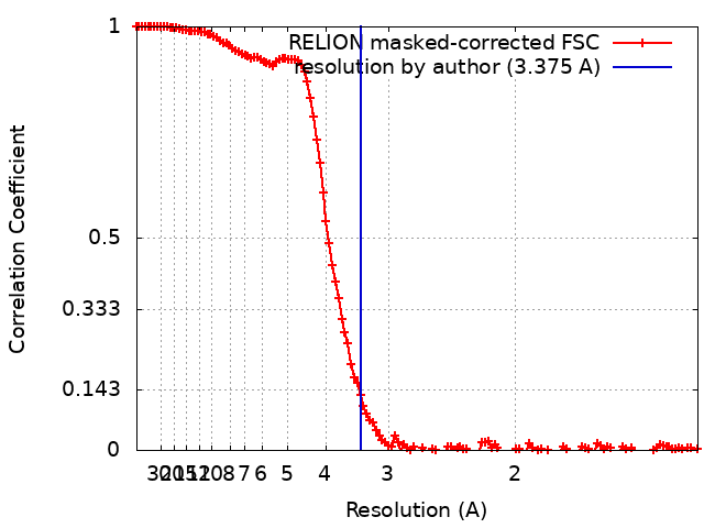
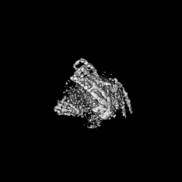
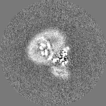
Journal: Cell Rep / Year: 2022 Title: Structural insights into the G protein selectivity revealed by the human EP3-G signaling complex. Authors: Ryoji Suno / Yukihiko Sugita / Kazushi Morimoto / Hiroko Takazaki / Hirokazu Tsujimoto / Mika Hirose / Chiyo Suno-Ikeda / Norimichi Nomura / Tomoya Hino / Asuka Inoue / Kenji Iwasaki / ...Authors: Ryoji Suno / Yukihiko Sugita / Kazushi Morimoto / Hiroko Takazaki / Hirokazu Tsujimoto / Mika Hirose / Chiyo Suno-Ikeda / Norimichi Nomura / Tomoya Hino / Asuka Inoue / Kenji Iwasaki / Takayuki Kato / So Iwata / Takuya Kobayashi / Abstract: Prostaglandin receptors have been implicated in a wide range of functions, including inflammation, immune response, reproduction, and cancer. Our group has previously determined the crystal structure ...Prostaglandin receptors have been implicated in a wide range of functions, including inflammation, immune response, reproduction, and cancer. Our group has previously determined the crystal structure of the active-like EP3 bound to its endogenous agonist, prostaglandin E. Here, we present the single-particle cryoelectron microscopy (cryo-EM) structure of the human EP3-G signaling complex at a resolution of 3.4 Å. The structure reveals the binding mode of G to EP3 and the structural changes induced in EP3 by G binding. In addition, we compare the structure of the EP3-G complex with other subtypes of prostaglandin receptors (EP2 and EP4) bound to G that have been previously reported and examine the differences in amino acid composition at the receptor-G protein interface. Mutational analysis reveals that the selectivity of the G protein depends on specific amino acid residues in the second intracellular loop and TM5.
In the structure databanks used in Yorodumi, some data are registered as the other names, "COVID-19 virus" and "2019-nCoV". Here are the details of the virus and the list of structure data.
Jan 31, 2019. EMDB accession codes are about to change! (news from PDBe EMDB page)
EMDB accession codes are about to change! (news from PDBe EMDB page)
The allocation of 4 digits for EMDB accession codes will soon come to an end. Whilst these codes will remain in use, new EMDB accession codes will include an additional digit and will expand incrementally as the available range of codes is exhausted. The current 4-digit format prefixed with “EMD-” (i.e. EMD-XXXX) will advance to a 5-digit format (i.e. EMD-XXXXX), and so on. It is currently estimated that the 4-digit codes will be depleted around Spring 2019, at which point the 5-digit format will come into force.
The EM Navigator/Yorodumi systems omit the EMD- prefix.
Related info.:Q: What is EMD? / ID/Accession-code notation in Yorodumi/EM Navigator
Yorodumi is a browser for structure data from EMDB, PDB, SASBDB, etc.
This page is also the successor to EM Navigator detail page, and also detail information page/front-end page for Omokage search.
The word "yorodu" (or yorozu) is an old Japanese word meaning "ten thousand". "mi" (miru) is to see.
Related info.:EMDB / PDB / SASBDB / Comparison of 3 databanks / Yorodumi Search / Aug 31, 2016. New EM Navigator & Yorodumi / Yorodumi Papers / Jmol/JSmol / Function and homology information / Changes in new EM Navigator and Yorodumi
 Movie
Movie Controller


 Open data
 Basic information
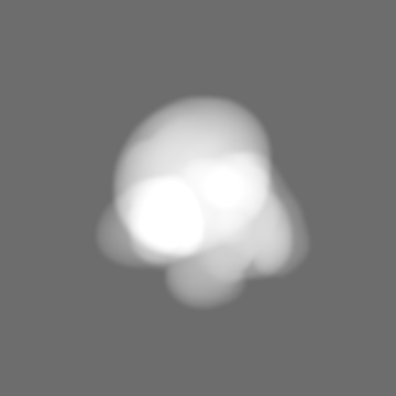
 Map data
 Sample
 Keywords
 Function and homology information
 Homo sapiens (human) /
 Authors
 Japan, 6 items
  Citation
 Structure visualization
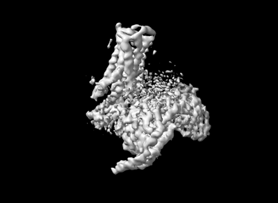
 Downloads & links
 emd_32824.png
 http://ftp.pdbj.org/pub/emdb/structures/EMD-32824
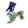


 Z (Sec.)
 Y (Row.)
 X (Col.)


 Sample components
 Spodoptera frugiperda (fall armyworm)
 Brevibacillus agri (bacteria)
 Processing
 Electron microscopy
 FIELD EMISSION GUN