National Institutes of Health/National Institute of General Medical Sciences (NIH/NIGMS)
GM131922
米国
National Institutes of Health/National Institute of General Medical Sciences (NIH/NIGMS)
GM118524
米国
National Institutes of Health/National Institute of General Medical Sciences (NIH/NIGMS)
S10OD020011
米国
National Institutes of Health/National Institute of General Medical Sciences (NIH/NIGMS)
S10OD030275
米国
引用
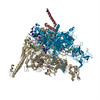
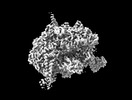
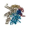
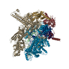
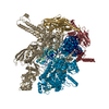
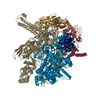
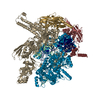
ジャーナル: Nat Struct Mol Biol / 年: 2023 タイトル: Structural basis for control of bacterial RNA polymerase pausing by a riboswitch and its ligand. 著者: Adrien Chauvier / Jason C Porta / Indrajit Deb / Emily Ellinger / Katarina Meze / Aaron T Frank / Melanie D Ohi / Nils G Walter / 要旨: Folding of nascent transcripts can be modulated by the RNA polymerase (RNAP) that carries out their transcription, and vice versa. A pause of RNAP during transcription of a preQ riboswitch (termed ...Folding of nascent transcripts can be modulated by the RNA polymerase (RNAP) that carries out their transcription, and vice versa. A pause of RNAP during transcription of a preQ riboswitch (termed que-PEC) is stabilized by a previously characterized template consensus sequence and the ligand-free conformation of the nascent RNA. Ligand binding to the riboswitch induces RNAP pause release and downstream transcription termination; however, the mechanism by which riboswitch folding modulates pausing is unclear. Here, we report single-particle cryo-electron microscopy reconstructions of que-PEC in ligand-free and ligand-bound states. In the absence of preQ, the RNA transcript is in an unexpected hyper-translocated state, preventing downstream nucleotide incorporation. Strikingly, on ligand binding, the riboswitch rotates around its helical axis, expanding the surrounding RNAP exit channel and repositioning the transcript for elongation. Our study reveals the tight coupling by which nascent RNA structures and their ligands can functionally regulate the macromolecular transcription machinery.
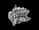
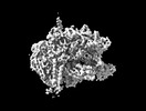
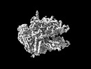
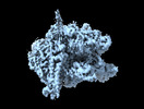
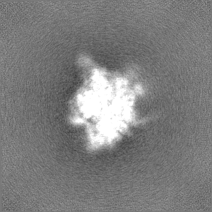
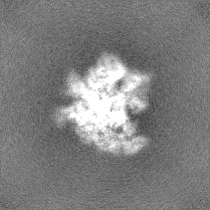
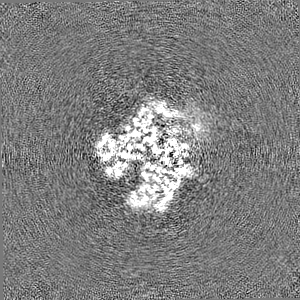
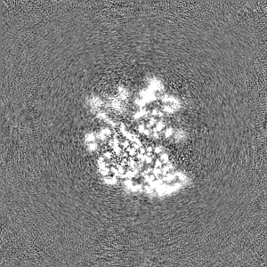
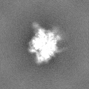
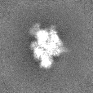
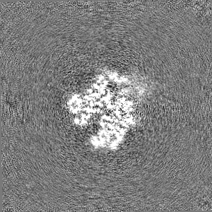
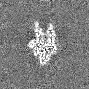
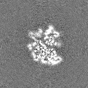
詳細: This is component 0 of 3DVA analysis of a particle stack of Cryo-EM consensus structure of Escherichia coli que-PEC (paused elongation complex) RNA Polymerase plus preQ1 ligand. Particles were already selected.
初期モデル
モデルのタイプ: NONE / 詳細: Ab initio
最終 再構成
解像度のタイプ: BY AUTHOR / 解像度: 3.9 Å / 解像度の算出法: FSC 0.143 CUT-OFF / 使用した粒子像数: 31355
 ムービー
ムービー コントローラー


 データを開く
 基本情報
基本情報
 マップデータ
 試料
試料 キーワード
キーワード 機能・相同性情報
 データ登録者
 米国, 4件
  引用
引用
 構造の表示
構造の表示
 ダウンロードとリンク
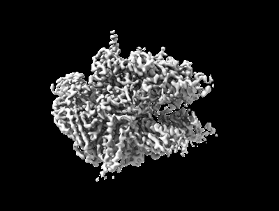 emd_29812.png
 http://ftp.pdbj.org/pub/emdb/structures/EMD-29812
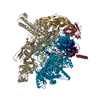


 Z
Z Y
Y X
X


 試料の構成要素
 解析
解析 電子顕微鏡法
 FIELD EMISSION GUN