 Movie
Movie Controller


+ Open data

- Basic information
| Entry |  | |||||||||
|---|---|---|---|---|---|---|---|---|---|---|
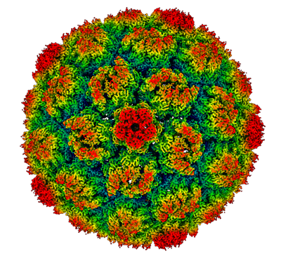
| Title | Prohead I ico symmetry | |||||||||
 Map data Map data | icosahedral symmetry | |||||||||
 Sample Sample |
| |||||||||
 Keywords Keywords | Prohead I / icosahedral symmetry / HK97 / phage / capsid / VIRUS | |||||||||
| Function / homology | : / Phage capsid / Phage capsid family / viral procapsid maturation / T=7 icosahedral viral capsid / viral capsid / identical protein binding / Major capsid protein Function and homology information Function and homology information | |||||||||
| Biological species |  Escherichia phage HK97 (virus) Escherichia phage HK97 (virus) | |||||||||
| Method | single particle reconstruction / cryo EM / Resolution: 3.5 Å | |||||||||
 Authors Authors | Huet A / Oh B / Maurer J / Duda RL / Conway JF | |||||||||
| Funding support |  United States, 2 items United States, 2 items
| |||||||||
 Citation Citation | Journal: Sci Adv / Year: 2023 Title: A symmetry mismatch unraveled: How phage HK97 scaffold flexibly accommodates a 12-fold pore at a 5-fold viral capsid vertex. Authors: Alexis Huet / Bonnie Oh / Josh Maurer / Robert L Duda / James F Conway / Abstract: Tailed bacteriophages and herpesviruses use a transient scaffold to assemble icosahedral capsids with hexameric capsomers on the faces and pentameric capsomers at all but one vertex where a 12-fold ...Tailed bacteriophages and herpesviruses use a transient scaffold to assemble icosahedral capsids with hexameric capsomers on the faces and pentameric capsomers at all but one vertex where a 12-fold portal is thought to nucleate the assembly. How does the scaffold orchestrate this step? We have determined the portal vertex structure of the bacteriophage HK97 procapsid, where the scaffold is a domain of the major capsid protein. The scaffold forms rigid helix-turn-strand structures on the interior surfaces of all capsomers and is further stabilized around the portal, forming trimeric coiled-coil towers, two per surrounding capsomer. These 10 towers bind identically to 10 of 12 portal subunits, adopting a pseudo-12-fold organization that explains how the symmetry mismatch is managed at this early step. | |||||||||
| History |
|
- Structure visualization
| Supplemental images |
|---|
- Downloads & links
-EMDB archive
| Map data | emd_29390.map.gz | 1.2 GB | EMDB map data format | |
|---|---|---|---|---|
| Header (meta data) | emd-29390-v30.xmlemd-29390.xml | 17.2 KB 17.2 KB | Display Display | EMDB header |
| FSC (resolution estimation) | emd_29390_fsc.xml | 24.7 KB | Display | FSC data file |
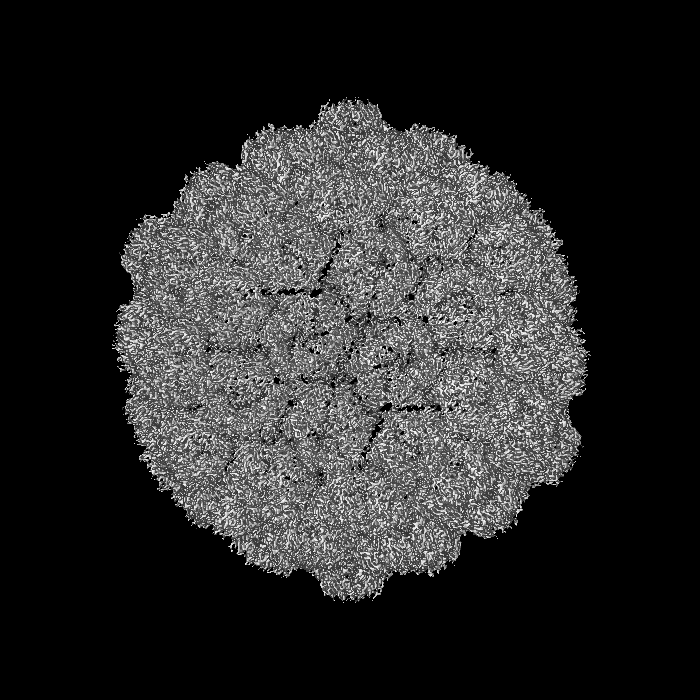
| Images |  emd_29390.png emd_29390.png | 213 KB | ||
| Filedesc metadata | emd-29390.cif.gz | 5.9 KB | ||
| Others | emd_29390_half_map_1.map.gzemd_29390_half_map_2.map.gz | 1 GB 1 GB | ||
| Archive directory |  http://ftp.pdbj.org/pub/emdb/structures/EMD-29390ftp://ftp.pdbj.org/pub/emdb/structures/EMD-29390 http://ftp.pdbj.org/pub/emdb/structures/EMD-29390ftp://ftp.pdbj.org/pub/emdb/structures/EMD-29390 | HTTPS FTP |
-Related structure data
| Related structure data |  8fqkMC  8fqlC M: atomic model generated by this map C: citing same article ( |
|---|---|
| Similar structure data |


-Links
| EMDB pages | EMDB (EBI/PDBe) / EMDataResource |
|---|---|
| Related items in Molecule of the Month |


-Map
| File | Download / File: emd_29390.map.gz / Format: CCP4 / Size: 1.3 GB / Type: IMAGE STORED AS FLOATING POINT NUMBER (4 BYTES) | ||||||||||||||||||||||||||||||||||||
|---|---|---|---|---|---|---|---|---|---|---|---|---|---|---|---|---|---|---|---|---|---|---|---|---|---|---|---|---|---|---|---|---|---|---|---|---|---|
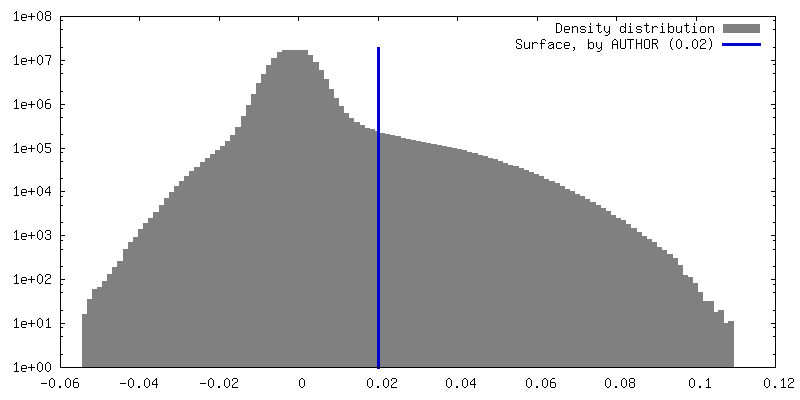
| Annotation | icosahedral symmetry | ||||||||||||||||||||||||||||||||||||
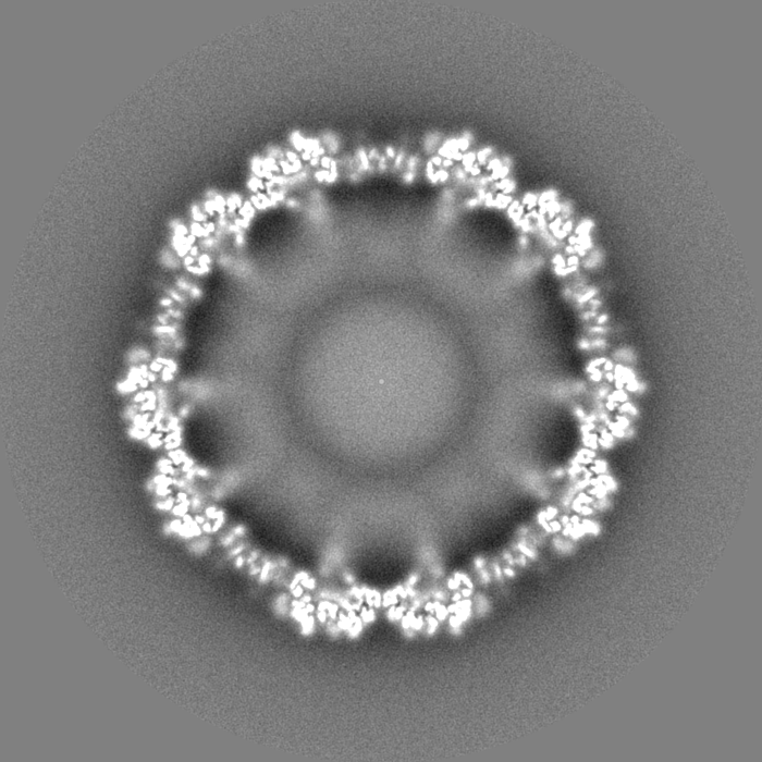
| Projections & slices | Image control
Images are generated by Spider. | ||||||||||||||||||||||||||||||||||||
| Voxel size | X=Y=Z: 1.08 Å | ||||||||||||||||||||||||||||||||||||
| Density |
| ||||||||||||||||||||||||||||||||||||
| Symmetry | Space group: 1 | ||||||||||||||||||||||||||||||||||||
| Details | EMDB XML:
|
 Z (Sec.)
 Y (Row.)
 X (Col.)


-Supplemental data
-Half map: #2
| File | emd_29390_half_map_1.map | ||||||||||||
|---|---|---|---|---|---|---|---|---|---|---|---|---|---|
| Projections & Slices |
| ||||||||||||
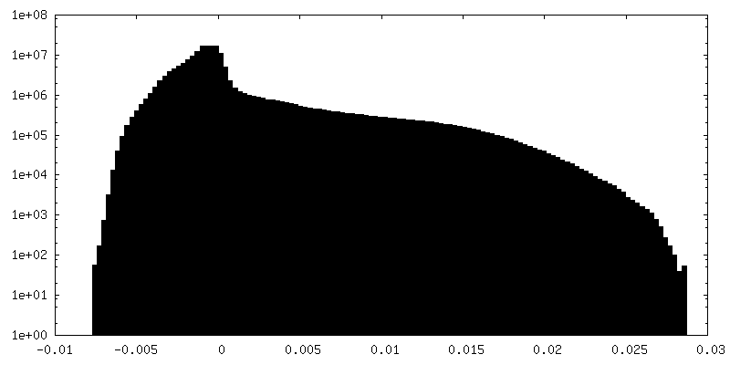
| Density Histograms |


-Half map: #1
| File | emd_29390_half_map_2.map | ||||||||||||
|---|---|---|---|---|---|---|---|---|---|---|---|---|---|
| Projections & Slices |
| ||||||||||||
| Density Histograms |


- Sample components
-Entire : Escherichia phage HK97
| Entire | Name: Escherichia phage HK97 (virus) |
|---|---|
| Components |
|
-Supramolecule #1: Escherichia phage HK97
| Supramolecule | Name: Escherichia phage HK97 / type: virus / ID: 1 / Parent: 0 / Macromolecule list: all / Details: Prohead I expressed from plasmid in E.Coli / NCBI-ID: 2681617 / Sci species name: Escherichia phage HK97 / Virus type: VIRUS-LIKE PARTICLE / Virus isolate: OTHER / Virus enveloped: No / Virus empty: Yes |
|---|---|
| Host (natural) | Organism:  |
| Molecular weight | Theoretical: 17.7 MDa |
| Virus shell | Shell ID: 1 / Name: Prohead I / Diameter: 500.0 Å / T number (triangulation number): 7 |
-Macromolecule #1: Scaffolding domain delta
| Macromolecule | Name: Scaffolding domain delta / type: protein_or_peptide / ID: 1 / Number of copies: 7 / Enantiomer: LEVO |
|---|---|
| Source (natural) | Organism: Escherichia phage HK97 (virus) |
| Molecular weight | Theoretical: 42.286453 KDa |
| Recombinant expression | Organism: |
| Sequence | String: MSELALIQKA IEESQQKMTQ LFDAQKAEIE STGQVSKQLQ SDLMKVQEEL TKSGTRLFDL EQKLASGAEN PGEKKSFSER AAEELIKSW DGKQGTFGAK TFNKSLGSDA DSAGSLIQPM QIPGIIMPGL RRLTIRDLLA QGRTSSNALE YVREEVFTNN A DVVAEKAL ...String: MSELALIQKA IEESQQKMTQ LFDAQKAEIE STGQVSKQLQ SDLMKVQEEL TKSGTRLFDL EQKLASGAEN PGEKKSFSER AAEELIKSW DGKQGTFGAK TFNKSLGSDA DSAGSLIQPM QIPGIIMPGL RRLTIRDLLA QGRTSSNALE YVREEVFTNN A DVVAEKAL KPESDITFSK QTANVKTIAH WVQASRQVMD DAPMLQSYIN NRLMYGLALK EEGQLLNGDG TGDNLEGLNK VA TAYDTSL NATGDTRADI IAHAIYQVTE SEFSASGIVL NPRDWHNIAL LKDNEGRYIF GGPQAFTSNI MWGLPVVPTK AQA AGTFTV GGFDMASQVW DRMDATVEVS REDRDNFVKN MLTILCEERL ALAHYRPTAI IKGTFSSGS UniProtKB: Major capsid protein |
-Experimental details
-Structure determination
| Method | cryo EM |
|---|---|
 Processing Processing | single particle reconstruction |
| Aggregation state | particle |
-Sample preparation
| Concentration | 10 mg/mL | |||||||||
|---|---|---|---|---|---|---|---|---|---|---|
| Buffer | pH: 7.5 Component:
| |||||||||
| Grid | Model: Quantifoil R2/1 / Material: COPPER / Mesh: 400 / Support film - Material: CARBON / Support film - topology: HOLEY / Pretreatment - Type: GLOW DISCHARGE / Pretreatment - Time: 10 sec. | |||||||||
| Vitrification | Cryogen name: ETHANE-PROPANE / Chamber humidity: 100 % / Chamber temperature: 293 K / Instrument: FEI VITROBOT MARK IV |
- Electron microscopy
| Microscope | FEI TITAN KRIOS |
|---|---|
| Image recording | Film or detector model: FEI FALCON III (4k x 4k) / Detector mode: COUNTING / Number grids imaged: 2 / Number real images: 2909 / Average electron dose: 60.0 e/Å2 |
| Electron beam | Acceleration voltage: 300 kV / Electron source:  FIELD EMISSION GUN FIELD EMISSION GUN |
| Electron optics | Calibrated defocus max: 4.0 µm / Calibrated defocus min: 1.0 µm / Illumination mode: FLOOD BEAM / Imaging mode: BRIGHT FIELD / Nominal defocus max: 4.0 µm / Nominal defocus min: 1.0 µm / Nominal magnification: 75000 |
| Sample stage | Specimen holder model: FEI TITAN KRIOS AUTOGRID HOLDER / Cooling holder cryogen: NITROGEN |
| Experimental equipment |  Model: Titan Krios / Image courtesy: FEI Company |
+Image processing

-Atomic model buiding 1
| Refinement | Space: REAL / Protocol: FLEXIBLE FIT |
|---|---|
| Output model | PDB-8fqk: |
