 Movie
Movie Controller

[English] 日本語
 Yorodumi
- EMDB-28254: Composite 70S ribosome structure for "Atomistic simulations of th... -
+ Open data

- Basic information
| Entry |  | |||||||||
|---|---|---|---|---|---|---|---|---|---|---|
| Title | Composite 70S ribosome structure for "Atomistic simulations of the E. coli ribosome provide selection criteria for translationally active substrates | |||||||||
 Map data Map data | Composite 70S map from 50S- and 30S-focused refinements | |||||||||
 Sample Sample |
| |||||||||
 Keywords Keywords | ribosome / tRNA / e. coli | |||||||||
| Function / homology |  Function and homology information Function and homology informationnegative regulation of cytoplasmic translational initiation / positive regulation of ribosome biogenesis / DnaA-L2 complex / negative regulation of translational initiation / negative regulation of DNA-templated DNA replication initiation / mRNA regulatory element binding translation repressor activity / cytosolic ribosome assembly / ribosome assembly / assembly of large subunit precursor of preribosome / transcription antitermination ...negative regulation of cytoplasmic translational initiation / positive regulation of ribosome biogenesis / DnaA-L2 complex / negative regulation of translational initiation / negative regulation of DNA-templated DNA replication initiation / mRNA regulatory element binding translation repressor activity / cytosolic ribosome assembly / ribosome assembly / assembly of large subunit precursor of preribosome / transcription antitermination / translational initiation / regulation of cell growth / DNA-templated transcription termination / maintenance of translational fidelity / mRNA 5'-UTR binding / large ribosomal subunit / transferase activity / ribosomal small subunit assembly / ribosome binding / ribosomal small subunit biogenesis / 5S rRNA binding / small ribosomal subunit / ribosomal large subunit assembly / small ribosomal subunit rRNA binding / cytosolic small ribosomal subunit / large ribosomal subunit rRNA binding / cytosolic large ribosomal subunit / cytoplasmic translation / tRNA binding / negative regulation of translation / rRNA binding / structural constituent of ribosome / ribosome / translation / ribonucleoprotein complex / response to antibiotic / mRNA binding / RNA binding / zinc ion binding / membrane / cytoplasm / cytosol Similarity search - Function | |||||||||
| Biological species |  | |||||||||
| Method | single particle reconstruction / cryo EM / Resolution: 2.1 Å | |||||||||
 Authors Authors | Watson ZL / Cate JHD | |||||||||
| Funding support |  United States, 1 items United States, 1 items
| |||||||||
 Citation Citation | Journal: Nat Chem / Year: 2023 Title: Atomistic simulations of the Escherichia coli ribosome provide selection criteria for translationally active substrates. Authors: Zoe L Watson / Isaac J Knudson / Fred R Ward / Scott J Miller / Jamie H D Cate / Alanna Schepartz / Ara M Abramyan / Abstract: As genetic code expansion advances beyond L-α-amino acids to backbone modifications and new polymerization chemistries, delineating what substrates the ribosome can accommodate remains a challenge. ...As genetic code expansion advances beyond L-α-amino acids to backbone modifications and new polymerization chemistries, delineating what substrates the ribosome can accommodate remains a challenge. The Escherichia coli ribosome tolerates non-L-α-amino acids in vitro, but few structural insights that explain how are available, and the boundary conditions for efficient bond formation are so far unknown. Here we determine a high-resolution cryogenic electron microscopy structure of the E. coli ribosome containing α-amino acid monomers and use metadynamics simulations to define energy surface minima and understand incorporation efficiencies. Reactive monomers across diverse structural classes favour a conformational space where the aminoacyl-tRNA nucleophile is <4 Å from the peptidyl-tRNA carbonyl with a Bürgi-Dunitz angle of 76-115°. Monomers with free energy minima that fall outside this conformational space do not react efficiently. This insight should accelerate the in vivo and in vitro ribosomal synthesis of sequence-defined, non-peptide heterooligomers. | |||||||||
| History |
|
- Structure visualization
| Supplemental images |
|---|

- Downloads & links
-EMDB archive
| Map data | emd_28254.map.gz | 35.5 MB | EMDB map data format | |
|---|---|---|---|---|
| Header (meta data) | emd-28254-v30.xmlemd-28254.xml | 67.1 KB 67.1 KB | Display Display | EMDB header |
| Images | 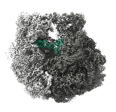 emd_28254.png emd_28254.png | 191 KB | ||
| Filedesc metadata | emd-28254.cif.gz | 14.9 KB | ||
| Archive directory |  http://ftp.pdbj.org/pub/emdb/structures/EMD-28254ftp://ftp.pdbj.org/pub/emdb/structures/EMD-28254 http://ftp.pdbj.org/pub/emdb/structures/EMD-28254ftp://ftp.pdbj.org/pub/emdb/structures/EMD-28254 | HTTPS FTP |
-Related structure data
| Related structure data |  8emmMC C: citing same article ( M: atomic model generated by this map |
|---|---|
| Similar structure data |


-Links
| EMDB pages | EMDB (EBI/PDBe) / EMDataResource |
|---|---|
| Related items in Molecule of the Month |


-Map
| File | Download / File: emd_28254.map.gz / Format: CCP4 / Size: 361.7 MB / Type: IMAGE STORED AS FLOATING POINT NUMBER (4 BYTES) | ||||||||||||||||||||||||||||||||||||
|---|---|---|---|---|---|---|---|---|---|---|---|---|---|---|---|---|---|---|---|---|---|---|---|---|---|---|---|---|---|---|---|---|---|---|---|---|---|
| Annotation | Composite 70S map from 50S- and 30S-focused refinements | ||||||||||||||||||||||||||||||||||||
| Projections & slices | Image control
Images are generated by Spider. | ||||||||||||||||||||||||||||||||||||
| Voxel size | X=Y=Z: 0.8296 Å | ||||||||||||||||||||||||||||||||||||
| Density |
| ||||||||||||||||||||||||||||||||||||
| Symmetry | Space group: 1 | ||||||||||||||||||||||||||||||||||||
| Details | EMDB XML:
|
 Z (Sec.)
 Y (Row.)
 X (Col.)


-Supplemental data
- Sample components
+Entire : 70S ribosome complex with mRNA, A- and P-site Met-NH-tRNAs
+Supramolecule #1: 70S ribosome complex with mRNA, A- and P-site Met-NH-tRNAs
+Macromolecule #1: 23S rRNA
+Macromolecule #2: 5S rRNA
+Macromolecule #32: Met-NH-tRNA
+Macromolecule #33: 16S rRNA
+Macromolecule #54: mRNA
+Macromolecule #3: 50S ribosomal protein L2
+Macromolecule #4: 50S ribosomal protein L3
+Macromolecule #5: 50S ribosomal protein L4
+Macromolecule #6: 50S ribosomal protein L5
+Macromolecule #7: 50S ribosomal protein L6
+Macromolecule #8: 50S ribosomal protein L9
+Macromolecule #9: 50S ribosomal protein L13
+Macromolecule #10: 50S ribosomal protein L14
+Macromolecule #11: 50S ribosomal protein L15
+Macromolecule #12: 50S ribosomal protein L16
+Macromolecule #13: 50S ribosomal protein L17
+Macromolecule #14: 50S ribosomal protein L18
+Macromolecule #15: 50S ribosomal protein L19
+Macromolecule #16: 50S ribosomal protein L20
+Macromolecule #17: Ribosomal protein L21
+Macromolecule #18: 50S ribosomal protein L22
+Macromolecule #19: 50S ribosomal protein L23
+Macromolecule #20: 50S ribosomal protein L24
+Macromolecule #21: 50S ribosomal protein L25
+Macromolecule #22: 50S ribosomal protein L27
+Macromolecule #23: 50S ribosomal protein L28
+Macromolecule #24: 50S ribosomal protein L29
+Macromolecule #25: 50S ribosomal protein L30
+Macromolecule #26: 50S ribosomal protein L32
+Macromolecule #27: 50S ribosomal protein L33
+Macromolecule #28: 50S ribosomal protein L34
+Macromolecule #29: 50S ribosomal protein L35
+Macromolecule #30: 50S ribosomal protein L36
+Macromolecule #31: 50S ribosomal protein L31
+Macromolecule #34: 30S ribosomal protein S2
+Macromolecule #35: 30S ribosomal protein S3
+Macromolecule #36: 30S ribosomal protein S4
+Macromolecule #37: 30S ribosomal protein S5
+Macromolecule #38: 30S ribosomal protein S6
+Macromolecule #39: 30S ribosomal protein S7
+Macromolecule #40: 30S ribosomal protein S8
+Macromolecule #41: 30S ribosomal protein S9
+Macromolecule #42: 30S ribosomal protein S10
+Macromolecule #43: 30S ribosomal protein S11
+Macromolecule #44: 30S ribosomal protein S12
+Macromolecule #45: 30S ribosomal protein S13
+Macromolecule #46: 30S ribosomal protein S14
+Macromolecule #47: 30S ribosomal protein S15
+Macromolecule #48: 30S ribosomal protein S16
+Macromolecule #49: 30S ribosomal protein S17
+Macromolecule #50: 30S ribosomal protein S18
+Macromolecule #51: 30S ribosomal protein S19
+Macromolecule #52: 30S ribosomal protein S20
+Macromolecule #53: 30S ribosomal protein S21
+Macromolecule #55: MAGNESIUM ION
+Macromolecule #56: SPERMIDINE

+Macromolecule #57: SPERMINE

+Macromolecule #58: POTASSIUM ION
+Macromolecule #59: ZINC ION
+Macromolecule #60: METHIONINE

+Macromolecule #61: PAROMOMYCIN
+Macromolecule #62: water

-Experimental details
-Structure determination
| Method | cryo EM |
|---|---|
 Processing Processing | single particle reconstruction |
| Aggregation state | particle |
-Sample preparation
| Buffer | pH: 7.5 |
|---|---|
| Vitrification | Cryogen name: ETHANE |
- Electron microscopy
| Microscope | FEI TITAN KRIOS |
|---|---|
| Image recording | Film or detector model: GATAN K3 (6k x 4k) / Average electron dose: 40.0 e/Å2 |
| Electron beam | Acceleration voltage: 300 kV / Electron source:  FIELD EMISSION GUN FIELD EMISSION GUN |
| Electron optics | Illumination mode: OTHER / Imaging mode: BRIGHT FIELD / Nominal defocus max: 2.0 µm / Nominal defocus min: 0.5 µm |
| Experimental equipment |  Model: Titan Krios / Image courtesy: FEI Company |
-Image processing
| Startup model | Type of model: PDB ENTRY PDB model - PDB ID: |
|---|---|
| Final reconstruction | Resolution.type: BY AUTHOR / Resolution: 2.1 Å / Resolution method: OTHER Details: This is a composite map from 50S- and 30S-focused maps Number images used: 129455 |
| Initial angle assignment | Type: MAXIMUM LIKELIHOOD |
| Final angle assignment | Type: MAXIMUM LIKELIHOOD |

