 Movie
Movie Controller


+ Open data

- Basic information
| Entry | 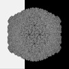 | |||||||||
|---|---|---|---|---|---|---|---|---|---|---|
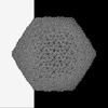
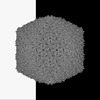
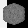
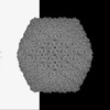
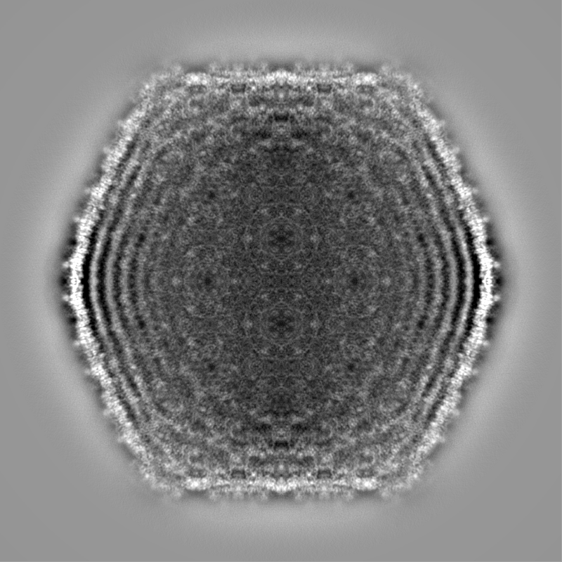
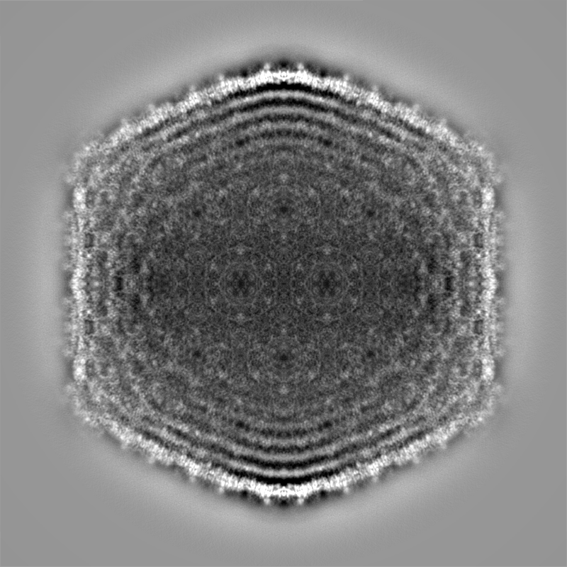
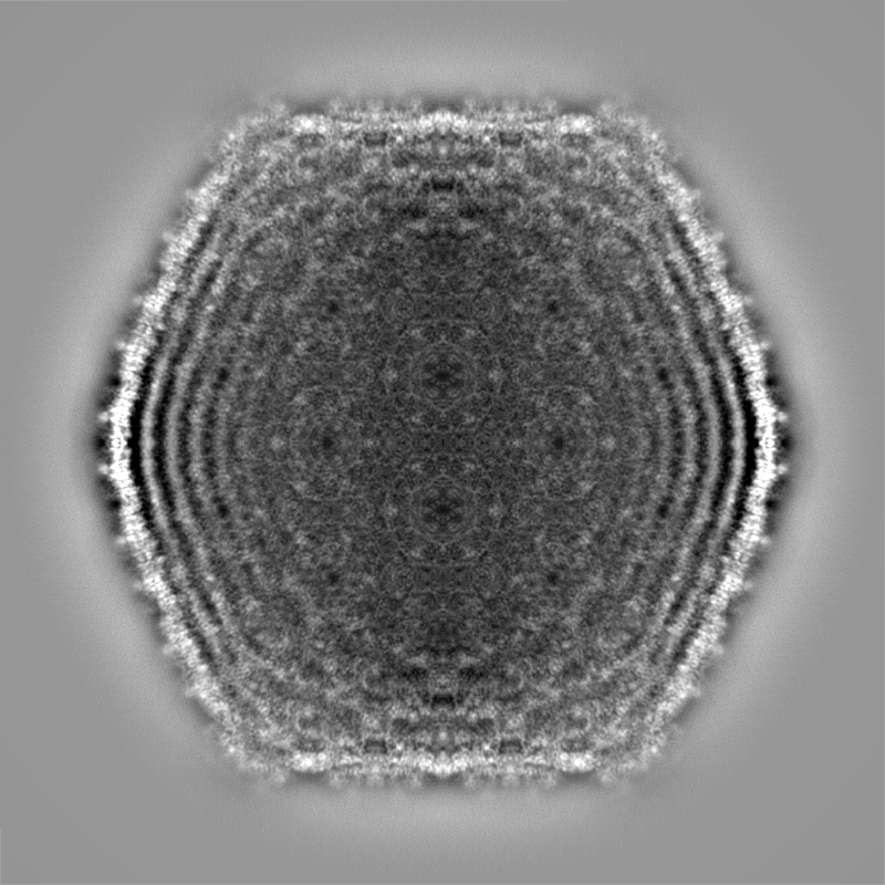
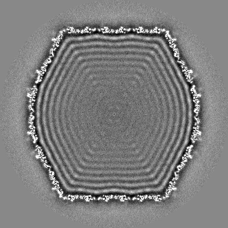
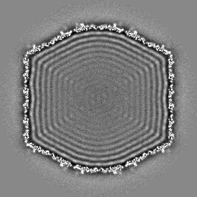
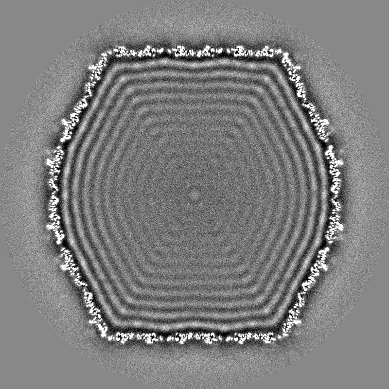
| Title | Mycobacterium phage Ogopogo | |||||||||
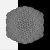
 Map data Map data | Sharpened map of ewald sphere corrected map. | |||||||||
 Sample Sample |
| |||||||||
 Keywords Keywords | HK97-fold / T=9 / tailed bacteriophage / VIRUS | |||||||||
| Function / homology | : / Phage capsid family / Major capsid protein Function and homology information Function and homology information | |||||||||
| Biological species |  Mycobacterium phage Ogopogo (virus) Mycobacterium phage Ogopogo (virus) | |||||||||
| Method | single particle reconstruction / cryo EM / Resolution: 2.7 Å | |||||||||
 Authors Authors | Podgorski JM / White SJ | |||||||||
| Funding support | 1 items
| |||||||||
 Citation Citation | Journal: Structure / Year: 2023 Title: A structural dendrogram of the actinobacteriophage major capsid proteins provides important structural insights into the evolution of capsid stability. Authors: Jennifer M Podgorski / Krista Freeman / Sophia Gosselin / Alexis Huet / James F Conway / Mary Bird / John Grecco / Shreya Patel / Deborah Jacobs-Sera / Graham Hatfull / Johann Peter Gogarten ...Authors: Jennifer M Podgorski / Krista Freeman / Sophia Gosselin / Alexis Huet / James F Conway / Mary Bird / John Grecco / Shreya Patel / Deborah Jacobs-Sera / Graham Hatfull / Johann Peter Gogarten / Janne Ravantti / Simon J White /   Abstract: Many double-stranded DNA viruses, including tailed bacteriophages (phages) and herpesviruses, use the HK97-fold in their major capsid protein to make the capsomers of the icosahedral viral capsid. ...Many double-stranded DNA viruses, including tailed bacteriophages (phages) and herpesviruses, use the HK97-fold in their major capsid protein to make the capsomers of the icosahedral viral capsid. After the genome packaging at near-crystalline densities, the capsid is subjected to a major expansion and stabilization step that allows it to withstand environmental stresses and internal high pressure. Several different mechanisms for stabilizing the capsid have been structurally characterized, but how these mechanisms have evolved is still not understood. Using cryo-EM structure determination of 10 capsids, structural comparisons, phylogenetic analyses, and Alphafold predictions, we have constructed a detailed structural dendrogram describing the evolution of capsid structural stability within the actinobacteriophages. We show that the actinobacteriophage major capsid proteins can be classified into 15 groups based upon their HK97-fold. | |||||||||
| History |
|
- Structure visualization
| Supplemental images |
|---|

- Downloads & links
-EMDB archive
| Map data | emd_28020.map.gz | 1.7 GB | EMDB map data format | |
|---|---|---|---|---|
| Header (meta data) | emd-28020-v30.xmlemd-28020.xml | 22.1 KB 22.1 KB | Display Display | EMDB header |
| Images | 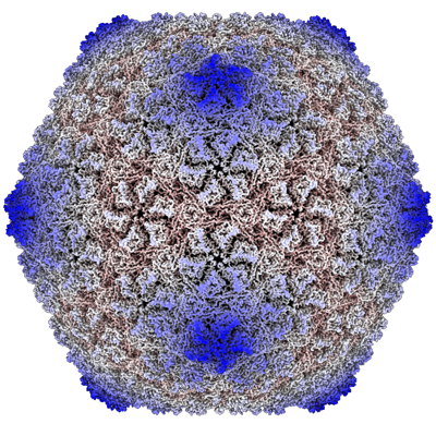 emd_28020.png emd_28020.png | 313.5 KB | ||
| Masks | emd_28020_msk_1.map | 1.9 GB | Mask map | |
| Filedesc metadata | emd-28020.cif.gz | 5.7 KB | ||
| Others | emd_28020_additional_1.map.gzemd_28020_additional_2.map.gzemd_28020_half_map_1.map.gzemd_28020_half_map_2.map.gz | 1.5 GB 1.6 GB 1.6 GB 1.6 GB | ||
| Archive directory |  http://ftp.pdbj.org/pub/emdb/structures/EMD-28020ftp://ftp.pdbj.org/pub/emdb/structures/EMD-28020 http://ftp.pdbj.org/pub/emdb/structures/EMD-28020ftp://ftp.pdbj.org/pub/emdb/structures/EMD-28020 | HTTPS FTP |
-Validation report
| Summary document | emd_28020_validation.pdf.gz | 1.1 MB | Display | EMDB validaton report |
|---|---|---|---|---|
| Full document | emd_28020_full_validation.pdf.gz | 1.1 MB | Display | |
| Data in XML | emd_28020_validation.xml.gz | 26.1 KB | Display | |
| Data in CIF | emd_28020_validation.cif.gz | 30.9 KB | Display | |
| Arichive directory | https://ftp.pdbj.org/pub/emdb/validation_reports/EMD-28020ftp://ftp.pdbj.org/pub/emdb/validation_reports/EMD-28020 | HTTPS FTP |
-Related structure data
| Related structure data |  8ecnMC  8e16C  8eb4C 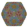 8ec2C 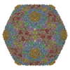 8ec8C 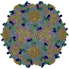 8eciC 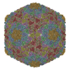 8ecjC 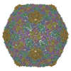 8eckC 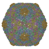 8ecoC 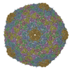 8eduC C: citing same article ( M: atomic model generated by this map |
|---|---|
| Similar structure data |

-Links
| EMDB pages | EMDB (EBI/PDBe) / EMDataResource |
|---|---|
| Related items in Molecule of the Month |


-Map
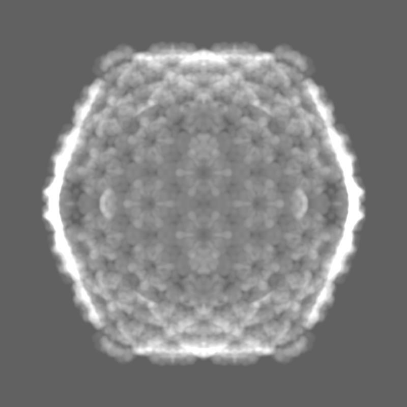
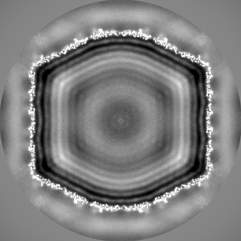
| File | Download / File: emd_28020.map.gz / Format: CCP4 / Size: 1.9 GB / Type: IMAGE STORED AS FLOATING POINT NUMBER (4 BYTES) | ||||||||||||||||||||||||||||||||||||
|---|---|---|---|---|---|---|---|---|---|---|---|---|---|---|---|---|---|---|---|---|---|---|---|---|---|---|---|---|---|---|---|---|---|---|---|---|---|
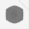
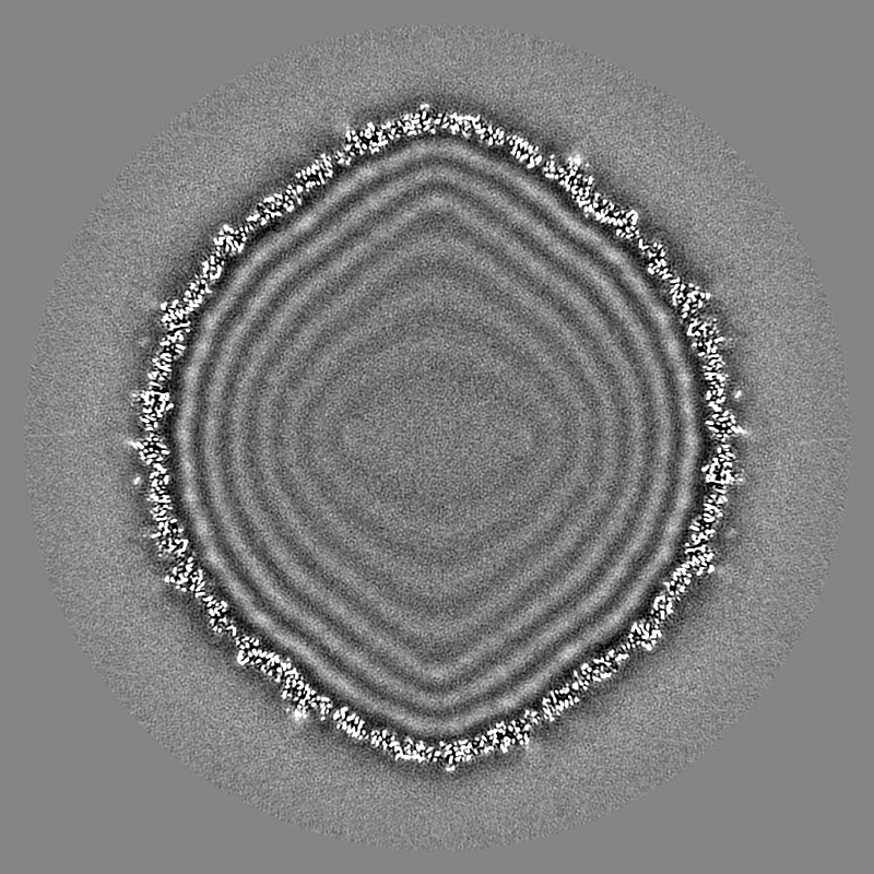
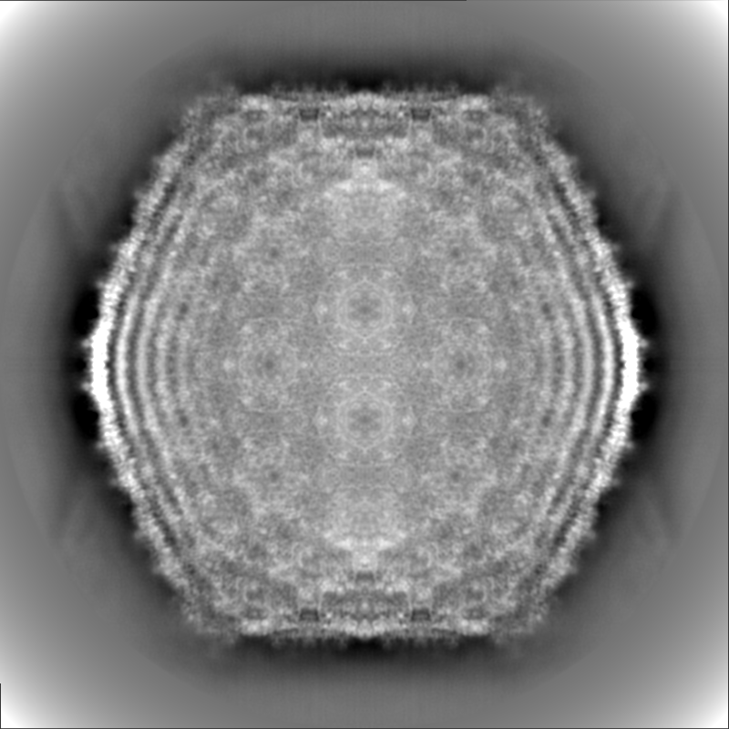
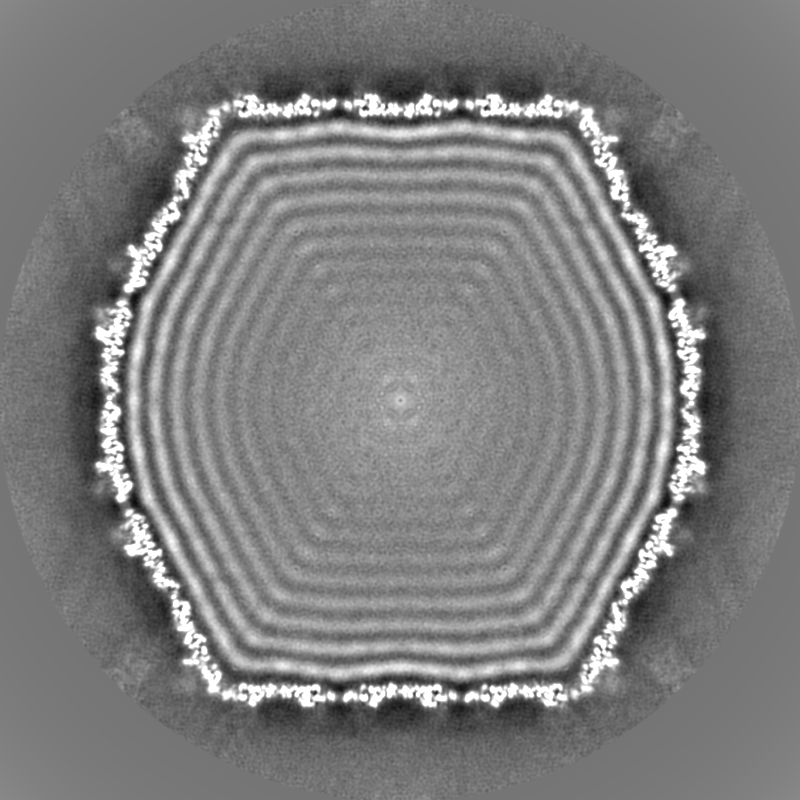
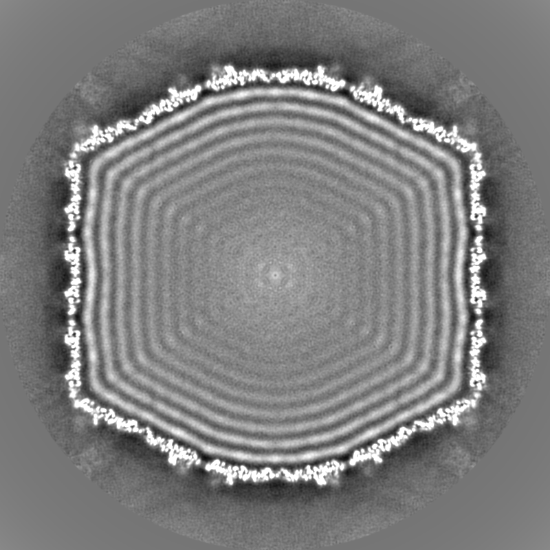
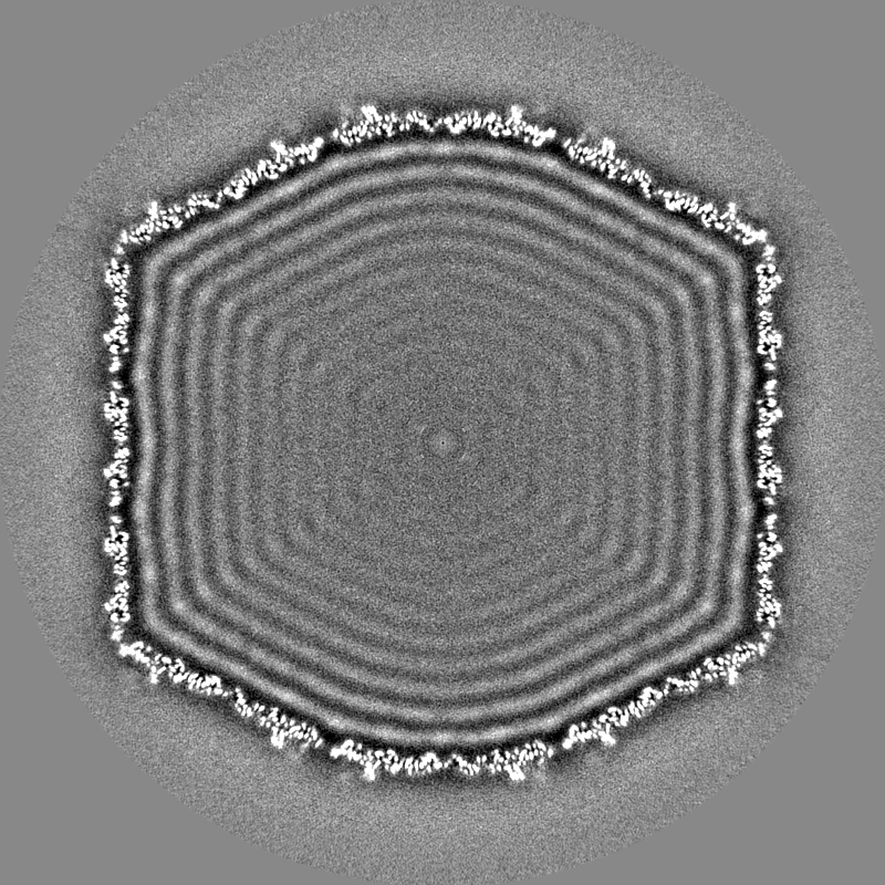
| Annotation | Sharpened map of ewald sphere corrected map. | ||||||||||||||||||||||||||||||||||||
| Projections & slices | Image control
Images are generated by Spider. | ||||||||||||||||||||||||||||||||||||
| Voxel size | X=Y=Z: 1.0624 Å | ||||||||||||||||||||||||||||||||||||
| Density |
| ||||||||||||||||||||||||||||||||||||
| Symmetry | Space group: 1 | ||||||||||||||||||||||||||||||||||||
| Details | EMDB XML:
|
 X (Sec.)
 Y (Row.)
 Z (Col.)


-Supplemental data
-Mask #1
| File | emd_28020_msk_1.map | ||||||||||||
|---|---|---|---|---|---|---|---|---|---|---|---|---|---|
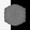
| Projections & Slices |
| ||||||||||||
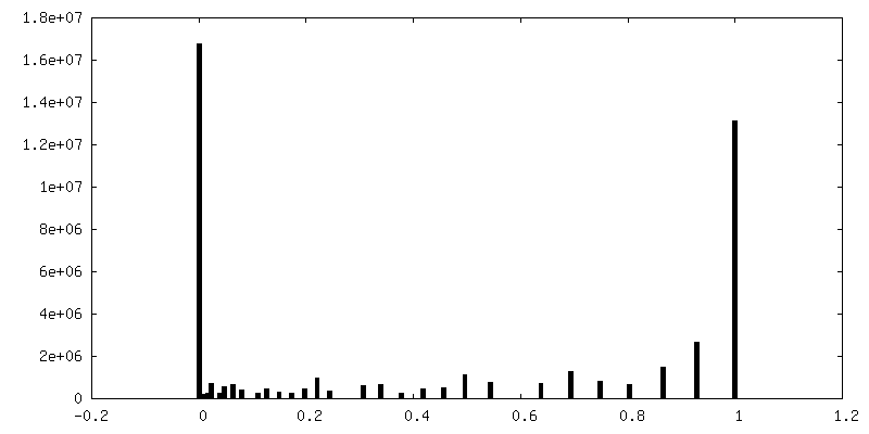
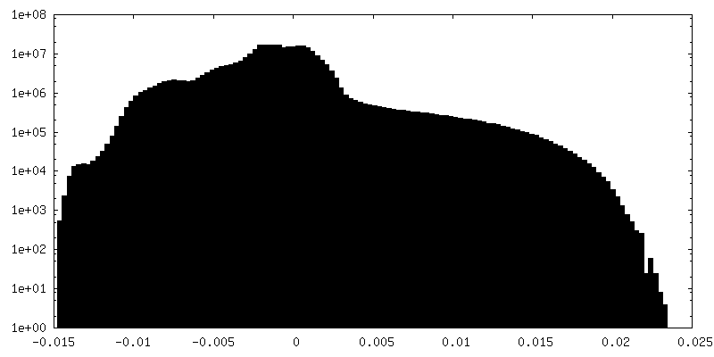
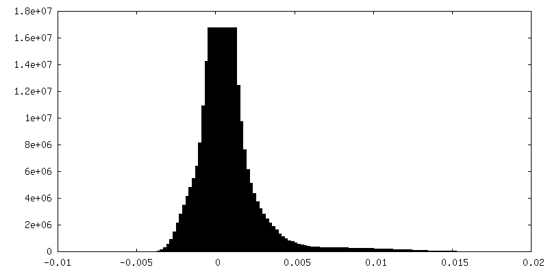
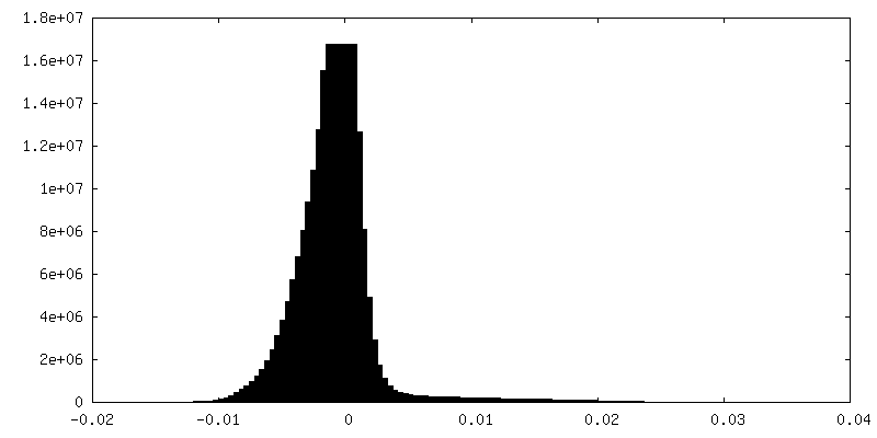
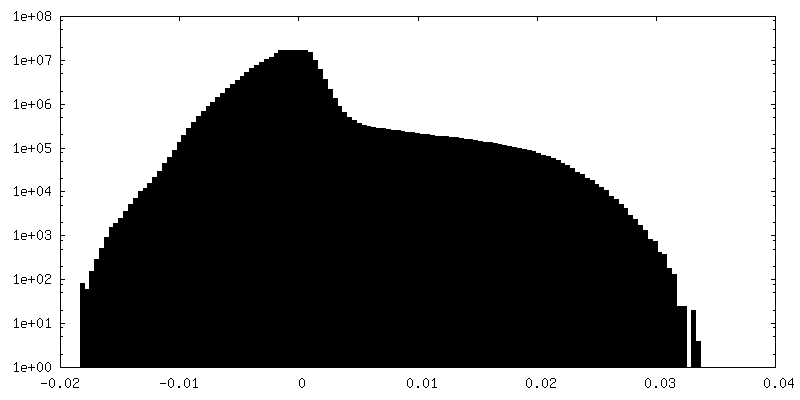
| Density Histograms |


-Additional map: Map after CTF Refinement.
| File | emd_28020_additional_1.map | ||||||||||||
|---|---|---|---|---|---|---|---|---|---|---|---|---|---|
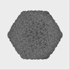
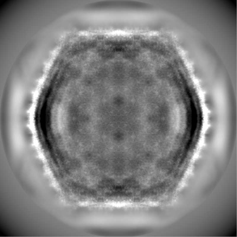
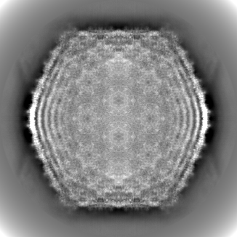
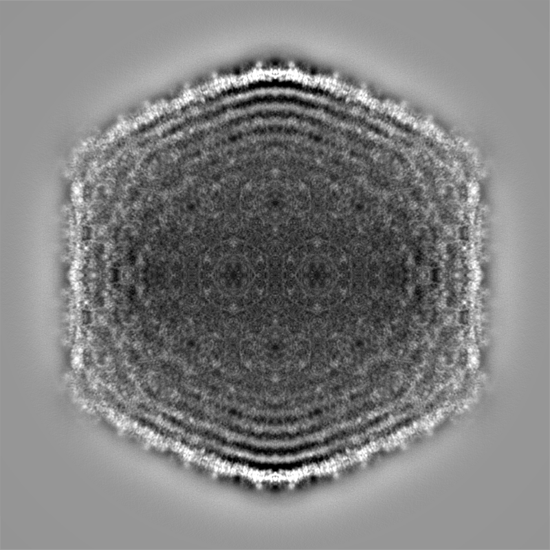
| Annotation | Map after CTF Refinement. | ||||||||||||
| Projections & Slices |
| ||||||||||||
| Density Histograms |


-Additional map: Map before CTF Refinement.
| File | emd_28020_additional_2.map | ||||||||||||
|---|---|---|---|---|---|---|---|---|---|---|---|---|---|
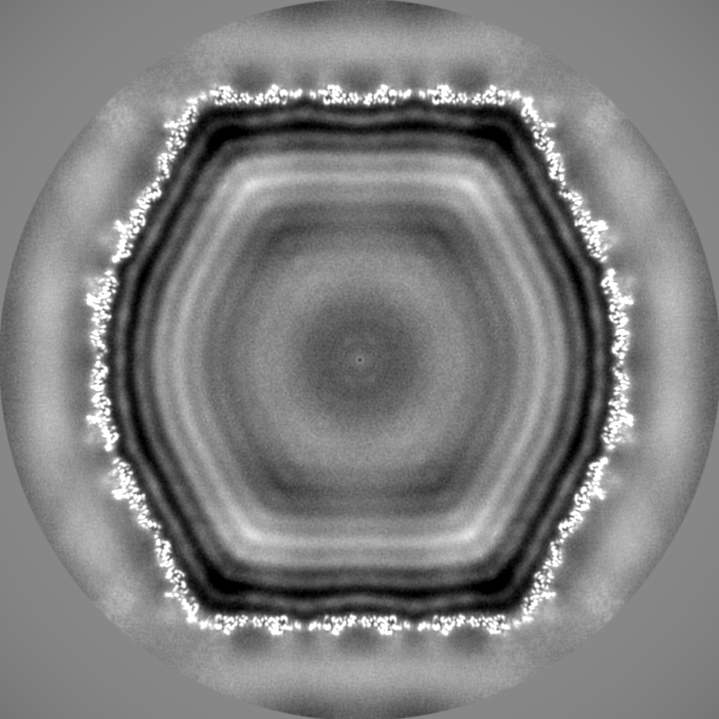
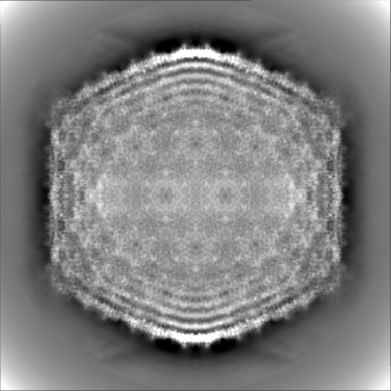
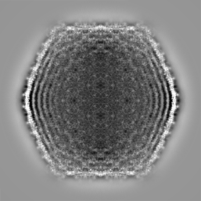
| Annotation | Map before CTF Refinement. | ||||||||||||
| Projections & Slices |
| ||||||||||||
| Density Histograms |


-Half map: Half map of ewald sphere corrected map.
| File | emd_28020_half_map_1.map | ||||||||||||
|---|---|---|---|---|---|---|---|---|---|---|---|---|---|
| Annotation | Half map of ewald sphere corrected map. | ||||||||||||
| Projections & Slices |
| ||||||||||||
| Density Histograms |


-Half map: Half map of ewald sphere corrected map.
| File | emd_28020_half_map_2.map | ||||||||||||
|---|---|---|---|---|---|---|---|---|---|---|---|---|---|
| Annotation | Half map of ewald sphere corrected map. | ||||||||||||
| Projections & Slices |
| ||||||||||||
| Density Histograms |


- Sample components
-Entire : Mycobacterium phage Ogopogo
| Entire | Name: Mycobacterium phage Ogopogo (virus) |
|---|---|
| Components |
|
-Supramolecule #1: Mycobacterium phage Ogopogo
| Supramolecule | Name: Mycobacterium phage Ogopogo / type: virus / ID: 1 / Parent: 0 / Macromolecule list: all / NCBI-ID: 2099640 / Sci species name: Mycobacterium phage Ogopogo / Virus type: VIRION / Virus isolate: STRAIN / Virus enveloped: No / Virus empty: No |
|---|---|
| Host (natural) | Organism:  Mycolicibacterium smegmatis MC2 155 (bacteria) Mycolicibacterium smegmatis MC2 155 (bacteria) |
| Virus shell | Shell ID: 1 / Diameter: 750.0 Å / T number (triangulation number): 9 |
-Macromolecule #1: Major capsid protein
| Macromolecule | Name: Major capsid protein / type: protein_or_peptide / ID: 1 / Number of copies: 9 / Enantiomer: LEVO |
|---|---|
| Source (natural) | Organism: Mycobacterium phage Ogopogo (virus) |
| Molecular weight | Theoretical: 33.033039 KDa |
| Sequence | String: MAVLKTSSFQ LPRHLVPGVW QKAQGQSVLA RLSNAEPQEF GEQQYMTLTA PPRGEVVGES AEKSESTATF APVTSLVRKV QVTQRFSQE VKWADESRQL GVLQTMADLS GVALGRALDL IGIHGINPLT GAALAGTPPK IIDTTNVVEL TTETLGLPDQ A IEAAVGLV ...String: MAVLKTSSFQ LPRHLVPGVW QKAQGQSVLA RLSNAEPQEF GEQQYMTLTA PPRGEVVGES AEKSESTATF APVTSLVRKV QVTQRFSQE VKWADESRQL GVLQTMADLS GVALGRALDL IGIHGINPLT GAALAGTPPK IIDTTNVVEL TTETLGLPDQ A IEAAVGLV LDDSISPNGL ALDNSFAFKL ATQRHPTTGQ KLYPELGFGT DISGFMSLGA AVSDTVRGGP EAVTPSTGAY RT TNPNIKA VVGDFSAFRW GVQANIPLTL IEYGDPDGSG DLQRKNELAI RAEVVYGVGI LSTDAFAVVR DADES UniProtKB: Major capsid protein |
-Experimental details
-Structure determination
| Method | cryo EM |
|---|---|
 Processing Processing | single particle reconstruction |
| Aggregation state | particle |
-Sample preparation
| Concentration | 10 mg/mL | ||||||||||||
|---|---|---|---|---|---|---|---|---|---|---|---|---|---|
| Buffer | pH: 7.5 Component:
| ||||||||||||
| Vitrification | Cryogen name: ETHANE / Chamber humidity: 100 % / Chamber temperature: 283 K / Instrument: FEI VITROBOT MARK IV |
- Electron microscopy
| Microscope | FEI TITAN KRIOS |
|---|---|
| Image recording | Film or detector model: FEI FALCON III (4k x 4k) / Detector mode: COUNTING / Number real images: 2220 / Average electron dose: 30.0 e/Å2 |
| Electron beam | Acceleration voltage: 300 kV / Electron source:  FIELD EMISSION GUN FIELD EMISSION GUN |
| Electron optics | Illumination mode: FLOOD BEAM / Imaging mode: BRIGHT FIELD / Cs: 2.7 mm / Nominal defocus max: 3.0 µm / Nominal defocus min: 1.0 µm |
| Sample stage | Specimen holder model: FEI TITAN KRIOS AUTOGRID HOLDER |
| Experimental equipment |  Model: Titan Krios / Image courtesy: FEI Company |
+Image processing
-Atomic model buiding 1
| Details | Amino acid sequence built into the map for a single major capsid protein and refined with Phenix. Model then used for rest of asymmetric unit and refined with Phenix. Final step involved using Isolde. |
|---|---|
| Refinement | Protocol: AB INITIO MODEL |
| Output model | PDB-8ecn: |
