: / : / Mitochondrial ribosomal protein L48 / 39S ribosomal protein L40, mitochondrial / Ribosomal protein S30, mitochondrial / Ribosomal protein L53, mitochondrial / 39S ribosomal protein L53/MRP-L53 / 39S ribosomal protein L42, mitochondrial / Mitochondrial 28S ribosomal protein S32 / Ribosomal protein 63, mitochondrial ...: / : / Mitochondrial ribosomal protein L48 / 39S ribosomal protein L40, mitochondrial / Ribosomal protein S30, mitochondrial / Ribosomal protein L53, mitochondrial / 39S ribosomal protein L53/MRP-L53 / 39S ribosomal protein L42, mitochondrial / Mitochondrial 28S ribosomal protein S32 / Ribosomal protein 63, mitochondrial / Growth arrest/ DNA-damage-inducible protein-interacting protein 1 / Ribosomal protein L35, mitochondrial / Ribosomal protein L51, mitochondrial / Growth arrest and DNA-damage-inducible proteins-interacting protein 1 / Mitochondrial ribosomal subunit / Mitochondrial ribosome protein 63 / Ribosomal protein L37/S30 / Growth arrest and DNA damage-inducible proteins-interacting protein 1 domain superfamily / : / : / Mitochondrial 28S ribosomal protein S30 (PDCD9) / 39S ribosomal protein L52, mitochondrial / Mitoribosomal protein mL52 / : / : / Large ribosomal subunit protein mL44, endonuclease domain / Ribosomal protein L28/L40, mitochondrial / Mitochondrial ribosomal protein L28 / : / : / Large ribosomal subunit protein bL9m C-terminal domain / Large ribosomal subunit protein bL9m N-terminal domain / Tim44-like domain / Tim44-like domain / Tim44 / : / : / Mitochondrial ribosomal protein L46 NUDIX / Ribosomal protein L50, mitochondria / MRPL44, double-stranded RNA binding domain / Ribosomal subunit 39S / MRPL44 dsRNA-binding domain / Ribosomal protein L46, N-terminal / 39S mitochondrial ribosomal protein L46 / Ribosomal protein L49/IMG2 / Mitochondrial large subunit ribosomal protein (Img2) / Ribosomal protein L27/L41, mitochondrial / : / Mitochondrial ribosomal protein L27 / 39S ribosomal protein L46, mitochondrial / Ribosomal protein L47, mitochondrial / MRP-L47 superfamily, mitochondrial / 39S ribosomal protein L43/54S ribosomal protein L51 / Mitochondrial 39-S ribosomal protein L47 (MRP-L47) / Phosphatidylethanolamine-binding protein, eukaryotic / Phosphatidylethanolamine-binding protein / Phosphatidylethanolamine-binding protein / PEBP-like superfamily / Threonyl/alanyl tRNA synthetase, class II-like, putative editing domain superfamily / TGS-like / TGS domain profile. / TGS / Peptide chain release factor class I / RF-1 domain / Ribonuclease III, endonuclease domain superfamily / Double stranded RNA-binding domain (dsRBD) profile. / Double-stranded RNA-binding domain / : / Beta-grasp domain superfamily / NTF2-like domain superfamily / Mitochondrial ribosomal protein L51 / S25 / CI-B8 domain / Ribosomal protein/NADH dehydrogenase domain / Mitochondrial ribosomal protein L51 / S25 / CI-B8 domain / NUDIX hydrolase-like domain superfamily / Ribosomal protein L11, bacterial-type / Ribosomal protein L10-like domain superfamily / Ribosomal protein L10P / Ribosomal protein L10 / Ribosomal protein L11, N-terminal / Ribosomal protein L11, N-terminal domain / Ribosomal protein L11/L12 / Ribosomal protein L11, C-terminal / Ribosomal protein L11, C-terminal domain superfamily / Ribosomal protein L11/L12, N-terminal domain superfamily / Ribosomal protein L11/L12 / Ribosomal protein L11, RNA binding domain / Ribosomal protein L28/L24 superfamily / Ribosomal protein L9 / Ribosomal protein L9, N-terminal domain superfamily / Ribosomal protein L9, N-terminal / Ribosomal protein L9, N-terminal domain / Ribosomal protein L9/RNase H1, N-terminal / : / Ribosomal protein L36 / Ribosomal protein L36 superfamily / Ribosomal protein L36 / Ribosomal protein L35 / Ribosomal protein L35 superfamily / Ribosomal protein L35 / Ribosomal protein L33 Similarity search - Domain/homology
Large ribosomal subunit protein mL52 / Large ribosomal subunit protein uL18m / Large ribosomal subunit protein uL22m / Large ribosomal subunit protein bL33m / Large ribosomal subunit protein uL3m / Large ribosomal subunit protein bL19m / Large ribosomal subunit protein bL28m / Large ribosomal subunit protein mL49 / Large ribosomal subunit protein mL62 / Large ribosomal subunit protein uL23m ...Large ribosomal subunit protein mL52 / Large ribosomal subunit protein uL18m / Large ribosomal subunit protein uL22m / Large ribosomal subunit protein bL33m / Large ribosomal subunit protein uL3m / Large ribosomal subunit protein bL19m / Large ribosomal subunit protein bL28m / Large ribosomal subunit protein mL49 / Large ribosomal subunit protein mL62 / Large ribosomal subunit protein uL23m / : / Large ribosomal subunit protein mL51 / Large ribosomal subunit protein uL2m / Large ribosomal subunit protein uL14m / Large ribosomal subunit protein bL21m / Large ribosomal subunit protein uL10m / Large ribosomal subunit protein mL52 / Large ribosomal subunit protein mL41 / Large ribosomal subunit protein mL50 / Large ribosomal subunit protein mL43 / Large ribosomal subunit protein mL64 / Large ribosomal subunit protein uL30m / Large ribosomal subunit protein uL24m / Large ribosomal subunit protein mL38 / Large ribosomal subunit protein mL53 / Large ribosomal subunit protein mL48 / Large ribosomal subunit protein bL34m / Large ribosomal subunit protein mL63 / Large ribosomal subunit protein mL45 / Large ribosomal subunit protein bL32m / Large ribosomal subunit protein bL20m / Large ribosomal subunit protein uL13m / Large ribosomal subunit protein bL9m / Large ribosomal subunit protein uL4m / Large ribosomal subunit protein mL37 / Large ribosomal subunit protein uL18m / Large ribosomal subunit protein mL46 / Large ribosomal subunit protein mL44 / Large ribosomal subunit protein uL29m / Large ribosomal subunit protein mL65 / Large ribosomal subunit protein mL40 / Large ribosomal subunit protein bL17m / Large ribosomal subunit protein mL66 / Large ribosomal subunit protein uL22m / Large ribosomal subunit protein uL16m / Large ribosomal subunit protein mL39 / Large ribosomal subunit protein bL35m / Large ribosomal subunit protein uL15m / Large ribosomal subunit protein bL36m / Large ribosomal subunit protein bL27m / Large ribosomal subunit protein uL11m / Large ribosomal subunit protein mL42 Similarity search - Component
Biological species
Homo sapiens (human)
Method
single particle reconstruction / cryo EM / Resolution: 3.4 Å
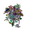
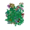
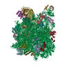
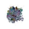
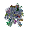
Journal: Science / Year: 2014 Title: Structure of the large ribosomal subunit from human mitochondria. Authors: Alan Brown / Alexey Amunts / Xiao-Chen Bai / Yoichiro Sugimoto / Patricia C Edwards / Garib Murshudov / Sjors H W Scheres / V Ramakrishnan / Abstract: Human mitochondrial ribosomes are highly divergent from all other known ribosomes and are specialized to exclusively translate membrane proteins. They are linked with hereditary mitochondrial ...Human mitochondrial ribosomes are highly divergent from all other known ribosomes and are specialized to exclusively translate membrane proteins. They are linked with hereditary mitochondrial diseases and are often the unintended targets of various clinically useful antibiotics. Using single-particle cryogenic electron microscopy, we have determined the structure of its large subunit to 3.4 angstrom resolution, revealing 48 proteins, 21 of which are specific to mitochondria. The structure unveils an adaptation of the exit tunnel for hydrophobic nascent peptides, extensive remodeling of the central protuberance, including recruitment of mitochondrial valine transfer RNA (tRNA(Val)) to play an integral structural role, and changes in the tRNA binding sites related to the unusual characteristics of mitochondrial tRNAs.
History
Deposition
Aug 26, 2014
-
Header (metadata) release
Sep 10, 2014
-
Map release
Oct 15, 2014
-
Update
Nov 19, 2014
-
Current status
Nov 19, 2014
Processing site: PDBe / Status: Released
-
Structure visualization
Movie
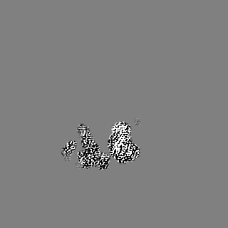
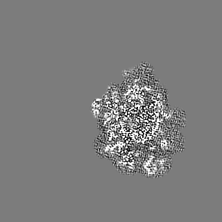
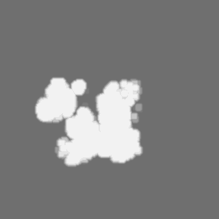
Surface view with section colored by density value
Organelle or cellular component: Large subunit of the human mitochondrial ribosome
-
Supramolecule #1000: Human mitochondrial ribosome
Supramolecule
Name: Human mitochondrial ribosome / type: sample / ID: 1000 / Oligomeric state: Multimer / Number unique components: 1
Molecular weight
Theoretical: 1.7 MDa
-
Supramolecule #1: Large subunit of the human mitochondrial ribosome
Supramolecule
Name: Large subunit of the human mitochondrial ribosome / type: organelle_or_cellular_component / ID: 1 / Name.synonym: 39S / Number of copies: 1 / Oligomeric state: Monomer / Recombinant expression: No
Source (natural)
Organism: Homo sapiens (human) / synonym: Human / Tissue: Kidney / Cell: HEK293 / Organelle: Mitochondria / Location in cell: Inner mitochondrial membrane
Molecular weight
Theoretical: 1.7 MDa
-
Experimental details
-
Structure determination
Method
cryo EM
Processing
single particle reconstruction
Aggregation state
particle
-
Sample preparation
Concentration
0.23 mg/mL
Buffer
pH: 7.45 Details: 20 mM Hepes-KOH pH 7.45, 100 mM KCl, 20 mM MgOAc, 2 mM DTT
Grid
Details: 30 s on glow-discharged holey carbon grids (Quantifoil R2/2), onto which a home-made continuous carbon film
Vitrification
Cryogen name: ETHANE / Chamber humidity: 100 % / Chamber temperature: 90 K / Instrument: FEI VITROBOT MARK II / Method: Blot 2.5 seconds before plunging
-
Electron microscopy
Microscope
FEI TITAN KRIOS
Temperature
Min: 80 K / Max: 90 K / Average: 85 K
Alignment procedure
Legacy - Astigmatism: Objective lens astigmatism was corrected at 59,000 times magnification
Date
Apr 12, 2014
Image recording
Category: CCD / Film or detector model: FEI FALCON II (4k x 4k) / Number real images: 1521 / Average electron dose: 25 e/Å2
Electron beam
Acceleration voltage: 300 kV / Electron source: FIELD EMISSION GUN
Data were processed to compensate for beam-induced movement.
CTF correction
Details: Each particle
Final reconstruction
Applied symmetry - Point group: C1 (asymmetric) / Resolution.type: BY AUTHOR / Resolution: 3.4 Å / Resolution method: OTHER / Software - Name: CTFFIND3, RELION Details: Final resolution was calculated using a soft mask over the large subunit Number images used: 107679
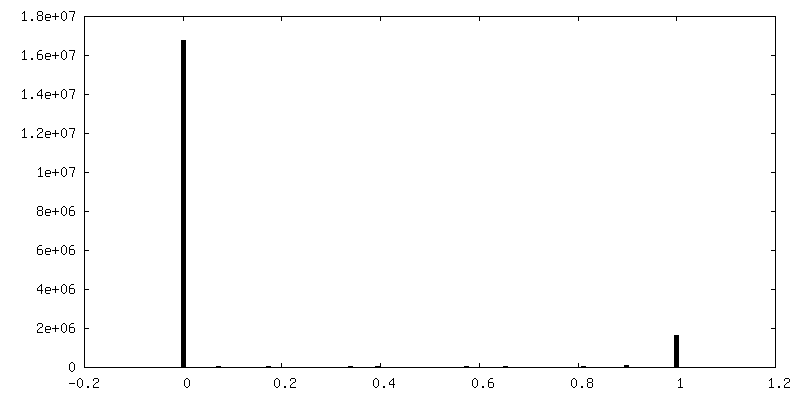
FSC plot (resolution estimation)
+
About Yorodumi
-
News
-
Feb 9, 2022. New format data for meta-information of EMDB entries
New format data for meta-information of EMDB entries
Version 3 of the EMDB header file is now the official format.
The previous official version 1.9 will be removed from the archive.
In the structure databanks used in Yorodumi, some data are registered as the other names, "COVID-19 virus" and "2019-nCoV". Here are the details of the virus and the list of structure data.
Jan 31, 2019. EMDB accession codes are about to change! (news from PDBe EMDB page)
EMDB accession codes are about to change! (news from PDBe EMDB page)
The allocation of 4 digits for EMDB accession codes will soon come to an end. Whilst these codes will remain in use, new EMDB accession codes will include an additional digit and will expand incrementally as the available range of codes is exhausted. The current 4-digit format prefixed with “EMD-” (i.e. EMD-XXXX) will advance to a 5-digit format (i.e. EMD-XXXXX), and so on. It is currently estimated that the 4-digit codes will be depleted around Spring 2019, at which point the 5-digit format will come into force.
The EM Navigator/Yorodumi systems omit the EMD- prefix.
Related info.:Q: What is EMD? / ID/Accession-code notation in Yorodumi/EM Navigator
Yorodumi is a browser for structure data from EMDB, PDB, SASBDB, etc.
This page is also the successor to EM Navigator detail page, and also detail information page/front-end page for Omokage search.
The word "yorodu" (or yorozu) is an old Japanese word meaning "ten thousand". "mi" (miru) is to see.
Related info.:EMDB / PDB / SASBDB / Comparison of 3 databanks / Yorodumi Search / Aug 31, 2016. New EM Navigator & Yorodumi / Yorodumi Papers / Jmol/JSmol / Function and homology information / Changes in new EM Navigator and Yorodumi
 Movie
Movie Controller

 Yorodumi
 Open data
 Basic information
 Map data
 Sample
 Keywords
 Function and homology information
 Homo sapiens (human)
 Authors
 Citation
 Structure visualization
 Downloads & links
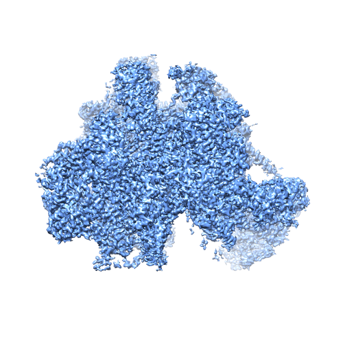 EMD_2762_map_image.png
 http://ftp.pdbj.org/pub/emdb/structures/EMD-2762
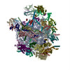


 Z (Sec.)
 Y (Row.)
 X (Col.)


 Sample components
 Processing
 Electron microscopy
 FIELD EMISSION GUN

