 Movie
Movie Controller


+ Open data

- Basic information
| Entry |  | ||||||||||||
|---|---|---|---|---|---|---|---|---|---|---|---|---|---|
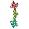
| Title | VWF tubule derived from dimeric D1-A1 | ||||||||||||
 Map data Map data | Single central bead, autosharpened in phenix, stitched into repeating unit of tubule. | ||||||||||||
 Sample Sample |
| ||||||||||||
 Keywords Keywords | VWF / tubule / blood clotting | ||||||||||||
| Function / homology |  Function and homology information Function and homology informationDefective VWF binding to collagen type I / Enhanced cleavage of VWF variant by ADAMTS13 / Defective VWF cleavage by ADAMTS13 variant / Defective F8 binding to von Willebrand factor / Enhanced binding of GP1BA variant to VWF multimer:collagen / Defective binding of VWF variant to GPIb:IX:V / Weibel-Palade body / hemostasis / platelet alpha granule / Platelet Adhesion to exposed collagen ...Defective VWF binding to collagen type I / Enhanced cleavage of VWF variant by ADAMTS13 / Defective VWF cleavage by ADAMTS13 variant / Defective F8 binding to von Willebrand factor / Enhanced binding of GP1BA variant to VWF multimer:collagen / Defective binding of VWF variant to GPIb:IX:V / Weibel-Palade body / hemostasis / platelet alpha granule / Platelet Adhesion to exposed collagen / GP1b-IX-V activation signalling / p130Cas linkage to MAPK signaling for integrins / Defective F8 cleavage by thrombin / Platelet Aggregation (Plug Formation) / cell-substrate adhesion / GRB2:SOS provides linkage to MAPK signaling for Integrins / positive regulation of intracellular signal transduction / immunoglobulin binding / Integrin cell surface interactions / collagen binding / Intrinsic Pathway of Fibrin Clot Formation / Integrin signaling / platelet alpha granule lumen / Signaling by high-kinase activity BRAF mutants / MAP2K and MAPK activation / platelet activation / response to wounding / : / integrin binding / extracellular matrix / blood coagulation / Signaling by RAF1 mutants / Signaling by moderate kinase activity BRAF mutants / Paradoxical activation of RAF signaling by kinase inactive BRAF / Signaling downstream of RAS mutants / Signaling by BRAF and RAF1 fusions / Platelet degranulation / protein-folding chaperone binding / protease binding / cell adhesion / endoplasmic reticulum / extracellular space / extracellular exosome / extracellular region / identical protein binding Similarity search - Function | ||||||||||||
| Biological species |  Homo sapiens (human) Homo sapiens (human) | ||||||||||||
| Method | helical reconstruction / cryo EM / Resolution: 3.2 Å | ||||||||||||
 Authors Authors | Anderson JR / Li J / Springer TA / Brown A | ||||||||||||
| Funding support |  United States, 3 items United States, 3 items
| ||||||||||||
 Citation Citation | Journal: Blood / Year: 2022 Title: Structures of VWF tubules before and after concatemerization reveal a mechanism of disulfide bond exchange. Authors: Jacob R Anderson / Jing Li / Timothy A Springer / Alan Brown / Abstract: von Willebrand factor (VWF) is an adhesive glycoprotein that circulates in the blood as disulfide-linked concatemers and functions in primary hemostasis. The loss of long VWF concatemers is ...von Willebrand factor (VWF) is an adhesive glycoprotein that circulates in the blood as disulfide-linked concatemers and functions in primary hemostasis. The loss of long VWF concatemers is associated with the excessive bleeding of type 2A von Willebrand disease (VWD). Formation of the disulfide bonds that concatemerize VWF requires VWF to self-associate into helical tubules, yet how the helical tubules template intermolecular disulfide bonds is not known. Here, we report electron cryomicroscopy (cryo-EM) structures of VWF tubules before and after intermolecular disulfide bond formation. The structures provide evidence that VWF tubulates through a charge-neutralization mechanism and that the A1 domain enhances tubule length by crosslinking successive helical turns. In addition, the structures reveal disulfide states before and after disulfide bond-mediated concatemerization. The structures and proposed assembly mechanism provide a foundation to rationalize VWD-causing mutations. | ||||||||||||
| History |
|
- Structure visualization
| Supplemental images |
|---|
- Downloads & links
-EMDB archive
| Map data | emd_27157.map.gz | 83.5 MB | EMDB map data format | |
|---|---|---|---|---|
| Header (meta data) | emd-27157-v30.xmlemd-27157.xml | 19.2 KB 19.2 KB | Display Display | EMDB header |
| FSC (resolution estimation) | emd_27157_fsc.xmlemd_27157_fsc_2.xml | 13.6 KB 13.6 KB | Display Display | FSC data file |
| Images |  emd_27157.png emd_27157.png | 82.1 KB | ||
| Masks | emd_27157_msk_1.map | 216 MB | Mask map | |
| Filedesc metadata | emd-27157.cif.gz | 7.1 KB | ||
| Others | emd_27157_half_map_1.map.gzemd_27157_half_map_2.map.gz | 171 MB 171 MB | ||
| Archive directory |  http://ftp.pdbj.org/pub/emdb/structures/EMD-27157ftp://ftp.pdbj.org/pub/emdb/structures/EMD-27157 http://ftp.pdbj.org/pub/emdb/structures/EMD-27157ftp://ftp.pdbj.org/pub/emdb/structures/EMD-27157 | HTTPS FTP |
-Related structure data
| Related structure data |  8d3dMC  8d3cC C: citing same article ( M: atomic model generated by this map |
|---|---|
| Similar structure data |


-Links
| EMDB pages | EMDB (EBI/PDBe) / EMDataResource |
|---|---|
| Related items in Molecule of the Month |


-Map
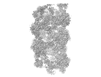
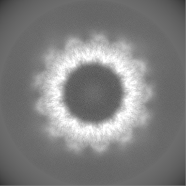
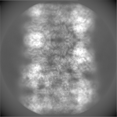
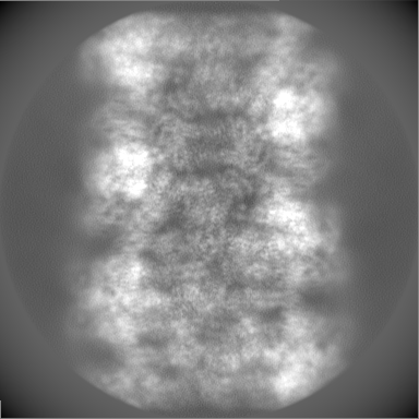
| File | Download / File: emd_27157.map.gz / Format: CCP4 / Size: 216 MB / Type: IMAGE STORED AS FLOATING POINT NUMBER (4 BYTES) | ||||||||||||||||||||||||||||||||||||
|---|---|---|---|---|---|---|---|---|---|---|---|---|---|---|---|---|---|---|---|---|---|---|---|---|---|---|---|---|---|---|---|---|---|---|---|---|---|
| Annotation | Single central bead, autosharpened in phenix, stitched into repeating unit of tubule. | ||||||||||||||||||||||||||||||||||||
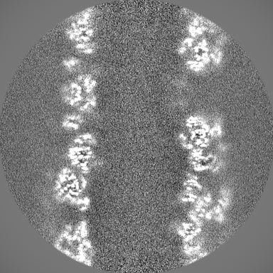
| Projections & slices | Image control
Images are generated by Spider. | ||||||||||||||||||||||||||||||||||||
| Voxel size | X=Y=Z: 1.06 Å | ||||||||||||||||||||||||||||||||||||
| Density |
| ||||||||||||||||||||||||||||||||||||
| Symmetry | Space group: 1 | ||||||||||||||||||||||||||||||||||||
| Details | EMDB XML:
|
 Z (Sec.)
 Y (Row.)
 X (Col.)


-Supplemental data
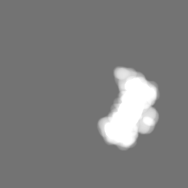
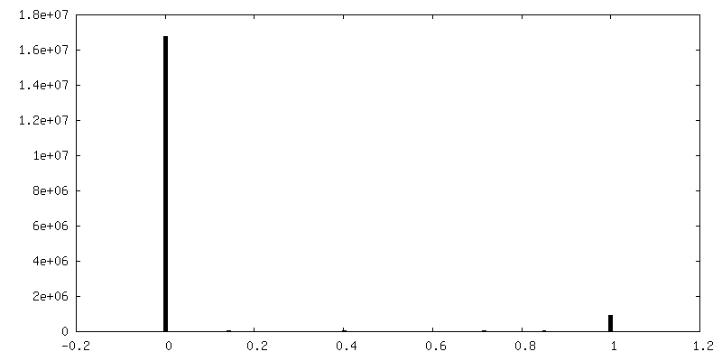
-Mask #1
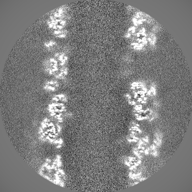
| File | emd_27157_msk_1.map | ||||||||||||
|---|---|---|---|---|---|---|---|---|---|---|---|---|---|
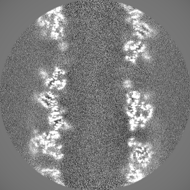
| Projections & Slices |
| ||||||||||||
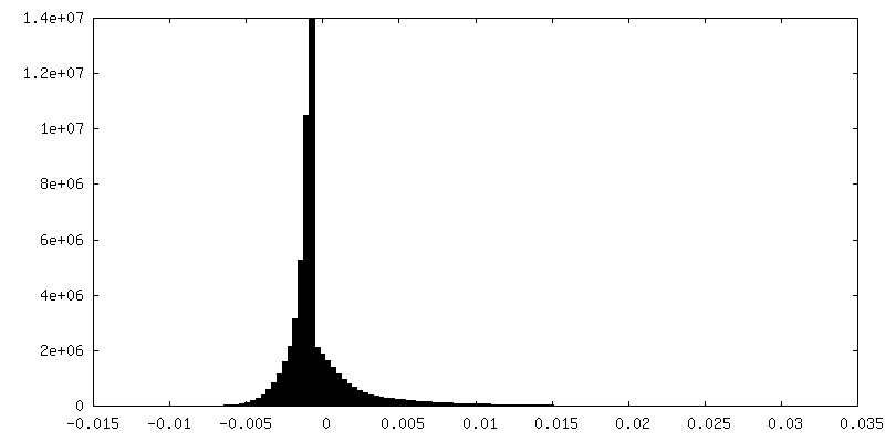
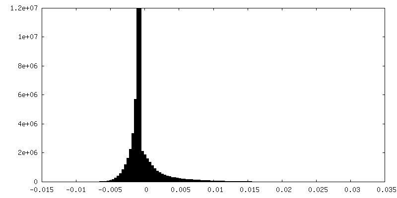
| Density Histograms |


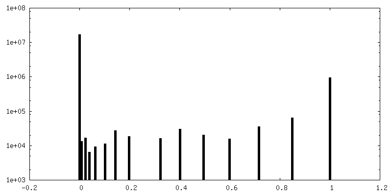
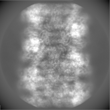
-Half map: 1 of 2 Half-Map of D1A1 dimer composed VWF tubule.
| File | emd_27157_half_map_1.map | ||||||||||||
|---|---|---|---|---|---|---|---|---|---|---|---|---|---|
| Annotation | 1 of 2 Half-Map of D1A1 dimer composed VWF tubule. | ||||||||||||
| Projections & Slices |
| ||||||||||||
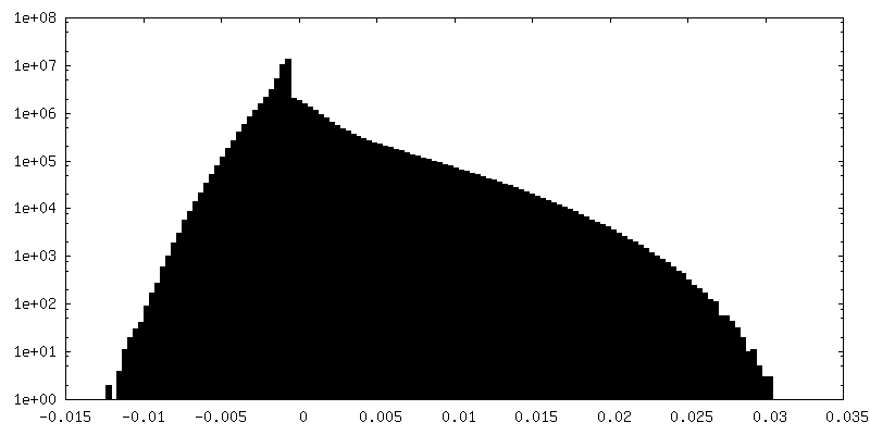
| Density Histograms |
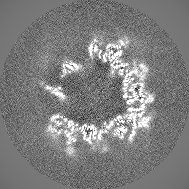
-Half map: 2 of 2 Half-Map of D1A1 dimer composed VWF tubule.
| File | emd_27157_half_map_2.map | ||||||||||||
|---|---|---|---|---|---|---|---|---|---|---|---|---|---|
| Annotation | 2 of 2 Half-Map of D1A1 dimer composed VWF tubule. | ||||||||||||
| Projections & Slices |
| ||||||||||||
| Density Histograms |


- Sample components
-Entire : Von Willebrand Factor tubule derived from dimeric D1-A1
| Entire | Name: Von Willebrand Factor tubule derived from dimeric D1-A1 |
|---|---|
| Components |
|
-Supramolecule #1: Von Willebrand Factor tubule derived from dimeric D1-A1
| Supramolecule | Name: Von Willebrand Factor tubule derived from dimeric D1-A1 type: complex / ID: 1 / Parent: 0 / Macromolecule list: #1 |
|---|---|
| Source (natural) | Organism: Homo sapiens (human) |
| Molecular weight | Theoretical: 12.4 kDa/nm |
-Macromolecule #1: von Willebrand factor
| Macromolecule | Name: von Willebrand factor / type: protein_or_peptide / ID: 1 / Number of copies: 16 / Enantiomer: LEVO |
|---|---|
| Source (natural) | Organism: Homo sapiens (human) |
| Molecular weight | Theoretical: 162.300984 KDa |
| Recombinant expression | Organism: Homo sapiens (human) |
| Sequence | String: MIPARFAGVL LALALILPGT LCAEGTRGRS STARCSLFGS DFVNTFDGSM YSFAGYCSYL LAGGCQKRSF SIIGDFQNGK RVSLSVYLG EFFDIHLFVN GTVTQGDQRV SMPYASKGLY LETEAGYYKL SGEAYGFVAR IDGSGNFQVL LSDRYFNKTC G LCGNFNIF ...String: MIPARFAGVL LALALILPGT LCAEGTRGRS STARCSLFGS DFVNTFDGSM YSFAGYCSYL LAGGCQKRSF SIIGDFQNGK RVSLSVYLG EFFDIHLFVN GTVTQGDQRV SMPYASKGLY LETEAGYYKL SGEAYGFVAR IDGSGNFQVL LSDRYFNKTC G LCGNFNIF AEDDFMTQEG TLTSDPYDFA NSWALSSGEQ WCERASPPSS SCNISSGEMQ KGLWEQCQLL KSTSVFARCH PL VDPEPFV ALCEKTLCEC AGGLECACPA LLEYARTCAQ EGMVLYGWTD HSACSPVCPA GMEYRQCVSP CARTCQSLHI NEM CQERCV DGCSCPEGQL LDEGLCVEST ECPCVHSGKR YPPGTSLSRD CNTCICRNSQ WICSNEECPG ECLVTGQSHF KSFD NRYFT FSGICQYLLA RDCQDHSFSI VIETVQCADD RDAVCTRSVT VRLPGLHNSL VKLKHGAGVA MDGQDVQLPL LKGDL RIQH TVTASVRLSY GEDLQMDWDG RGRLLVKLSP VYAGKTCGLC GNYNGNQGDD FLTPSGLAEP RVEDFGNAWK LHGDCQ DLQ KQHSDPCALN PRMTRFSEEA CAVLTSPTFE ACHRAVSPLP YLRNCRYDVC SCSDGRECLC GALASYAAAC AGRGVRV AW REPGRCELNC PKGQVYLQCG TPCNLTCRSL SYPDEECNEA CLEGCFCPPG LYMDERGDCV PKAQCPCYYD GEIFQPED I FSDHHTMCYC EDGFMHCTMS GVPGSLLPDA VLSSPLSHAS ASLSCRPPMV KLVCPADNLR AEGLECAKTC QNYDLECMS MGCVSGCLCP PGMVRHENRC VALERCPCFH QGKEYAPGET VKIGCNTCVC RDRKWNCTDH VCDATCSTIG MAHYLTFDGL KYLFPGECQ YVLVQDYCGS NPGTFRILVG NKGCSHPSVK CKKRVTILVE GGEIELFDGE VNVKRPMKDE THFEVVESGR Y IILLLGKA LSVVWDRHLS ISVVLKQTYQ EKVCGLCGNF DGIQNNDLTS SNLQVEEDPV DFGNSWKVSS QCADTRKVPL DS SPATCHN NIMKQTMVDS SCRILTSDVF QDCNKLVDPE PYLDVCIYDT CSCESIGDCA CFCDTIAAYA HVCAQHGKVV TWR TATLCP QSCEERNLRE NGYECEWRYN SCAPACQVTC QHPEPLACPV QCVEGCHAHC PPGKILDELL QTCVDPEDCP VCEV AGRRF ASGKKVTLNP SDPEHCQICH CDVVNLTCEA CQEPGGLVVP PTDAPVSPTT LYVEDISEPP LHDFYCSRLL DLVFL LDGS SRLSEAEFEV LKAFVVDMME RLRISQKWVR VAVVEYHDGS HAYIGLKDRK RPSELRRIAS QVKYAGSQVA STSEVL KYT LFQIFSKIDR PEASRIALLL MASQEPQRMS RNFVRYVQGL KKKKVIVIPV GIGPHANLKQ IRLIEKQAPE NKAFVLS SV DELEQQRDEI VSYLCDLAPE AHHHHHH UniProtKB: von Willebrand factor |
-Macromolecule #2: CALCIUM ION
| Macromolecule | Name: CALCIUM ION / type: ligand / ID: 2 / Number of copies: 48 / Formula: CA |
|---|---|
| Molecular weight | Theoretical: 40.078 Da |
-Macromolecule #3: 2-acetamido-2-deoxy-beta-D-glucopyranose
| Macromolecule | Name: 2-acetamido-2-deoxy-beta-D-glucopyranose / type: ligand / ID: 3 / Number of copies: 80 / Formula: NAG |
|---|---|
| Molecular weight | Theoretical: 221.208 Da |
| Chemical component information |  ChemComp-NAG: |
-Experimental details
-Structure determination
| Method | cryo EM |
|---|---|
 Processing Processing | helical reconstruction |
| Aggregation state | helical array |
-Sample preparation
| Concentration | 0.6 mg/mL | ||||||||||||
|---|---|---|---|---|---|---|---|---|---|---|---|---|---|
| Buffer | pH: 5.2 Component:
Details: 100 mM Sodium Cacodylate at pH 5.2, 10 mM CaCl2, and 100 mM NaCl | ||||||||||||
| Grid | Model: Quantifoil R2/1 / Material: GOLD / Mesh: 300 / Support film - Material: CARBON / Support film - topology: CONTINUOUS / Support film - Film thickness: 2 / Pretreatment - Type: GLOW DISCHARGE | ||||||||||||
| Vitrification | Cryogen name: ETHANE / Chamber humidity: 100 % / Chamber temperature: 277 K / Instrument: FEI VITROBOT MARK IV |
- Electron microscopy
| Microscope | FEI TITAN KRIOS |
|---|---|
| Image recording | Film or detector model: GATAN K3 (6k x 4k) / Average electron dose: 55.0 e/Å2 |
| Electron beam | Acceleration voltage: 300 kV / Electron source:  FIELD EMISSION GUN FIELD EMISSION GUN |
| Electron optics | Illumination mode: OTHER / Imaging mode: OTHER / Nominal defocus max: 2.0 µm / Nominal defocus min: 1.0 µm |
| Sample stage | Specimen holder model: FEI TITAN KRIOS AUTOGRID HOLDER |
| Experimental equipment |  Model: Titan Krios / Image courtesy: FEI Company |
-Image processing
| Final reconstruction | Applied symmetry - Helical parameters - Δz: 26.8 Å Applied symmetry - Helical parameters - Δ&Phi: 83.3 ° Applied symmetry - Helical parameters - Axial symmetry: C1 (asymmetric) Resolution.type: BY AUTHOR / Resolution: 3.2 Å / Resolution method: FSC 0.143 CUT-OFF / Software - Name: RELION (ver. 3.1.3) Details: Resolution of tubule is 3.3. Resolution of masked central bead 3.2. Number images used: 250388 |
|---|---|
| Startup model | Type of model: PDB ENTRY PDB model - PDB ID: |
| Final angle assignment | Type: NOT APPLICABLE / Software - Name: RELION (ver. 3.1.3) |
| FSC plot (resolution estimation) |  |
-Atomic model buiding 1
| Refinement | Space: REAL |
|---|---|
| Output model | PDB-8d3d: |
