 Movie
Movie Controller

[English] 日本語
 Yorodumi
- EMDB-26977: CryoEM structure of human S-OPA1 assembled on lipid membrane in m... -
+ Open data

- Basic information
| Entry |  | |||||||||
|---|---|---|---|---|---|---|---|---|---|---|
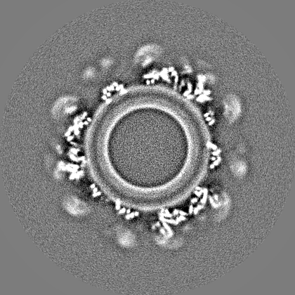
| Title | CryoEM structure of human S-OPA1 assembled on lipid membrane in membrane-adjacent state | |||||||||
 Map data Map data | Refine3D map | |||||||||
 Sample Sample |
| |||||||||
 Keywords Keywords | GTPase / polymer / filament / membrane / remodeling / fusion / mitochondria / MEMBRANE PROTEIN | |||||||||
| Function / homology |  Function and homology information Function and homology informationRegulation of Apoptosis / mitochondrial inner membrane fusion / membrane tubulation / GTPase-dependent fusogenic activity / membrane bending activity / inner mitochondrial membrane organization / dynamin GTPase / cristae formation / phosphatidic acid binding / mitochondrial genome maintenance ...Regulation of Apoptosis / mitochondrial inner membrane fusion / membrane tubulation / GTPase-dependent fusogenic activity / membrane bending activity / inner mitochondrial membrane organization / dynamin GTPase / cristae formation / phosphatidic acid binding / mitochondrial genome maintenance / cardiolipin binding / mitochondrial fission / GTP metabolic process / mitochondrial fusion / axonal transport of mitochondrion / negative regulation of release of cytochrome c from mitochondria / positive regulation of interleukin-17 production / mitochondrial crista / negative regulation of endoplasmic reticulum stress-induced intrinsic apoptotic signaling pathway / positive regulation of T-helper 17 cell lineage commitment / axon cytoplasm / Mitochondrial protein degradation / visual perception / mitochondrion organization / neural tube closure / mitochondrial membrane / protein complex oligomerization / mitochondrial intermembrane space / cellular senescence / microtubule binding / mitochondrial outer membrane / microtubule / mitochondrial inner membrane / GTPase activity / dendrite / GTP binding / negative regulation of apoptotic process / apoptotic process / magnesium ion binding / mitochondrion / nucleoplasm / membrane / cytosol / cytoplasm Similarity search - Function | |||||||||
| Biological species |  Homo sapiens (human) Homo sapiens (human) | |||||||||
| Method | helical reconstruction / cryo EM / Resolution: 4.8 Å | |||||||||
 Authors Authors | Du Pont KE / Aydin H | |||||||||
| Funding support |  United States, 1 items United States, 1 items
| |||||||||
 Citation Citation | Journal: Nature / Year: 2023 Title: Structural mechanism of mitochondrial membrane remodelling by human OPA1. Authors: Alexander von der Malsburg / Gracie M Sapp / Kelly E Zuccaro / Alexander von Appen / Frank R Moss / Raghav Kalia / Jeremy A Bennett / Luciano A Abriata / Matteo Dal Peraro / Martin van der ...Authors: Alexander von der Malsburg / Gracie M Sapp / Kelly E Zuccaro / Alexander von Appen / Frank R Moss / Raghav Kalia / Jeremy A Bennett / Luciano A Abriata / Matteo Dal Peraro / Martin van der Laan / Adam Frost / Halil Aydin /   Abstract: Distinct morphologies of the mitochondrial network support divergent metabolic and regulatory processes that determine cell function and fate. The mechanochemical GTPase optic atrophy 1 (OPA1) ...Distinct morphologies of the mitochondrial network support divergent metabolic and regulatory processes that determine cell function and fate. The mechanochemical GTPase optic atrophy 1 (OPA1) influences the architecture of cristae and catalyses the fusion of the mitochondrial inner membrane. Despite its fundamental importance, the molecular mechanisms by which OPA1 modulates mitochondrial morphology are unclear. Here, using a combination of cellular and structural analyses, we illuminate the molecular mechanisms that are key to OPA1-dependent membrane remodelling and fusion. Human OPA1 embeds itself into cardiolipin-containing membranes through a lipid-binding paddle domain. A conserved loop within the paddle domain inserts deeply into the bilayer, further stabilizing the interactions with cardiolipin-enriched membranes. OPA1 dimerization through the paddle domain promotes the helical assembly of a flexible OPA1 lattice on the membrane, which drives mitochondrial fusion in cells. Moreover, the membrane-bending OPA1 oligomer undergoes conformational changes that pull the membrane-inserting loop out of the outer leaflet and contribute to the mechanics of membrane remodelling. Our findings provide a structural framework for understanding how human OPA1 shapes mitochondrial morphology and show us how human disease mutations compromise OPA1 functions. | |||||||||
| History |
|
- Structure visualization
| Supplemental images |
|---|

- Downloads & links
-EMDB archive
| Map data | emd_26977.map.gz | 225.9 MB | EMDB map data format | |
|---|---|---|---|---|
| Header (meta data) | emd-26977-v30.xmlemd-26977.xml | 18 KB 18 KB | Display Display | EMDB header |
| FSC (resolution estimation) | emd_26977_fsc.xml | 14.9 KB | Display | FSC data file |
| Images | 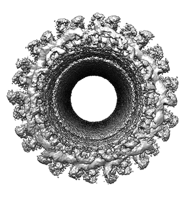 emd_26977.png emd_26977.png | 168.4 KB | ||
| Masks | emd_26977_msk_1.map | 282.6 MB | Mask map | |
| Others | emd_26977_additional_1.map.gzemd_26977_half_map_1.map.gzemd_26977_half_map_2.map.gz | 263.6 MB 226 MB 226 MB | ||
| Archive directory |  http://ftp.pdbj.org/pub/emdb/structures/EMD-26977ftp://ftp.pdbj.org/pub/emdb/structures/EMD-26977 http://ftp.pdbj.org/pub/emdb/structures/EMD-26977ftp://ftp.pdbj.org/pub/emdb/structures/EMD-26977 | HTTPS FTP |
-Validation report
| Summary document | emd_26977_validation.pdf.gz | 1 MB | Display | EMDB validaton report |
|---|---|---|---|---|
| Full document | emd_26977_full_validation.pdf.gz | 1 MB | Display | |
| Data in XML | emd_26977_validation.xml.gz | 21.9 KB | Display | |
| Data in CIF | emd_26977_validation.cif.gz | 28.8 KB | Display | |
| Arichive directory | https://ftp.pdbj.org/pub/emdb/validation_reports/EMD-26977ftp://ftp.pdbj.org/pub/emdb/validation_reports/EMD-26977 | HTTPS FTP |
-Related structure data
| Related structure data |  8ct1MC  8ct9C M: atomic model generated by this map C: citing same article ( |
|---|---|
| Similar structure data |

-Links
| EMDB pages | EMDB (EBI/PDBe) / EMDataResource |
|---|
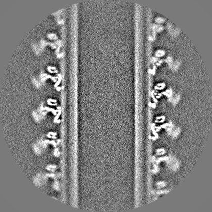
-Map
| File | Download / File: emd_26977.map.gz / Format: CCP4 / Size: 282.6 MB / Type: IMAGE STORED AS FLOATING POINT NUMBER (4 BYTES) | ||||||||||||||||||||||||||||||||||||
|---|---|---|---|---|---|---|---|---|---|---|---|---|---|---|---|---|---|---|---|---|---|---|---|---|---|---|---|---|---|---|---|---|---|---|---|---|---|
| Annotation | Refine3D map | ||||||||||||||||||||||||||||||||||||
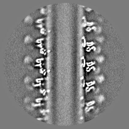
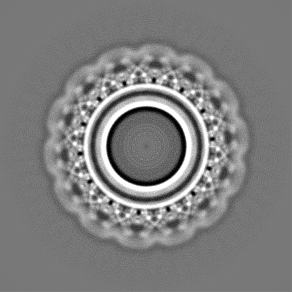
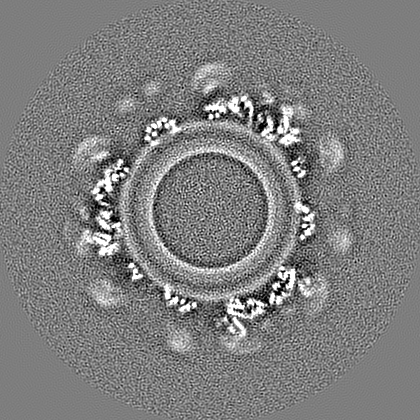
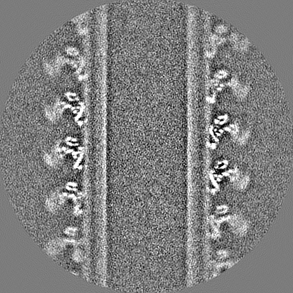
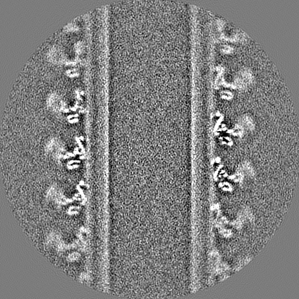
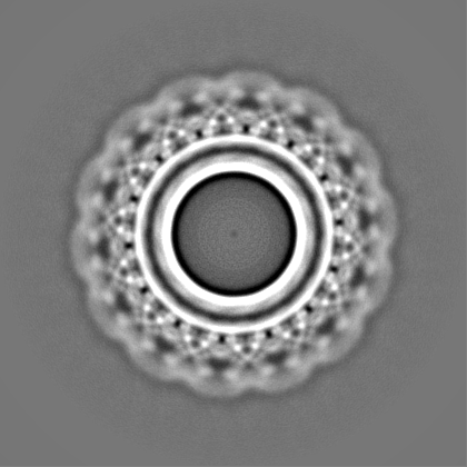
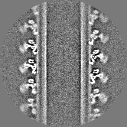
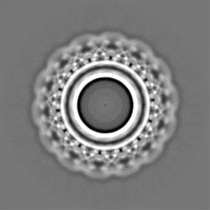
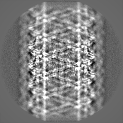
| Projections & slices | Image control
Images are generated by Spider. | ||||||||||||||||||||||||||||||||||||
| Voxel size | X=Y=Z: 1.666 Å | ||||||||||||||||||||||||||||||||||||
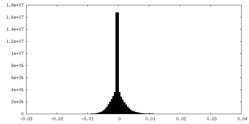
| Density |
| ||||||||||||||||||||||||||||||||||||
| Symmetry | Space group: 1 | ||||||||||||||||||||||||||||||||||||
| Details | EMDB XML:
|
 Z (Sec.)
 Y (Row.)
 X (Col.)


-Supplemental data
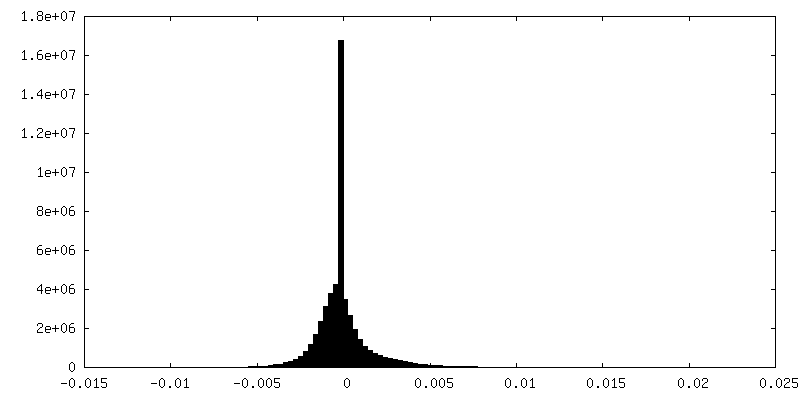
-Mask #1
| File | emd_26977_msk_1.map | ||||||||||||
|---|---|---|---|---|---|---|---|---|---|---|---|---|---|
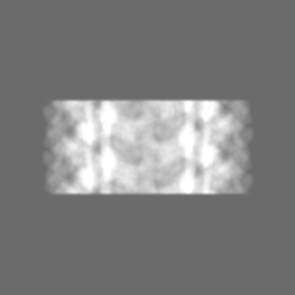
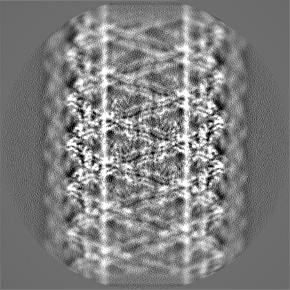
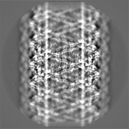
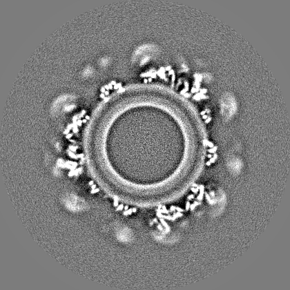
| Projections & Slices |
| ||||||||||||
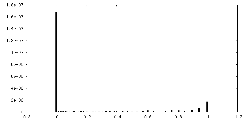
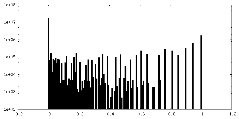
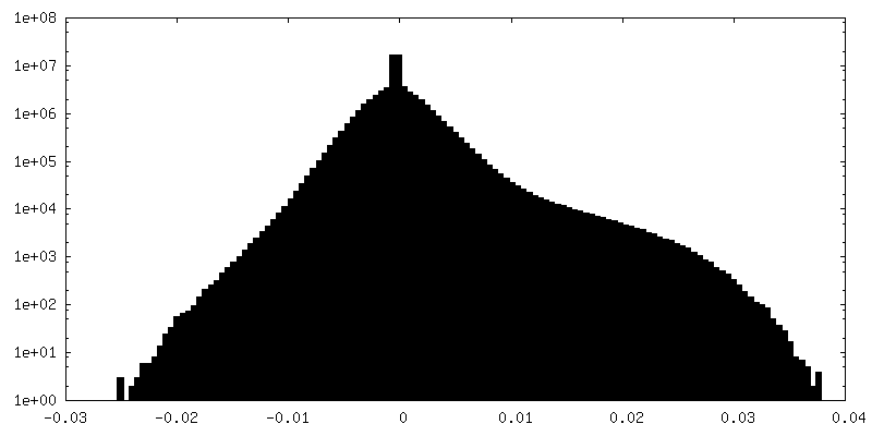
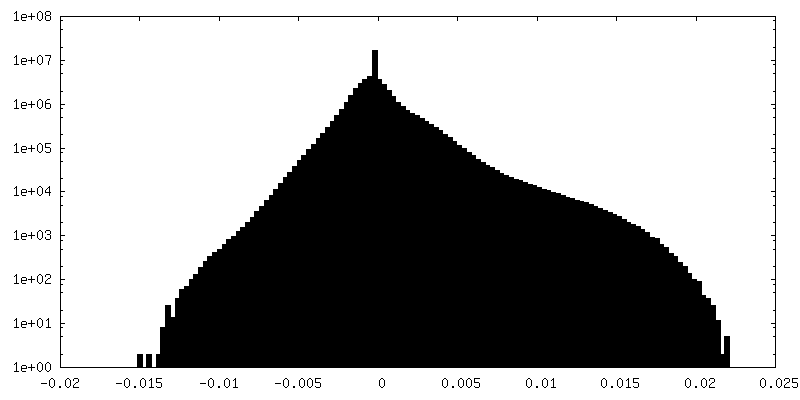
| Density Histograms |


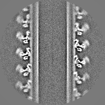
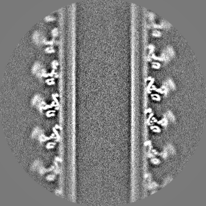
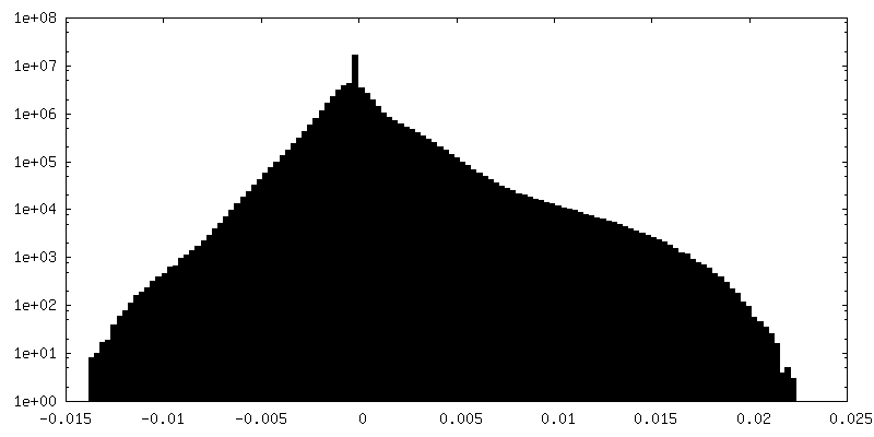
-Additional map: Postprocess map
| File | emd_26977_additional_1.map | ||||||||||||
|---|---|---|---|---|---|---|---|---|---|---|---|---|---|
| Annotation | Postprocess map | ||||||||||||
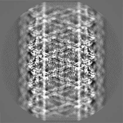
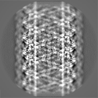
| Projections & Slices |
| ||||||||||||
| Density Histograms |
-Half map: Refine3D half map 1
| File | emd_26977_half_map_1.map | ||||||||||||
|---|---|---|---|---|---|---|---|---|---|---|---|---|---|
| Annotation | Refine3D half map 1 | ||||||||||||
| Projections & Slices |
| ||||||||||||
| Density Histograms |
-Half map: Refine3D half map 2
| File | emd_26977_half_map_2.map | ||||||||||||
|---|---|---|---|---|---|---|---|---|---|---|---|---|---|
| Annotation | Refine3D half map 2 | ||||||||||||
| Projections & Slices |
| ||||||||||||
| Density Histograms |

- Sample components
-Entire : OPA1
| Entire | Name: OPA1 |
|---|---|
| Components |
|
-Supramolecule #1: OPA1
| Supramolecule | Name: OPA1 / type: complex / ID: 1 / Parent: 0 / Macromolecule list: all Details: Uniprot ID: O60313 - HUMAN OPA1, Dynamin-like 120 kDa protein, mitochondrial |
|---|---|
| Source (natural) | Organism: Homo sapiens (human) |
| Molecular weight | Theoretical: 820 KDa |
-Macromolecule #1: Dynamin-like 120 kDa protein, mitochondrial
| Macromolecule | Name: Dynamin-like 120 kDa protein, mitochondrial / type: protein_or_peptide / ID: 1 / Number of copies: 34 / Enantiomer: LEVO / EC number: dynamin GTPase |
|---|---|
| Source (natural) | Organism: Homo sapiens (human) |
| Molecular weight | Theoretical: 111.804789 KDa |
| Recombinant expression | Organism:  |
| Sequence | String: MWRLRRAAVA CEVCQSLVKH SSGIKGSLPL QKLHLVSRSI YHSHHPTLKL QRPQLRTSFQ QFSSLTNLPL RKLKFSPIKY GYQPRRNFW PARLATRLLK LRYLILGSAV GGGYTAKKTF DQWKDMIPDL SEYKWIVPDI VWEIDEYIDF EKIRKALPSS E DLVKLAPD ...String: MWRLRRAAVA CEVCQSLVKH SSGIKGSLPL QKLHLVSRSI YHSHHPTLKL QRPQLRTSFQ QFSSLTNLPL RKLKFSPIKY GYQPRRNFW PARLATRLLK LRYLILGSAV GGGYTAKKTF DQWKDMIPDL SEYKWIVPDI VWEIDEYIDF EKIRKALPSS E DLVKLAPD FDKIVESLSL LKDFFTSGSP EETAFRATDR GSESDKHFRK VSDKEKIDQL QEELLHTQLK YQRILERLEK EN KELRKLV LQKDDKGIHH RKLKKSLIDM YSEVLDVLSD YDASYNTQDH LPRVVVVGDQ SAGKTSVLEM IAQARIFPRG SGE MMTRSP VKVTLSEGPH HVALFKDSSR EFDLTKEEDL AALRHEIELR MRKNVKEGCT VSPETISLNV KGPGLQRMVL VDLP GVINT VTSGMAPDTK ETIFSISKAY MQNPNAIILC IQDGSVDAER SIVTDLVSQM DPHGRRTIFV LTKVDLAEKN VASPS RIQQ IIEGKLFPMK ALGYFAVVTG KGNSSESIEA IREYEEEFFQ NSKLLKTSML KAHQVTTRNL SLAVSDCFWK MVRESV EQQ ADSFKATRFN LETEWKNNYP RLRELDRNEL FEKAKNEILD EVISLSQVTP KHWEEILQQS LWERVSTHVI ENIYLPA AQ TMNSGTFNTT VDIKLKQWTD KQLPNKAVEV AWETLQEEFS RFMTEPKGKE HDDIFDKLKE AVKEESIKRH KWNDFAED S LRVIQHNALE DRSISDKQQW DAAIYFMEEA LQARLKDTEN AIENMVGPDW KKRWLYWKNR TQEQCVHNET KNELEKMLK CNEEHPAYLA SDEITTVRKN LESRGVEVDP SLIKDTWHQV YRRHFLKTAL NHCNLCRRGF YYYQRHFVDS ELECNDVVLF WRIQRMLAI TANTLRQQLT NTEVRRLEKN VKEVLEDFAE DGEKKIKLLT GKRVQLAEDL KKVREIQEKL DAFIEALHQE K UniProtKB: Dynamin-like GTPase OPA1, mitochondrial |
-Experimental details
-Structure determination
| Method | cryo EM |
|---|---|
 Processing Processing | helical reconstruction |
| Aggregation state | filament |
-Sample preparation
| Concentration | 1.0 mg/mL |
|---|---|
| Buffer | pH: 7.5 |
| Grid | Model: Quantifoil R1.2/1.3 / Material: COPPER / Mesh: 200 |
| Vitrification | Cryogen name: ETHANE |
- Electron microscopy
| Microscope | FEI TITAN KRIOS |
|---|---|
| Specialist optics | Energy filter - Name: GIF Bioquantum / Energy filter - Slit width: 20 eV |
| Image recording | Film or detector model: GATAN K3 (6k x 4k) / Average exposure time: 2.0 sec. / Average electron dose: 82.0 e/Å2 |
| Electron beam | Acceleration voltage: 300 kV / Electron source:  FIELD EMISSION GUN FIELD EMISSION GUN |
| Electron optics | Illumination mode: FLOOD BEAM / Imaging mode: BRIGHT FIELD / Nominal defocus max: 1.2 µm / Nominal defocus min: 0.5 µm |
| Experimental equipment |  Model: Titan Krios / Image courtesy: FEI Company |
-Image processing
| Final reconstruction | Applied symmetry - Helical parameters - Δz: 7.73 Å Applied symmetry - Helical parameters - Δ&Phi: 128.634 ° Applied symmetry - Helical parameters - Axial symmetry: C1 (asymmetric) Resolution.type: BY AUTHOR / Resolution: 4.8 Å / Resolution method: FSC 0.143 CUT-OFF / Software - Name: RELION (ver. 3.1) / Number images used: 139018 |
|---|---|
| Startup model | Type of model: OTHER / Details: Featureless cylinder |
| Final angle assignment | Type: NOT APPLICABLE |
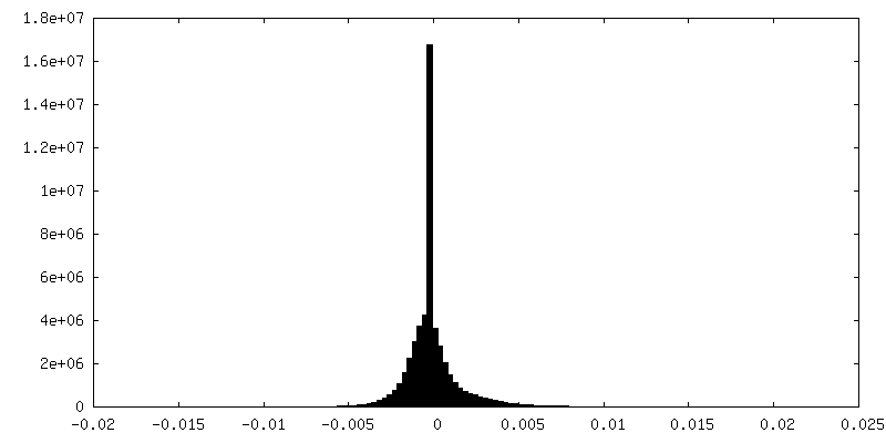
| FSC plot (resolution estimation) | 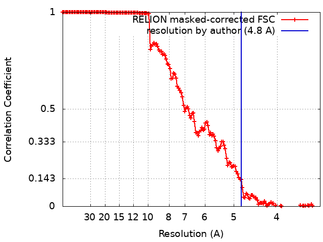 |
