 Movie
Movie Controller


+ Open data

- Basic information
| Entry | 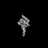 | |||||||||
|---|---|---|---|---|---|---|---|---|---|---|
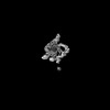
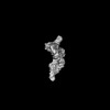
| Title | E.coli RNaseP Holoenzyme with Mg2+ | |||||||||
 Map data Map data | ||||||||||
 Sample Sample |
| |||||||||
 Keywords Keywords | ribozyme / protein-RNA complex / divalent ion / RNA / HYDROLASE-RNA complex | |||||||||
| Function / homology |  Function and homology information Function and homology information3'-tRNA processing endoribonuclease activity / ribonuclease P complex / ribonuclease P / ribonuclease P activity / tRNA 5'-leader removal / tRNA binding Similarity search - Function | |||||||||
| Biological species |  | |||||||||
| Method | single particle reconstruction / cryo EM / Resolution: 3.4 Å | |||||||||
 Authors Authors | Huang W / Taylor DJ | |||||||||
| Funding support |  United States, 1 items United States, 1 items
| |||||||||
 Citation Citation | Journal: Nat Commun / Year: 2022 Title: Structural and mechanistic basis for recognition of alternative tRNA precursor substrates by bacterial ribonuclease P. Authors: Jiaqiang Zhu / Wei Huang / Jing Zhao / Loc Huynh / Derek J Taylor / Michael E Harris / Abstract: Binding of precursor tRNAs (ptRNAs) by bacterial ribonuclease P (RNase P) involves an encounter complex (ES) that isomerizes to a catalytic conformation (ES*). However, the structures of ...Binding of precursor tRNAs (ptRNAs) by bacterial ribonuclease P (RNase P) involves an encounter complex (ES) that isomerizes to a catalytic conformation (ES*). However, the structures of intermediates and the conformational changes that occur during binding are poorly understood. Here, we show that pairing between the 5' leader and 3'RCCA extending the acceptor stem of ptRNA inhibits ES* formation. Cryo-electron microscopy single particle analysis reveals a dynamic enzyme that becomes ordered upon formation of ES* in which extended acceptor stem pairing is unwound. Comparisons of structures with alternative ptRNAs reveals that once unwinding is completed RNase P primarily uses stacking interactions and shape complementarity to accommodate alternative sequences at its cleavage site. Our study reveals active site interactions and conformational changes that drive molecular recognition by RNase P and lays the foundation for understanding how binding interactions are linked to helix unwinding and catalysis. | |||||||||
| History |
|
- Structure visualization
| Supplemental images |
|---|

- Downloads & links
-EMDB archive
| Map data | emd_26636.map.gz | 89.4 MB | EMDB map data format | |
|---|---|---|---|---|
| Header (meta data) | emd-26636-v30.xmlemd-26636.xml | 11.2 KB 11.2 KB | Display Display | EMDB header |
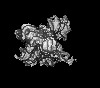
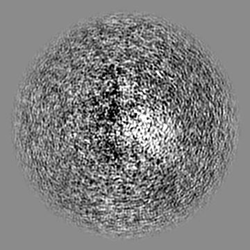
| Images | 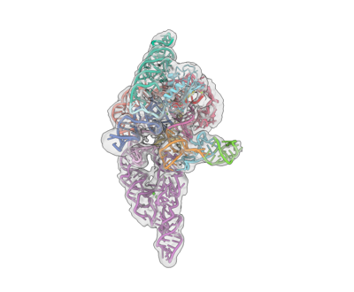 emd_26636.png emd_26636.png | 53.7 KB | ||
| Filedesc metadata | emd-26636.cif.gz | 5.4 KB | ||
| Archive directory |  http://ftp.pdbj.org/pub/emdb/structures/EMD-26636ftp://ftp.pdbj.org/pub/emdb/structures/EMD-26636 http://ftp.pdbj.org/pub/emdb/structures/EMD-26636ftp://ftp.pdbj.org/pub/emdb/structures/EMD-26636 | HTTPS FTP |
-Validation report
| Summary document | emd_26636_validation.pdf.gz | 411.3 KB | Display | EMDB validaton report |
|---|---|---|---|---|
| Full document | emd_26636_full_validation.pdf.gz | 410.8 KB | Display | |
| Data in XML | emd_26636_validation.xml.gz | 6.8 KB | Display | |
| Data in CIF | emd_26636_validation.cif.gz | 7.8 KB | Display | |
| Arichive directory | https://ftp.pdbj.org/pub/emdb/validation_reports/EMD-26636ftp://ftp.pdbj.org/pub/emdb/validation_reports/EMD-26636 | HTTPS FTP |
-Related structure data
| Related structure data | 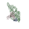 7uo0MC  7uo1C 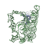 7uo2C 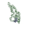 7uo5C M: atomic model generated by this map C: citing same article ( |
|---|---|
| Similar structure data |
-Links
| EMDB pages | EMDB (EBI/PDBe) / EMDataResource |
|---|---|
| Related items in Molecule of the Month |

-Map
| File | Download / File: emd_26636.map.gz / Format: CCP4 / Size: 178 MB / Type: IMAGE STORED AS FLOATING POINT NUMBER (4 BYTES) | ||||||||||||||||||||||||||||||||||||
|---|---|---|---|---|---|---|---|---|---|---|---|---|---|---|---|---|---|---|---|---|---|---|---|---|---|---|---|---|---|---|---|---|---|---|---|---|---|
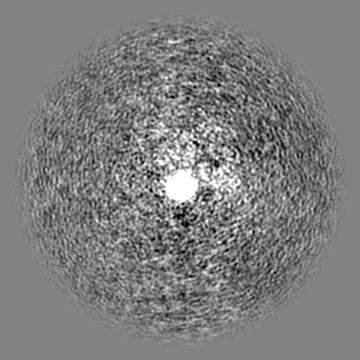
| Projections & slices | Image control
Images are generated by Spider. | ||||||||||||||||||||||||||||||||||||
| Voxel size | X=Y=Z: 1.058 Å | ||||||||||||||||||||||||||||||||||||
| Density |
| ||||||||||||||||||||||||||||||||||||
| Symmetry | Space group: 1 | ||||||||||||||||||||||||||||||||||||
| Details | EMDB XML:
|
 Z (Sec.)
 Y (Row.)
 X (Col.)


-Supplemental data
- Sample components
-Entire : E.coli RNase P holoenzyme with Mg2+
| Entire | Name: E.coli RNase P holoenzyme with Mg2+ |
|---|---|
| Components |
|
-Supramolecule #1: E.coli RNase P holoenzyme with Mg2+
| Supramolecule | Name: E.coli RNase P holoenzyme with Mg2+ / type: complex / ID: 1 / Parent: 0 / Macromolecule list: #1-#3 |
|---|---|
| Source (natural) | Organism: |
-Macromolecule #1: Ribonuclease P protein component
| Macromolecule | Name: Ribonuclease P protein component / type: protein_or_peptide / ID: 1 / Number of copies: 1 / Enantiomer: LEVO / EC number: ribonuclease P |
|---|---|
| Source (natural) | Organism: |
| Molecular weight | Theoretical: 13.818256 KDa |
| Recombinant expression | Organism: |
| Sequence | String: MVKLAFPREL RLLTPSQFTF VFQQPQRAGT PQITILGRLN SLGHPRIGLT VAKKNVRRAH ERNRIKRLTR ESFRLRQHEL PAMDFVVVA KKGVADLDNR ALSEALEKLW RRHCRLARGS UniProtKB: Ribonuclease P protein component |
-Macromolecule #2: RNase P RNA
| Macromolecule | Name: RNase P RNA / type: rna / ID: 2 / Number of copies: 1 |
|---|---|
| Source (natural) | Organism: |
| Molecular weight | Theoretical: 121.191891 KDa |
| Sequence | String: GAAGCUGACC AGACAGUCGC CGCUUCGUCG UCGUCCUCUU CGGGGGAGAC GGGCGGAGGG GAGGAAAGUC CGGGCUCCAU AGGGCAGGG UGCCAGGUAA CGCCUGGGGG GGAAACCCAC GACCAGUGCA ACAGAGAGCA AACCGCCGAU GGCCCGCGCA A GCGGGAUC ...String: GAAGCUGACC AGACAGUCGC CGCUUCGUCG UCGUCCUCUU CGGGGGAGAC GGGCGGAGGG GAGGAAAGUC CGGGCUCCAU AGGGCAGGG UGCCAGGUAA CGCCUGGGGG GGAAACCCAC GACCAGUGCA ACAGAGAGCA AACCGCCGAU GGCCCGCGCA A GCGGGAUC AGGUAAGGGU GAAAGGGUGC GGUAAGAGCG CACCGCGCGG CUGGUAACAG UCCGUGGCAC GGUAAACUCC AC CCGGAGC AAGGCCAAAU AGGGGUUCAU AAGGUACGGC CCGUACUGAA CCCGGGUAGG CUGCUUGAGC CAGUGAGCGA UUG CUGGCC UAGAUGAAUG ACUGUCCACG ACAGAACCCG GCUUAUCGGU CAGUUUC GENBANK: GENBANK: CP053595.1 |
-Macromolecule #3: Precursor tRNA substrate G(-1) G(-2)
| Macromolecule | Name: Precursor tRNA substrate G(-1) G(-2) / type: rna / ID: 3 / Number of copies: 1 |
|---|---|
| Source (natural) | Organism: |
| Molecular weight | Theoretical: 26.336613 KDa |
| Sequence | String: UCUGGGGCUA CGUAGCUCAG UUGGUUAGAG CACAUCACUC AUAAUGAUGG GGUCACAGGU UCGAAUCCCG UCGUAGCCAC CA |
-Macromolecule #4: CALCIUM ION
| Macromolecule | Name: CALCIUM ION / type: ligand / ID: 4 / Number of copies: 7 / Formula: CA |
|---|---|
| Molecular weight | Theoretical: 40.078 Da |
-Experimental details
-Structure determination
| Method | cryo EM |
|---|---|
 Processing Processing | single particle reconstruction |
| Aggregation state | particle |
-Sample preparation
| Buffer | pH: 7.1 |
|---|---|
| Vitrification | Cryogen name: ETHANE |
- Electron microscopy
| Microscope | FEI TITAN KRIOS |
|---|---|
| Image recording | Film or detector model: GATAN K3 BIOQUANTUM (6k x 4k) / Average electron dose: 40.0 e/Å2 |
| Electron beam | Acceleration voltage: 300 kV / Electron source:  FIELD EMISSION GUN FIELD EMISSION GUN |
| Electron optics | Illumination mode: FLOOD BEAM / Imaging mode: BRIGHT FIELD / Nominal defocus max: 3.0 µm / Nominal defocus min: 1.0 µm |
| Experimental equipment |  Model: Titan Krios / Image courtesy: FEI Company |
-Image processing
| Startup model | Type of model: OTHER / Details: ab initio map generated by cryosparc |
|---|---|
| Final reconstruction | Resolution.type: BY AUTHOR / Resolution: 3.4 Å / Resolution method: FSC 0.143 CUT-OFF / Number images used: 546283 |
| Initial angle assignment | Type: MAXIMUM LIKELIHOOD |
| Final angle assignment | Type: MAXIMUM LIKELIHOOD |
