Movie
Movie Controller


+ Open data

- Basic information
| Entry |  | |||||||||
|---|---|---|---|---|---|---|---|---|---|---|
| Title | Cryo-EM Structure of GAPDH | |||||||||
 Map data Map data | ||||||||||
 Sample Sample |
| |||||||||
 Keywords Keywords | GAPDH / glyceraldehyde-3-phosphate dehydrogenase / TRANSFERASE | |||||||||
| Function / homology |  Function and homology information Function and homology informationregulation of neurotransmitter loading into synaptic vesicle / Glycolysis / Gluconeogenesis / peptidyl-cysteine S-trans-nitrosylation / Transferases; Transferring nitrogenous groups; Transferring other nitrogenous groups / extrinsic component of synaptic vesicle membrane / glyceraldehyde-3-phosphate dehydrogenase (phosphorylating) / : / aspartic-type endopeptidase inhibitor activity / glyceraldehyde-3-phosphate dehydrogenase (NAD+) (phosphorylating) activity ...regulation of neurotransmitter loading into synaptic vesicle / Glycolysis / Gluconeogenesis / peptidyl-cysteine S-trans-nitrosylation / Transferases; Transferring nitrogenous groups; Transferring other nitrogenous groups / extrinsic component of synaptic vesicle membrane / glyceraldehyde-3-phosphate dehydrogenase (phosphorylating) / : / aspartic-type endopeptidase inhibitor activity / glyceraldehyde-3-phosphate dehydrogenase (NAD+) (phosphorylating) activity / GAIT complex / peptidyl-cysteine S-nitrosylase activity / defense response to fungus / positive regulation of type I interferon production / lipid droplet / glycolytic process / cellular response to type II interferon / microtubule cytoskeleton organization / glucose metabolic process / NAD binding / disordered domain specific binding / NADP binding / antimicrobial humoral immune response mediated by antimicrobial peptide / microtubule cytoskeleton / neuron apoptotic process / nuclear membrane / microtubule binding / positive regulation of canonical NF-kappaB signal transduction / negative regulation of translation / protein stabilization / ribonucleoprotein complex / identical protein binding / nucleus / plasma membrane / cytoplasm / cytosol Similarity search - Function | |||||||||
| Biological species |  | |||||||||
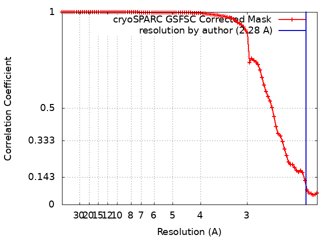
| Method | single particle reconstruction / cryo EM / Resolution: 2.28 Å | |||||||||
 Authors Authors | Morgan CE / Zhang Z / Yu EW | |||||||||
| Funding support |  United States, 1 items United States, 1 items
| |||||||||
 Citation Citation | Journal: Cell Rep / Year: 2022 Title: Toward structural-omics of the bovine retinal pigment epithelium. Authors: Christopher E Morgan / Zhemin Zhang / Masaru Miyagi / Marcin Golczak / Edward W Yu / Abstract: The use of an integrated systems biology approach to investigate tissues and organs has been thought to be impracticable in the field of structural biology, where the techniques mainly focus on ...The use of an integrated systems biology approach to investigate tissues and organs has been thought to be impracticable in the field of structural biology, where the techniques mainly focus on determining the structure of a particular biomacromolecule of interest. Here, we report the use of cryoelectron microscopy (cryo-EM) to define the composition of a raw bovine retinal pigment epithelium (RPE) lysate. From this sample, we simultaneously identify and solve cryo-EM structures of seven different RPE enzymes whose functions affect neurotransmitter recycling, iron metabolism, gluconeogenesis, glycolysis, axonal development, and energy homeostasis. Interestingly, dysfunction of these important proteins has been directly linked to several neurodegenerative disorders, including Huntington's disease, amyotrophic lateral sclerosis (ALS), Parkinson's disease, Alzheimer's disease, and schizophrenia. Our work underscores the importance of cryo-EM in facilitating tissue and organ proteomics at the atomic level. | |||||||||
| History |
|
- Structure visualization
| Supplemental images |
|---|

- Downloads & links
-EMDB archive
| Map data | emd_26355.map.gz | 3.8 MB | EMDB map data format | |
|---|---|---|---|---|
| Header (meta data) | emd-26355-v30.xmlemd-26355.xml | 19.8 KB 19.8 KB | Display Display | EMDB header |
| FSC (resolution estimation) | emd_26355_fsc.xml | 8.3 KB | Display | FSC data file |
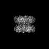
| Images | 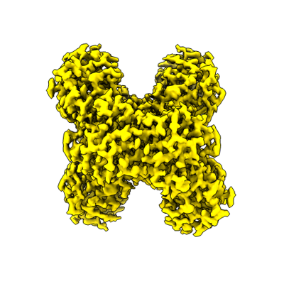 emd_26355.png emd_26355.png | 108.9 KB | ||
| Filedesc metadata | emd-26355.cif.gz | 5.9 KB | ||
| Others | emd_26355_additional_1.map.gzemd_26355_half_map_1.map.gzemd_26355_half_map_2.map.gz | 32.4 MB 59.1 MB 59.1 MB | ||
| Archive directory |  http://ftp.pdbj.org/pub/emdb/structures/EMD-26355ftp://ftp.pdbj.org/pub/emdb/structures/EMD-26355 http://ftp.pdbj.org/pub/emdb/structures/EMD-26355ftp://ftp.pdbj.org/pub/emdb/structures/EMD-26355 | HTTPS FTP |
-Related structure data
| Related structure data |  7u5mMC  7u5hC 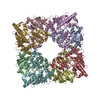 7u5iC 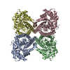 7u5jC 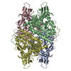 7u5kC 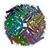 7u5lC 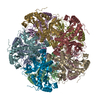 7u5nC M: atomic model generated by this map C: citing same article ( |
|---|---|
| Similar structure data |


-Links
| EMDB pages | EMDB (EBI/PDBe) / EMDataResource |
|---|---|
| Related items in Molecule of the Month |

-Map
| File | Download / File: emd_26355.map.gz / Format: CCP4 / Size: 64 MB / Type: IMAGE STORED AS FLOATING POINT NUMBER (4 BYTES) | ||||||||||||||||||||||||||||||||||||
|---|---|---|---|---|---|---|---|---|---|---|---|---|---|---|---|---|---|---|---|---|---|---|---|---|---|---|---|---|---|---|---|---|---|---|---|---|---|
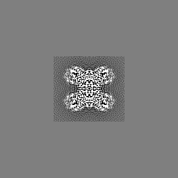
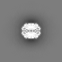
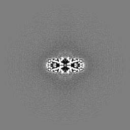
| Projections & slices | Image control
Images are generated by Spider. | ||||||||||||||||||||||||||||||||||||
| Voxel size | X=Y=Z: 1.07 Å | ||||||||||||||||||||||||||||||||||||
| Density |
| ||||||||||||||||||||||||||||||||||||
| Symmetry | Space group: 1 | ||||||||||||||||||||||||||||||||||||
| Details | EMDB XML:
|
 Z (Sec.)
 Y (Row.)
 X (Col.)


-Supplemental data
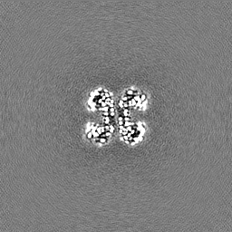
-Additional map: #1
| File | emd_26355_additional_1.map | ||||||||||||
|---|---|---|---|---|---|---|---|---|---|---|---|---|---|
| Projections & Slices |
| ||||||||||||
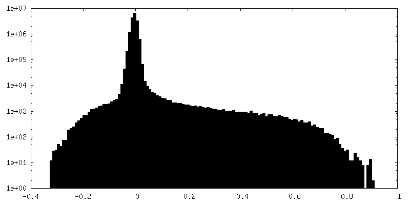
| Density Histograms |


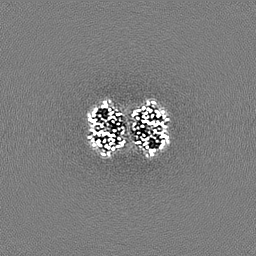
-Half map: #2
| File | emd_26355_half_map_1.map | ||||||||||||
|---|---|---|---|---|---|---|---|---|---|---|---|---|---|
| Projections & Slices |
| ||||||||||||
| Density Histograms |


-Half map: #1
| File | emd_26355_half_map_2.map | ||||||||||||
|---|---|---|---|---|---|---|---|---|---|---|---|---|---|
| Projections & Slices |
| ||||||||||||
| Density Histograms |


- Sample components
-Entire : Tetramer of GAPDH
| Entire | Name: Tetramer of GAPDH |
|---|---|
| Components |
|
-Supramolecule #1: Tetramer of GAPDH
| Supramolecule | Name: Tetramer of GAPDH / type: complex / ID: 1 / Parent: 0 / Macromolecule list: #1 |
|---|---|
| Source (natural) | Organism: |
-Macromolecule #1: Glyceraldehyde-3-phosphate dehydrogenase
| Macromolecule | Name: Glyceraldehyde-3-phosphate dehydrogenase / type: protein_or_peptide / ID: 1 / Number of copies: 4 / Enantiomer: LEVO EC number: glyceraldehyde-3-phosphate dehydrogenase (phosphorylating) |
|---|---|
| Source (natural) | Organism: |
| Molecular weight | Theoretical: 35.914043 KDa |
| Sequence | String: MVKVGVNGFG RIGRLVTRAA FNSGKVDIVA INDPFIDLHY MVYMFQYDST HGKFNGTVKA ENGKLVINGK AITIFQERDP ANIKWGDAG AEYVVESTGV FTTMEKAGAH LKGGAKRVII SAPSADAPMF VMGVNHEKYN NTLKIVSNAS CTTNCLAPLA K VIHDHFGI ...String: MVKVGVNGFG RIGRLVTRAA FNSGKVDIVA INDPFIDLHY MVYMFQYDST HGKFNGTVKA ENGKLVINGK AITIFQERDP ANIKWGDAG AEYVVESTGV FTTMEKAGAH LKGGAKRVII SAPSADAPMF VMGVNHEKYN NTLKIVSNAS CTTNCLAPLA K VIHDHFGI VEGLMTTVHA ITATQKTVDG PSGKLWRDGR GAAQNIIPAS TGAAKAVGKV IPELNGKLTG MAFRVPTPNV SV VDLTCRL EKPAKYDEIK KVVKQASEGP LKGILGYTED QVVSCDFNSD THSSTFDAGA GIALNDHFVK LISWYDNEFG YSN RVVDLM VHMASKE UniProtKB: Glyceraldehyde-3-phosphate dehydrogenase |
-Macromolecule #2: NICOTINAMIDE-ADENINE-DINUCLEOTIDE
| Macromolecule | Name: NICOTINAMIDE-ADENINE-DINUCLEOTIDE / type: ligand / ID: 2 / Number of copies: 4 / Formula: NAD |
|---|---|
| Molecular weight | Theoretical: 663.425 Da |
| Chemical component information |  ChemComp-NAD: |
-Macromolecule #3: water
| Macromolecule | Name: water / type: ligand / ID: 3 / Number of copies: 399 / Formula: HOH |
|---|---|
| Molecular weight | Theoretical: 18.015 Da |
| Chemical component information |  ChemComp-HOH: |
-Experimental details
-Structure determination
| Method | cryo EM |
|---|---|
 Processing Processing | single particle reconstruction |
| Aggregation state | particle |
-Sample preparation
| Buffer | pH: 7.5 |
|---|---|
| Vitrification | Cryogen name: ETHANE |
- Electron microscopy
| Microscope | TFS KRIOS |
|---|---|
| Image recording | Film or detector model: GATAN K3 (6k x 4k) / Average electron dose: 36.0 e/Å2 |
| Electron beam | Acceleration voltage: 300 kV / Electron source:  FIELD EMISSION GUN FIELD EMISSION GUN |
| Electron optics | Illumination mode: SPOT SCAN / Imaging mode: BRIGHT FIELD / Nominal defocus max: 2.5 µm / Nominal defocus min: 1.0 µm |
| Experimental equipment |  Model: Titan Krios / Image courtesy: FEI Company |