+ Open data
- Basic information
| Entry |  | |||||||||
|---|---|---|---|---|---|---|---|---|---|---|
| Title | Cryo-EM Structure of DPYSL2 | |||||||||
 Map data Map data | Density-modified map of DPYSL2 from bos taurus | |||||||||
 Sample Sample |
| |||||||||
 Keywords Keywords | DPYSL2 / dihydropyrimidinase-related protein 2 / NEUROPEPTIDE | |||||||||
| Function / homology |  Function and homology information Function and homology informationCRMPs in Sema3A signaling / hydrolase activity, acting on carbon-nitrogen (but not peptide) bonds, in cyclic amides / cytoskeleton organization / endocytosis / nervous system development / cell differentiation / cytoskeleton / membrane / cytosol Similarity search - Function | |||||||||
| Biological species |  | |||||||||
| Method | single particle reconstruction / cryo EM / Resolution: 2.78 Å | |||||||||
 Authors Authors | Morgan CE / Yu EW | |||||||||
| Funding support |  United States, 1 items United States, 1 items
| |||||||||
 Citation Citation |  Journal: Cell Rep / Year: 2022 Journal: Cell Rep / Year: 2022Title: Toward structural-omics of the bovine retinal pigment epithelium. Authors: Christopher E Morgan / Zhemin Zhang / Masaru Miyagi / Marcin Golczak / Edward W Yu /  Abstract: The use of an integrated systems biology approach to investigate tissues and organs has been thought to be impracticable in the field of structural biology, where the techniques mainly focus on ...The use of an integrated systems biology approach to investigate tissues and organs has been thought to be impracticable in the field of structural biology, where the techniques mainly focus on determining the structure of a particular biomacromolecule of interest. Here, we report the use of cryoelectron microscopy (cryo-EM) to define the composition of a raw bovine retinal pigment epithelium (RPE) lysate. From this sample, we simultaneously identify and solve cryo-EM structures of seven different RPE enzymes whose functions affect neurotransmitter recycling, iron metabolism, gluconeogenesis, glycolysis, axonal development, and energy homeostasis. Interestingly, dysfunction of these important proteins has been directly linked to several neurodegenerative disorders, including Huntington's disease, amyotrophic lateral sclerosis (ALS), Parkinson's disease, Alzheimer's disease, and schizophrenia. Our work underscores the importance of cryo-EM in facilitating tissue and organ proteomics at the atomic level. | |||||||||
| History |
|
- Structure visualization
| Supplemental images |
|---|
- Downloads & links
-EMDB archive
| Map data |  emd_26353.map.gz emd_26353.map.gz | 5.6 MB |  EMDB map data format EMDB map data format | |
|---|---|---|---|---|
| Header (meta data) |  emd-26353-v30.xml emd-26353-v30.xml emd-26353.xml emd-26353.xml | 19.7 KB 19.7 KB | Display Display |  EMDB header EMDB header |
| Images |  emd_26353.png emd_26353.png | 126.5 KB | ||
| Filedesc metadata |  emd-26353.cif.gz emd-26353.cif.gz | 5.9 KB | ||
| Others |  emd_26353_additional_1.map.gz emd_26353_additional_1.map.gz emd_26353_half_map_1.map.gz emd_26353_half_map_1.map.gz emd_26353_half_map_2.map.gz emd_26353_half_map_2.map.gz | 32.3 MB 59.3 MB 59.3 MB | ||
| Archive directory |  http://ftp.pdbj.org/pub/emdb/structures/EMD-26353 http://ftp.pdbj.org/pub/emdb/structures/EMD-26353 ftp://ftp.pdbj.org/pub/emdb/structures/EMD-26353 ftp://ftp.pdbj.org/pub/emdb/structures/EMD-26353 | HTTPS FTP |
-Validation report
| Summary document |  emd_26353_validation.pdf.gz emd_26353_validation.pdf.gz | 857.9 KB | Display |  EMDB validaton report EMDB validaton report |
|---|---|---|---|---|
| Full document |  emd_26353_full_validation.pdf.gz emd_26353_full_validation.pdf.gz | 857.5 KB | Display | |
| Data in XML |  emd_26353_validation.xml.gz emd_26353_validation.xml.gz | 12.5 KB | Display | |
| Data in CIF |  emd_26353_validation.cif.gz emd_26353_validation.cif.gz | 14.8 KB | Display | |
| Arichive directory |  https://ftp.pdbj.org/pub/emdb/validation_reports/EMD-26353 https://ftp.pdbj.org/pub/emdb/validation_reports/EMD-26353 ftp://ftp.pdbj.org/pub/emdb/validation_reports/EMD-26353 ftp://ftp.pdbj.org/pub/emdb/validation_reports/EMD-26353 | HTTPS FTP |
-Related structure data
| Related structure data | 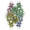 7u5kMC 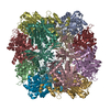 7u5hC  7u5iC 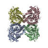 7u5jC 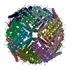 7u5lC 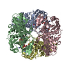 7u5mC 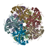 7u5nC M: atomic model generated by this map C: citing same article ( |
|---|---|
| Similar structure data | Similarity search - Function & homology  F&H Search F&H Search |
- Links
Links
| EMDB pages |  EMDB (EBI/PDBe) / EMDB (EBI/PDBe) /  EMDataResource EMDataResource |
|---|---|
| Related items in Molecule of the Month |
- Map
Map
| File |  Download / File: emd_26353.map.gz / Format: CCP4 / Size: 64 MB / Type: IMAGE STORED AS FLOATING POINT NUMBER (4 BYTES) Download / File: emd_26353.map.gz / Format: CCP4 / Size: 64 MB / Type: IMAGE STORED AS FLOATING POINT NUMBER (4 BYTES) | ||||||||||||||||||||||||||||||||||||
|---|---|---|---|---|---|---|---|---|---|---|---|---|---|---|---|---|---|---|---|---|---|---|---|---|---|---|---|---|---|---|---|---|---|---|---|---|---|
| Annotation | Density-modified map of DPYSL2 from bos taurus | ||||||||||||||||||||||||||||||||||||
| Projections & slices | Image control
Images are generated by Spider. | ||||||||||||||||||||||||||||||||||||
| Voxel size | X=Y=Z: 1.07 Å | ||||||||||||||||||||||||||||||||||||
| Density |
| ||||||||||||||||||||||||||||||||||||
| Symmetry | Space group: 1 | ||||||||||||||||||||||||||||||||||||
| Details | EMDB XML:
|
-Supplemental data
-Additional map: Unsharpened map of DPYSL2 from bos taurus
| File | emd_26353_additional_1.map | ||||||||||||
|---|---|---|---|---|---|---|---|---|---|---|---|---|---|
| Annotation | Unsharpened map of DPYSL2 from bos taurus | ||||||||||||
| Projections & Slices |
| ||||||||||||
| Density Histograms |
-Half map: Half-map A
| File | emd_26353_half_map_1.map | ||||||||||||
|---|---|---|---|---|---|---|---|---|---|---|---|---|---|
| Annotation | Half-map A | ||||||||||||
| Projections & Slices |
| ||||||||||||
| Density Histograms |
-Half map: Half-map B
| File | emd_26353_half_map_2.map | ||||||||||||
|---|---|---|---|---|---|---|---|---|---|---|---|---|---|
| Annotation | Half-map B | ||||||||||||
| Projections & Slices |
| ||||||||||||
| Density Histograms |
- Sample components
-Entire : Homo-tetramer of DPYSL2
| Entire | Name: Homo-tetramer of DPYSL2 |
|---|---|
| Components |
|
-Supramolecule #1: Homo-tetramer of DPYSL2
| Supramolecule | Name: Homo-tetramer of DPYSL2 / type: complex / ID: 1 / Parent: 0 / Macromolecule list: #1 |
|---|---|
| Source (natural) | Organism:  |
-Macromolecule #1: Dihydropyrimidinase-related protein 2
| Macromolecule | Name: Dihydropyrimidinase-related protein 2 / type: protein_or_peptide / ID: 1 / Number of copies: 4 / Enantiomer: LEVO |
|---|---|
| Source (natural) | Organism:  |
| Molecular weight | Theoretical: 62.349352 KDa |
| Sequence | String: MSYQGKKNIP RITSDRLLIK GGKIVNDDQS FYADIYMEDG LIKQIGENLI VPGGVKTIEA HSRMVIPGGI DVHTRFQMPD QGMTSADDF FQGTKAALAG GTTMIIDHVV PEPGTSLLAA FDQWREWADS KSCCDYSLHV DITEWHKGVQ EEMEALVKDH G VNSFLVYM ...String: MSYQGKKNIP RITSDRLLIK GGKIVNDDQS FYADIYMEDG LIKQIGENLI VPGGVKTIEA HSRMVIPGGI DVHTRFQMPD QGMTSADDF FQGTKAALAG GTTMIIDHVV PEPGTSLLAA FDQWREWADS KSCCDYSLHV DITEWHKGVQ EEMEALVKDH G VNSFLVYM AFKDRFQLTD SQIYEVLSVI RDIGAIAQVH AENGDIIAEE QQRILDLGIT GPEGHVLSRP EEVEAEAVNR SI TIANQTN CPLYITKVMS KSAAEVIAQA RKKGTVVYGE PITASLGTDG SHYWSKNWAK AAAFVTSPPL SPDPTTPDFL NSL LSCGDL QVTGSAHCTF NTAQKAVGKD NFTLIPEGTN GTEERMSVIW DKAVVTGKMD ENQFVAVTST NAAKVFNLYP RKGR IAVGS DADLVIWDPD SVKTISAKTH NSSLEYNIFE GMECRGSPLV VISQGKIVLE DGTLHVTEGS GRYIPRKPFP DFVYK RIKA RSRLAELRGV PRGLYDGPVC EVSVTPKTVT PASSAKTSPA KQQAPPVRNL HQSGFSLSGA QIDDNIPRRT TQRIVA PPG GRANITSLG UniProtKB: Dihydropyrimidinase-related protein 2 |
-Macromolecule #2: water
| Macromolecule | Name: water / type: ligand / ID: 2 / Number of copies: 148 / Formula: HOH |
|---|---|
| Molecular weight | Theoretical: 18.015 Da |
| Chemical component information |  ChemComp-HOH: |
-Experimental details
-Structure determination
| Method | cryo EM |
|---|---|
 Processing Processing | single particle reconstruction |
| Aggregation state | particle |
- Sample preparation
| Buffer | pH: 7.5 |
|---|---|
| Vitrification | Cryogen name: ETHANE |
- Electron microscopy
| Microscope | TFS KRIOS |
|---|---|
| Image recording | Film or detector model: GATAN K3 (6k x 4k) / Average electron dose: 37.0 e/Å2 |
| Electron beam | Acceleration voltage: 300 kV / Electron source:  FIELD EMISSION GUN FIELD EMISSION GUN |
| Electron optics | Illumination mode: SPOT SCAN / Imaging mode: BRIGHT FIELD / Nominal defocus max: 2.5 µm / Nominal defocus min: 1.0 µm |
| Experimental equipment |  Model: Titan Krios / Image courtesy: FEI Company |
 Movie
Movie Controller


 X (Sec.)
 Y (Row.)
 Z (Col.)


