chromosomal region / telomeric 3' overhang formation / telomere maintenance via telomere trimming / Mre11 complex / establishment of RNA localization to telomere / positive regulation of telomerase catalytic core complex assembly / cellular response to nitrosative stress / negative regulation of telomere capping / BRCA1-C complex / blastocyst growth ...chromosomal region / telomeric 3' overhang formation / telomere maintenance via telomere trimming / Mre11 complex / establishment of RNA localization to telomere / positive regulation of telomerase catalytic core complex assembly / cellular response to nitrosative stress / negative regulation of telomere capping / BRCA1-C complex / blastocyst growth / establishment of protein-containing complex localization to telomere / Sensing of DNA Double Strand Breaks / protection from non-homologous end joining at telomere / peptidyl-serine autophosphorylation / R-loop processing / positive regulation of telomere maintenance via telomere lengthening / meiotic telomere clustering / phosphorylation-dependent protein binding / DNA-dependent protein kinase activity / extrinsic component of synaptic vesicle membrane / pre-B cell allelic exclusion / histone mRNA catabolic process / histone H2AXS139 kinase activity / female meiotic nuclear division / regulation of telomere maintenance via telomerase / male meiotic nuclear division / DNA strand resection involved in replication fork processing / nuclear inclusion body / homologous recombination / lipoprotein catabolic process / t-circle formation / DNA double-strand break processing / regulation of autophagosome assembly / V(D)J recombination / cellular response to X-ray / pexophagy / double-strand break repair via alternative nonhomologous end joining / mitotic G2/M transition checkpoint / isotype switching / Impaired BRCA2 binding to PALB2 / HDR through MMEJ (alt-NHEJ) / protein localization to site of double-strand break / oocyte development / DNA repair complex / chromatin-protein adaptor activity / reciprocal meiotic recombination / regulation of DNA-templated DNA replication initiation / positive regulation of DNA damage response, signal transduction by p53 class mediator / 1-phosphatidylinositol-3-kinase activity / neuromuscular process controlling balance / Homologous DNA Pairing and Strand Exchange / Defective homologous recombination repair (HRR) due to BRCA1 loss of function / Defective HDR through Homologous Recombination Repair (HRR) due to PALB2 loss of BRCA1 binding function / Defective HDR through Homologous Recombination Repair (HRR) due to PALB2 loss of BRCA2/RAD51/RAD51C binding function / Resolution of D-loop Structures through Synthesis-Dependent Strand Annealing (SDSA) / negative regulation of B cell proliferation / TP53 Regulates Transcription of Caspase Activators and Caspases / HDR through Single Strand Annealing (SSA) / Resolution of D-loop Structures through Holliday Junction Intermediates / mitotic spindle assembly checkpoint signaling / positive regulation of double-strand break repair / cellular response to stress / response to ionizing radiation / Impaired BRCA2 binding to RAD51 / mitotic G2 DNA damage checkpoint signaling / TP53 Regulates Transcription of Genes Involved in Cytochrome C Release / peroxisomal matrix / telomere maintenance in response to DNA damage / positive regulation of telomere maintenance / neuroblast proliferation / replicative senescence / Presynaptic phase of homologous DNA pairing and strand exchange / Regulation of HSF1-mediated heat shock response / protein K63-linked ubiquitination / somitogenesis / ovarian follicle development / regulation of cellular response to heat / positive regulation of double-strand break repair via homologous recombination / cellular response to retinoic acid / signal transduction in response to DNA damage / positive regulation of telomere maintenance via telomerase / negative regulation of TORC1 signaling / intrinsic apoptotic signaling pathway / positive regulation of cell adhesion / telomere maintenance / Pexophagy / DNA damage checkpoint signaling / thymus development / regulation of signal transduction by p53 class mediator / replication fork / regulation of autophagy / determination of adult lifespan / protein serine/threonine kinase activator activity / post-embryonic development / meiotic cell cycle / cellular response to reactive oxygen species / TP53 Regulates Transcription of DNA Repair Genes / DNA damage response, signal transduction by p53 class mediator / Stabilization of p53 / Nonhomologous End-Joining (NHEJ) Similarity search - Function
Nibrin, C-terminal / Nibrin / DNA damage repair protein Nbs1 / DNA damage repair protein Nbs1 / Nibrin, second BRCT domain / Nibrin, second BRCT domain superfamily / Second BRCT domain on Nijmegen syndrome breakage protein / Nibrin-related / Telomere-length maintenance and DNA damage repair / Serine/threonine-protein kinase ATM, plant ...Nibrin, C-terminal / Nibrin / DNA damage repair protein Nbs1 / DNA damage repair protein Nbs1 / Nibrin, second BRCT domain / Nibrin, second BRCT domain superfamily / Second BRCT domain on Nijmegen syndrome breakage protein / Nibrin-related / Telomere-length maintenance and DNA damage repair / Serine/threonine-protein kinase ATM, plant / ATM, catalytic domain / Telomere-length maintenance and DNA damage repair / Telomere-length maintenance and DNA damage repair / Forkhead associated domain / Forkhead-associated (FHA) domain profile. / FHA domain / FATC domain / Forkhead-associated (FHA) domain / PIK-related kinase, FAT / FAT domain / FATC / FATC domain / PIK-related kinase / FAT domain profile. / FATC domain profile. / SMAD/FHA domain superfamily / BRCA1 C Terminus (BRCT) domain / Phosphatidylinositol 3- and 4-kinases signature 1. / Phosphatidylinositol 3/4-kinase, conserved site / Phosphatidylinositol 3- and 4-kinases signature 2. / Phosphatidylinositol 3-/4-kinase, catalytic domain superfamily / Phosphoinositide 3-kinase, catalytic domain / Phosphatidylinositol 3- and 4-kinase / Phosphatidylinositol 3- and 4-kinases catalytic domain profile. / Phosphatidylinositol 3-/4-kinase, catalytic domain / BRCT domain / BRCT domain superfamily / Armadillo-type fold / Protein kinase-like domain superfamily Similarity search - Domain/homology
National Institutes of Health/National Cancer Institute (NIH/NCI)
5F32CA247320
United States
National Institutes of Health/Eunice Kennedy Shriver National Institute of Child Health & Human Development (NIH/NICHD)
CA008748
United States
Citation
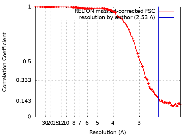
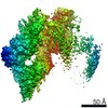
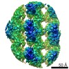
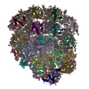
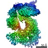
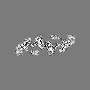
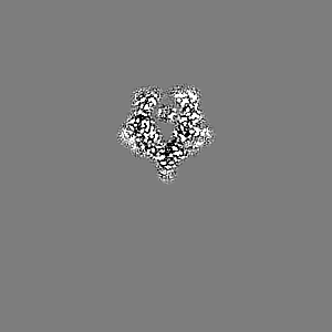
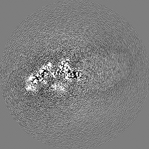
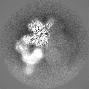
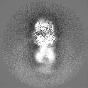
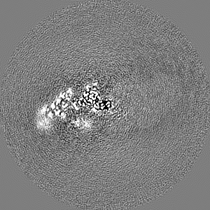
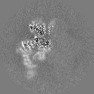
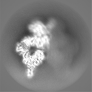
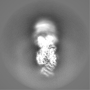
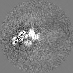
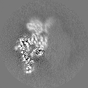
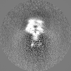
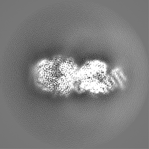
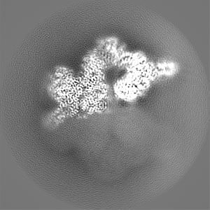
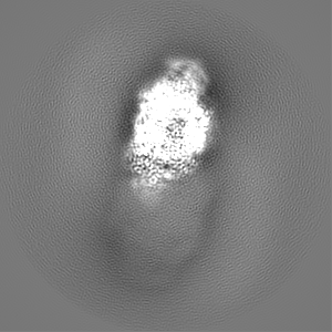
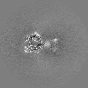
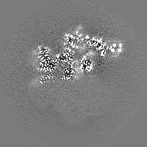
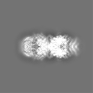
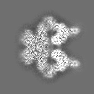
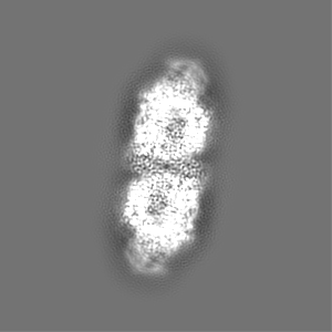
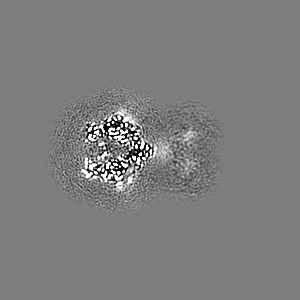
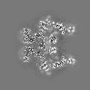
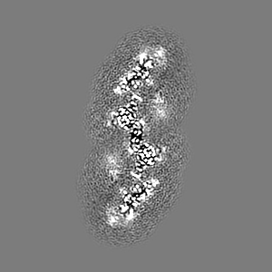
Journal: Elife / Year: 2022 Title: Structure of the human ATM kinase and mechanism of Nbs1 binding. Authors: Christopher Warren / Nikola P Pavletich / Abstract: DNA double-strand breaks (DSBs) can lead to mutations, chromosomal rearrangements, genome instability, and cancer. Central to the sensing of DSBs is the ATM (Ataxia-telangiectasia mutated) kinase, ...DNA double-strand breaks (DSBs) can lead to mutations, chromosomal rearrangements, genome instability, and cancer. Central to the sensing of DSBs is the ATM (Ataxia-telangiectasia mutated) kinase, which belongs to the phosphatidylinositol 3-kinase-related protein kinase (PIKK) family. In response to DSBs, ATM is activated by the MRN (Mre11-Rad50-Nbs1) protein complex through a poorly understood process that also requires double-stranded DNA. Previous studies indicate that the FxF/Y motif of Nbs1 directly binds to ATM, and is required to retain active ATM at sites of DNA damage. Here, we report the 2.5 Å resolution cryo-EM structures of human ATM and its complex with the Nbs1 FxF/Y motif. In keeping with previous structures of ATM and its yeast homolog Tel1, the dimeric human ATM kinase adopts a symmetric, butterfly-shaped structure. The conformation of the ATM kinase domain is most similar to the inactive states of other PIKKs, suggesting that activation may involve an analogous realigning of the N and C lobes along with relieving the blockage of the substrate-binding site. We also show that the Nbs1 FxF/Y motif binds to a conserved hydrophobic cleft within the Spiral domain of ATM, suggesting an allosteric mechanism of activation. We evaluate the importance of these structural findings with mutagenesis and biochemical assays.
History
Deposition
Oct 13, 2021
-
Header (metadata) release
Feb 2, 2022
-
Map release
Feb 2, 2022
-
Update
Jun 5, 2024
-
Current status
Jun 5, 2024
Processing site: RCSB / Status: Released
-
Structure visualization
Movie
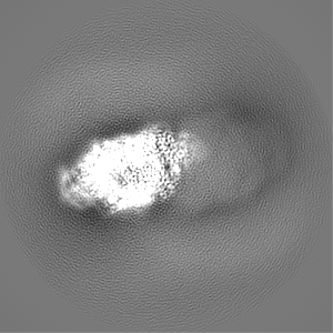
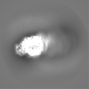
Surface view with section colored by density value
In the structure databanks used in Yorodumi, some data are registered as the other names, "COVID-19 virus" and "2019-nCoV". Here are the details of the virus and the list of structure data.
Jan 31, 2019. EMDB accession codes are about to change! (news from PDBe EMDB page)
EMDB accession codes are about to change! (news from PDBe EMDB page)
The allocation of 4 digits for EMDB accession codes will soon come to an end. Whilst these codes will remain in use, new EMDB accession codes will include an additional digit and will expand incrementally as the available range of codes is exhausted. The current 4-digit format prefixed with “EMD-” (i.e. EMD-XXXX) will advance to a 5-digit format (i.e. EMD-XXXXX), and so on. It is currently estimated that the 4-digit codes will be depleted around Spring 2019, at which point the 5-digit format will come into force.
The EM Navigator/Yorodumi systems omit the EMD- prefix.
Related info.:Q: What is EMD? / ID/Accession-code notation in Yorodumi/EM Navigator
Yorodumi is a browser for structure data from EMDB, PDB, SASBDB, etc.
This page is also the successor to EM Navigator detail page, and also detail information page/front-end page for Omokage search.
The word "yorodu" (or yorozu) is an old Japanese word meaning "ten thousand". "mi" (miru) is to see.
Related info.:EMDB / PDB / SASBDB / Comparison of 3 databanks / Yorodumi Search / Aug 31, 2016. New EM Navigator & Yorodumi / Yorodumi Papers / Jmol/JSmol / Function and homology information / Changes in new EM Navigator and Yorodumi
 Movie
Movie Controller


 Open data
 Basic information
 Map data
 Sample
 Keywords
 Function and homology information
 Homo sapiens (human)
 Authors
 United States, 2 items
  Citation
 Structure visualization
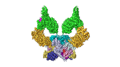
 Downloads & links
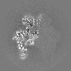
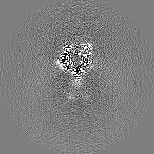
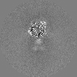
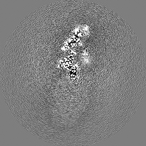
 emd_25141.png
 http://ftp.pdbj.org/pub/emdb/structures/EMD-25141


 Z (Sec.)
 Y (Row.)
 X (Col.)


 Sample components
 Processing
 Electron microscopy
 FIELD EMISSION GUN