 Movie
Movie Controller

[English] 日本語
 Yorodumi
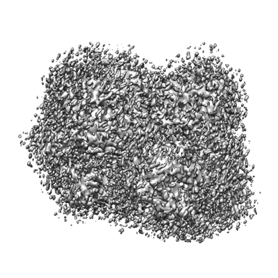
- EMDB-25037: Cryo-EM 3D map of the human Exostosin-1 and Exostosin-2 heterodim... -
+ Open data

- Basic information
| Entry |  | |||||||||
|---|---|---|---|---|---|---|---|---|---|---|
| Title | Cryo-EM 3D map of the human Exostosin-1 and Exostosin-2 heterodimer in complex with a 7-sugar oligosaccharide acceptor analog | |||||||||
 Map data Map data | ||||||||||
 Sample Sample |
| |||||||||
 Keywords Keywords | exostosin1 / exostosin2 / glycosyltransferase / heparan sulfate / acceptor / TRANSFERASE | |||||||||
| Function / homology |  Function and homology information Function and homology informationglucuronosyl-N-acetylglucosaminyl-proteoglycan 4-alpha-N-acetylglucosaminyltransferase / N-acetylglucosaminyl-proteoglycan 4-beta-glucuronosyltransferase / hypersensitivity / heart field specification / lymphocyte adhesion to endothelial cell of high endothelial venule / heparan sulfate N-acetylglucosaminyltransferase activity / glucuronosyl-N-acetylglucosaminyl-proteoglycan 4-alpha-N-acetylglucosaminyltransferase activity / N-acetylglucosaminyl-proteoglycan 4-beta-glucuronosyltransferase activity / chondroitin sulfate proteoglycan metabolic process / smoothened signaling pathway involved in lung development ...glucuronosyl-N-acetylglucosaminyl-proteoglycan 4-alpha-N-acetylglucosaminyltransferase / N-acetylglucosaminyl-proteoglycan 4-beta-glucuronosyltransferase / hypersensitivity / heart field specification / lymphocyte adhesion to endothelial cell of high endothelial venule / heparan sulfate N-acetylglucosaminyltransferase activity / glucuronosyl-N-acetylglucosaminyl-proteoglycan 4-alpha-N-acetylglucosaminyltransferase activity / N-acetylglucosaminyl-proteoglycan 4-beta-glucuronosyltransferase activity / chondroitin sulfate proteoglycan metabolic process / smoothened signaling pathway involved in lung development / developmental growth involved in morphogenesis / sweat gland development / perichondral bone morphogenesis / mesenchymal cell differentiation involved in bone development / response to leukemia inhibitory factor / UDP-N-acetylglucosamine transferase complex / chondrocyte hypertrophy / fluid transport / hematopoietic stem cell migration to bone marrow / chondrocyte differentiation involved in endochondral bone morphogenesis / glucuronosyltransferase activity / heparin proteoglycan biosynthetic process / tight junction organization / Defective EXT2 causes exostoses 2 / Defective EXT1 causes exostoses 1, TRPS2 and CHDS / sebaceous gland development / glomerular basement membrane development / response to heparin / stomach development / glandular epithelial cell differentiation / lymphocyte migration into lymphoid organs / glycosaminoglycan biosynthetic process / HS-GAG biosynthesis / sulfation / embryonic skeletal limb joint morphogenesis / chondrocyte proliferation / endochondral bone morphogenesis / acetylglucosaminyltransferase activity / dendritic cell migration / heparan sulfate proteoglycan biosynthetic process / dendrite self-avoidance / podocyte differentiation / endochondral bone growth / hematopoietic stem cell homeostasis / sodium ion homeostasis / limb bud formation / basement membrane organization / bone trabecula morphogenesis / olfactory bulb development / endochondral ossification / vacuole organization / multicellular organismal-level water homeostasis / cranial skeletal system development / endoderm development / leukocyte tethering or rolling / response to light intensity / vocalization behavior / fear response / protein N-linked glycosylation / polysaccharide biosynthetic process / regulation of tumor necrosis factor-mediated signaling pathway / mesodermal cell differentiation / optic nerve development / collagen fibril organization / cell adhesion mediated by integrin / ossification involved in bone maturation / hair follicle morphogenesis / stem cell division / neural crest cell differentiation / antigen processing and presentation / heart contraction / glycosyltransferase activity / epithelial tube branching involved in lung morphogenesis / motor behavior / outflow tract morphogenesis / mesoderm formation / fibroblast growth factor receptor signaling pathway / chondrocyte differentiation / cell fate commitment / blood vessel remodeling / bone resorption / hematopoietic stem cell differentiation / canonical Wnt signaling pathway / BMP signaling pathway / social behavior / catalytic complex / ERK1 and ERK2 cascade / cellular response to fibroblast growth factor stimulus / ossification / axon guidance / protein catabolic process / wound healing / synaptic transmission, glutamatergic / cellular response to virus / regulation of blood pressure / multicellular organism growth / vasodilation / protein-containing complex assembly / gene expression / protein heterodimerization activity Similarity search - Function | |||||||||
| Biological species |  Homo sapiens (human) Homo sapiens (human) | |||||||||
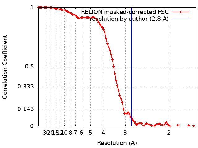
| Method | single particle reconstruction / cryo EM / Resolution: 2.8 Å | |||||||||
 Authors Authors | Li H | |||||||||
| Funding support |  United States, 1 items United States, 1 items
| |||||||||
 Citation Citation | Journal: Nat Chem Biol / Year: 2023 Title: Structural basis for heparan sulfate co-polymerase action by the EXT1-2 complex. Authors: Hua Li / Digantkumar Chapla / Robert A Amos / Annapoorani Ramiah / Kelley W Moremen / Huilin Li / Abstract: Heparan sulfate (HS) proteoglycans are extended (-GlcAβ1,4GlcNAcα1,4-) co-polymers containing decorations of sulfation and epimerization that are linked to cell surface and extracellular matrix ...Heparan sulfate (HS) proteoglycans are extended (-GlcAβ1,4GlcNAcα1,4-) co-polymers containing decorations of sulfation and epimerization that are linked to cell surface and extracellular matrix proteins. In mammals, HS repeat units are extended by an obligate heterocomplex of two exostosin family members, EXT1 and EXT2, where each protein monomer contains distinct GT47 (GT-B fold) and GT64 (GT-A fold) glycosyltransferase domains. In this study, we generated human EXT1-EXT2 (EXT1-2) as a functional heterocomplex and determined its structure in the presence of bound donor and acceptor substrates. Structural data and enzyme activity of catalytic site mutants demonstrate that only two of the four glycosyltransferase domains are major contributors to co-polymer syntheses: the EXT1 GT-B fold β1,4GlcA transferase domain and the EXT2 GT-A fold α1,4GlcNAc transferase domain. The two catalytic sites are over 90 Å apart, indicating that HS is synthesized by a dissociative process that involves a single catalytic site on each monomer. | |||||||||
| History |
|
- Structure visualization
| Supplemental images |
|---|
- Downloads & links
-EMDB archive
| Map data | emd_25037.map.gz | 56.8 MB | EMDB map data format | |
|---|---|---|---|---|
| Header (meta data) | emd-25037-v30.xmlemd-25037.xml | 22 KB 22 KB | Display Display | EMDB header |
| FSC (resolution estimation) | emd_25037_fsc.xml | 9.2 KB | Display | FSC data file |
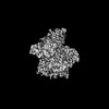
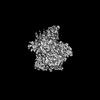
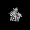
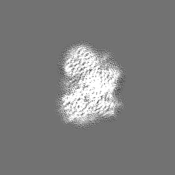
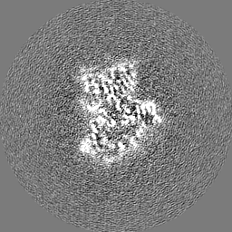
| Images |  emd_25037.png emd_25037.png | 162.6 KB | ||
| Masks | emd_25037_msk_1.map | 64 MB | Mask map | |
| Filedesc metadata | emd-25037.cif.gz | 7 KB | ||
| Others | emd_25037_additional_1.map.gzemd_25037_half_map_1.map.gzemd_25037_half_map_2.map.gz | 6.4 MB 49.7 MB 49.7 MB | ||
| Archive directory |  http://ftp.pdbj.org/pub/emdb/structures/EMD-25037ftp://ftp.pdbj.org/pub/emdb/structures/EMD-25037 http://ftp.pdbj.org/pub/emdb/structures/EMD-25037ftp://ftp.pdbj.org/pub/emdb/structures/EMD-25037 | HTTPS FTP |
-Related structure data
| Related structure data | 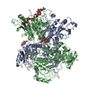 7sckMC 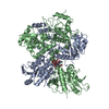 7schC 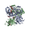 7scjC  7uqxC 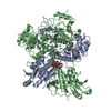 7uqyC M: atomic model generated by this map C: citing same article ( |
|---|---|
| Similar structure data |

-Links
| EMDB pages | EMDB (EBI/PDBe) / EMDataResource |
|---|---|
| Related items in Molecule of the Month |


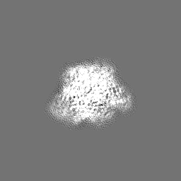
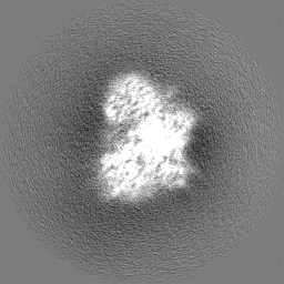
-Map
| File | Download / File: emd_25037.map.gz / Format: CCP4 / Size: 64 MB / Type: IMAGE STORED AS FLOATING POINT NUMBER (4 BYTES) | ||||||||||||||||||||||||||||||||||||
|---|---|---|---|---|---|---|---|---|---|---|---|---|---|---|---|---|---|---|---|---|---|---|---|---|---|---|---|---|---|---|---|---|---|---|---|---|---|
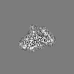
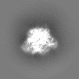
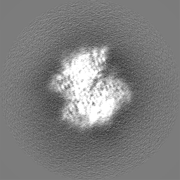
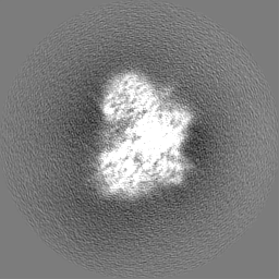
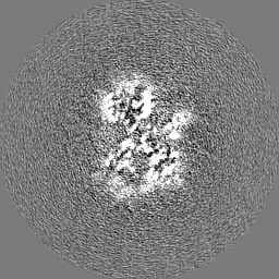
| Projections & slices | Image control
Images are generated by Spider. | ||||||||||||||||||||||||||||||||||||
| Voxel size | X=Y=Z: 0.828 Å | ||||||||||||||||||||||||||||||||||||
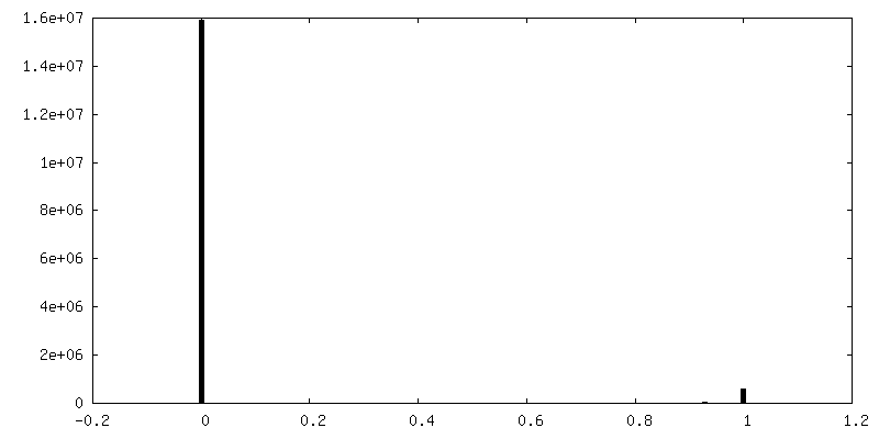
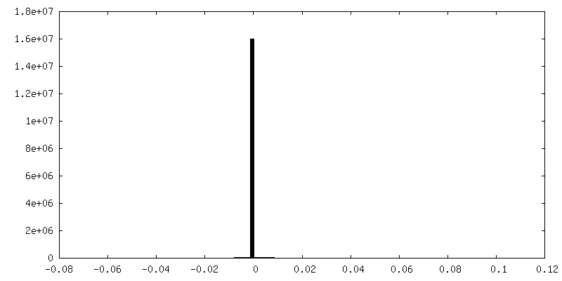
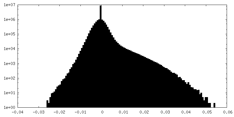
| Density |
| ||||||||||||||||||||||||||||||||||||
| Symmetry | Space group: 1 | ||||||||||||||||||||||||||||||||||||
| Details | EMDB XML:
|
 Z (Sec.)
 Y (Row.)
 X (Col.)


-Supplemental data
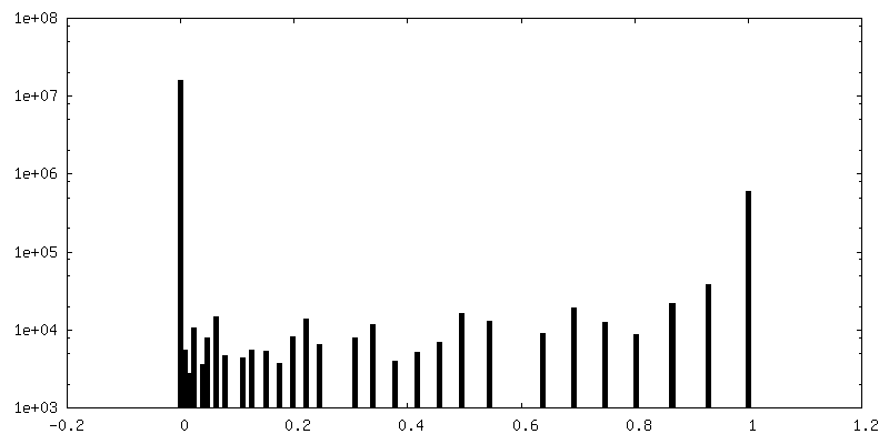
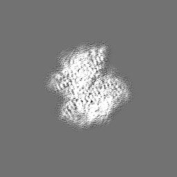
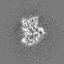
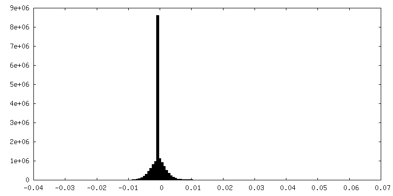
-Mask #1
| File | emd_25037_msk_1.map | ||||||||||||
|---|---|---|---|---|---|---|---|---|---|---|---|---|---|
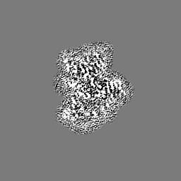
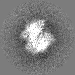
| Projections & Slices |
| ||||||||||||
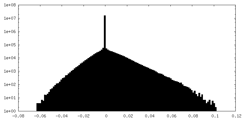
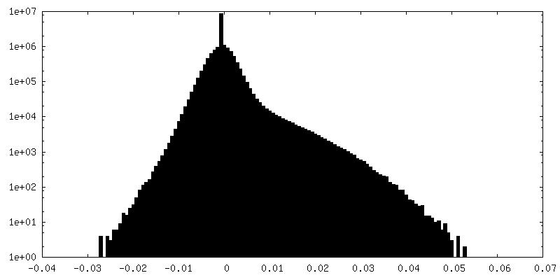
| Density Histograms |


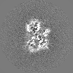
-Additional map: #1
| File | emd_25037_additional_1.map | ||||||||||||
|---|---|---|---|---|---|---|---|---|---|---|---|---|---|
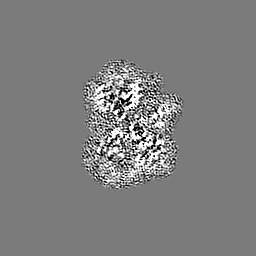
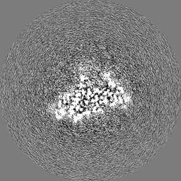
| Projections & Slices |
| ||||||||||||
| Density Histograms |
-Half map: #2
| File | emd_25037_half_map_1.map | ||||||||||||
|---|---|---|---|---|---|---|---|---|---|---|---|---|---|
| Projections & Slices |
| ||||||||||||
| Density Histograms |

-Half map: #1
| File | emd_25037_half_map_2.map | ||||||||||||
|---|---|---|---|---|---|---|---|---|---|---|---|---|---|
| Projections & Slices |
| ||||||||||||
| Density Histograms |


- Sample components
-Entire : human EXT1/2
| Entire | Name: human EXT1/2 |
|---|---|
| Components |
|
-Supramolecule #1: human EXT1/2
| Supramolecule | Name: human EXT1/2 / type: complex / ID: 1 / Parent: 0 / Macromolecule list: #1-#2 |
|---|---|
| Source (natural) | Organism: Homo sapiens (human) |
-Macromolecule #1: Exostosin-1
| Macromolecule | Name: Exostosin-1 / type: protein_or_peptide / ID: 1 / Number of copies: 1 / Enantiomer: LEVO EC number: glucuronosyl-N-acetylglucosaminyl-proteoglycan 4-alpha-N-acetylglucosaminyltransferase |
|---|---|
| Source (natural) | Organism: Homo sapiens (human) |
| Molecular weight | Theoretical: 83.404023 KDa |
| Recombinant expression | Organism: Homo sapiens (human) |
| Sequence | String: GFRASRSHSR REEHSGRNGL HHPSPDHFWP RFPDALRPFV PWDQLENEDS SVHISPRQKR DANSSIYKGK KCRMESCFDF TLCKKNGFK VYVYPQQKGE KIAESYQNIL AAIEGSRFYT SDPSQACLFV LSLDTLDRDQ LSPQYVHNLR SKVQSLHLWN N GRNHLIFN ...String: GFRASRSHSR REEHSGRNGL HHPSPDHFWP RFPDALRPFV PWDQLENEDS SVHISPRQKR DANSSIYKGK KCRMESCFDF TLCKKNGFK VYVYPQQKGE KIAESYQNIL AAIEGSRFYT SDPSQACLFV LSLDTLDRDQ LSPQYVHNLR SKVQSLHLWN N GRNHLIFN LYSGTWPDYT EDVGFDIGQA MLAKASISTE NFRPNFDVSI PLFSKDHPRT GGERGFLKFN TIPPLRKYML VF KGKRYLT GIGSDTRNAL YHVHNGEDVV LLTTCKHGKD WQKHKDSRCD RDNTEYEKYD YREMLHNATF CLVPRGRRLG SFR FLEALQ AACVPVMLSN GWELPFSEVI NWNQAAVIGD ERLLLQIPST IRSIHQDKIL ALRQQTQFLW EAYFSSVEKI VLTT LEIIQ DRIFKHISRN SLIWNKHPGG LFVLPQYSSY LGDFPYYYAN LGLKPPSKFT AVIHAVTPLV SQSQPVLKLL VAAAK SQYC AQIIVLWNCD KPLPAKHRWP ATAVPVVVIE GESKVMSSRF LPYDNIITDA VLSLDEDTVL STTEVDFAFT VWQSFP ERI VGYPARSHFW DNSKERWGYT SKWTNDYSMV LTGAAIYHKY YHYLYSHYLP ASLKNMVDQL ANCEDILMNF LVSAVTK LP PIKVTQKKQY KETMMGQTSR ASRWADPDHF AQRQSCMNTF ASWFGYMPLI HSQMRLDPVL FKDQVSILRK KYRDIERL UniProtKB: Exostosin-1 |
-Macromolecule #2: Exostosin-2
| Macromolecule | Name: Exostosin-2 / type: protein_or_peptide / ID: 2 / Number of copies: 1 / Enantiomer: LEVO EC number: glucuronosyl-N-acetylglucosaminyl-proteoglycan 4-alpha-N-acetylglucosaminyltransferase |
|---|---|
| Source (natural) | Organism: Homo sapiens (human) |
| Molecular weight | Theoretical: 77.238031 KDa |
| Recombinant expression | Organism: Homo sapiens (human) |
| Sequence | String: GWPHSIESSN DWNVEKRSIR DVPVVRLPAD SPIPERGDLS CRMHTCFDVY RCGFNPKNKI KVYIYALKKY VDDFGVSVSN TISREYNEL LMAISDSDYY TDDINRACLF VPSIDVLNQN TLRIKETAQA MAQLSRWDRG TNHLLFNMLP GGPPDYNTAL D VPRDRALL ...String: GWPHSIESSN DWNVEKRSIR DVPVVRLPAD SPIPERGDLS CRMHTCFDVY RCGFNPKNKI KVYIYALKKY VDDFGVSVSN TISREYNEL LMAISDSDYY TDDINRACLF VPSIDVLNQN TLRIKETAQA MAQLSRWDRG TNHLLFNMLP GGPPDYNTAL D VPRDRALL AGGGFSTWTY RQGYDVSIPV YSPLSAEVDL PEKGPGPRQY FLLSSQVGLH PEYREDLEAL QVKHGESVLV LD KCTNLSE GVLSVRKRCH KHQVFDYPQV LQEATFCVVL RGARLGQAVL SDVLQAGCVP VVIADSYILP FSEVLDWKRA SVV VPEEKM SDVYSILQSI PQRQIEEMQR QARWFWEAYF QSIKAIALAT LQIINDRIYP YAAISYEEWN DPPAVKWGSV SNPL FLPLI PPQSQGFTAI VLTYDRVESL FRVITEVSKV PSLSKLLVVW NNQNKNPPED SLWPKIRVPL KVVRTAENKL SNRFF PYDE IETEAVLAID DDIIMLTSDE LQFGYEVWRE FPDRLVGYPG RLHLWDHEMN KWKYESEWTN EVSMVLTGAA FYHKYF NYL YTYKMPGDIK NWVDAHMNCE DIAMNFLVAN VTGKAVIKVT PRKKFKCPEC TAIDGLSLDQ THMVERSECI NKFASVF GT MPLKVVEHRA DPVLYKDDFP EKLKSFPNIG SL UniProtKB: Exostosin-2 |
-Macromolecule #5: URIDINE-5'-DIPHOSPHATE
| Macromolecule | Name: URIDINE-5'-DIPHOSPHATE / type: ligand / ID: 5 / Number of copies: 2 / Formula: UDP |
|---|---|
| Molecular weight | Theoretical: 404.161 Da |
| Chemical component information |  ChemComp-UDP: |
-Macromolecule #6: 2-acetamido-2-deoxy-beta-D-glucopyranose
| Macromolecule | Name: 2-acetamido-2-deoxy-beta-D-glucopyranose / type: ligand / ID: 6 / Number of copies: 1 / Formula: NAG |
|---|---|
| Molecular weight | Theoretical: 221.208 Da |
| Chemical component information |  ChemComp-NAG: |
-Macromolecule #7: MANGANESE (II) ION
| Macromolecule | Name: MANGANESE (II) ION / type: ligand / ID: 7 / Number of copies: 1 / Formula: MN |
|---|---|
| Molecular weight | Theoretical: 54.938 Da |
-Experimental details
-Structure determination
| Method | cryo EM |
|---|---|
 Processing Processing | single particle reconstruction |
| Aggregation state | particle |
-Sample preparation
| Concentration | 0.3 mg/mL | |||||||||
|---|---|---|---|---|---|---|---|---|---|---|
| Buffer | pH: 7 Component:
| |||||||||
| Grid | Model: Quantifoil R2/1 / Material: GOLD / Mesh: 300 / Support film - Material: CARBON / Support film - topology: HOLEY | |||||||||
| Vitrification | Cryogen name: ETHANE / Chamber humidity: 95 % / Chamber temperature: 299 K / Instrument: FEI VITROBOT MARK IV |
- Electron microscopy
| Microscope | FEI TITAN KRIOS |
|---|---|
| Temperature | Min: 193.0 K / Max: 193.0 K |
| Alignment procedure | Coma free - Residual tilt: 0.05 mrad |
| Image recording | #0 - Image recording ID: 1 / #0 - Film or detector model: GATAN K3 (6k x 4k) / #0 - Number grids imaged: 1 / #0 - Number real images: 5108 / #0 - Average exposure time: 1.5 sec. / #0 - Average electron dose: 68.0 e/Å2 / #1 - Image recording ID: 2 / #1 - Film or detector model: GATAN K3 (6k x 4k) / #1 - Digitization - Dimensions - Width: 5760 pixel / #1 - Digitization - Dimensions - Height: 4092 pixel / #1 - Number grids imaged: 1 / #1 - Number real images: 7609 / #1 - Average exposure time: 1.5 sec. / #1 - Average electron dose: 72.0 e/Å2 |
| Electron beam | Acceleration voltage: 300 kV / Electron source:  FIELD EMISSION GUN FIELD EMISSION GUN |
| Electron optics | C2 aperture diameter: 70.0 µm / Illumination mode: OTHER / Imaging mode: BRIGHT FIELD / Cs: 2.7 mm / Nominal defocus max: 1.8 µm / Nominal defocus min: 0.9 µm / Nominal magnification: 105000 |
| Sample stage | Specimen holder model: FEI TITAN KRIOS AUTOGRID HOLDER / Cooling holder cryogen: NITROGEN |
| Experimental equipment |  Model: Titan Krios / Image courtesy: FEI Company |
+Image processing
-Atomic model buiding 1
| Refinement | Protocol: AB INITIO MODEL |
|---|---|
| Output model | PDB-7sck: |
